Antiphospholipid antibodies in critically ill patients
INTRODUCTION
Antiphospholipid antibodies (aPL) are a heterogeneous group of autoantibodies that work in against membrane phospholipids or antiphospholipid-binding proteins. he presence of a pathogenic aPL, such as anticardiolipin (aCL), lupus anticoagulant (LAC) or anti-β2GLP I (aβ2GLP I), is indicative of antiphospholipid antibody syndrome (APS), which is responsible for an increased risk of arterial, venous and microvascular thrombosis.(1-4)
he mechanism of aPL-mediated thrombosis is not completely understood,; however, because the presence of persistent or transient antibodies does not always generate thrombosis, additional risk factors, also called “second or multiple hits”, are required to initiate the thrombogenic process.(3,5) In patients with disseminated thrombosis, multiple organ dysfunction and circulating aPL triggering events can be identiied in up to 60% of cases, of which severe infections are the most common.(6-8)
Most patients with severe acute illness have activated coagulation systems,
resulting in thrombin and ibrin microvascular deposition.(9) his, in turn,
leads to poor tissue perfusion, increasing tissue damage and perpetuation of
Juliana Vassalo1, Nelson Spector1,2, Ernesto de Meis3,
Márcio Soares1,4,5, Jorge Ibrain Figueira Salluh1,4,5
1. Postgraduate Program, Universidade Federal do Rio de Janeiro - Rio de Janeiro (RJ), Brazil. 2. School of Medicine, Universidade Federal do Rio de Janeiro - Rio de Janeiro (RJ), Brazil. 3. Hospital do Câncer, Instituto Nacional de Câncer - Rio de Janeiro (RJ), Brazil.
4. Postgraduate Program, Instituto Nacional de Câncer - Rio de Janeiro (RJ), Brazil.
5. Instituto D’Or de Pesquisa e Ensino - Rio de Janeiro (RJ), Brazil.
Antiphospholipid antibodies are responsible for a wide spectrum of clinical manifestations. Venous, arterial and microvascular thrombosis and severe catastrophic cases account for a large morbidly/mortality. hrough the connection between the immune, inlammatory and hemostatic systems, it is possible that these antibodies may contribute to the development of organ dysfunction and are associated with poor short and long-term prognoses in critically ill patients. We performed a search of the PubMed/MedLine database for articles written during the
Conflicts of interest: None.
Submitted on March 17, 2014 Accepted on April 18, 2014
Corresponding author:
Juliana Vassalo
Hospital Universitário Clementino Fraga Filho Universidade Federal do Rio de Janeiro Rua Professor Rodolpho Paulo Rocco, 255 - Ilha do Fundão
Zip code: 21941-913 - Rio de Janeiro (RJ), Brazil E-mail: [email protected]
Anticorpos antifosfolipídeos em pacientes gravemente enfermos
ABSTRACT
Keywords: Antibodies, antiphospholipid; Prognosis; Critical illness; Catastrophic illness; Multiple organ failure; Antiphospholipid syndrome; Intensive care units
period from January 2000 to February 2013 to evaluate the frequency of antiphospholipid antibodies in critically ill patients and their impact on the outcomes of these patients. Only eight original studies involving critically ill patients were found. However, the development of antiphospholipid antibodies in critically ill patients seems to be frequent, but more studies are necessary to clarify their pathogenic role and implications for clinical practice.
the pro-inlammatory and pro-thrombotic cycle. he presence of aPL can further feed into this cycle and can be a link in the complex connection between inlammation, coagulation and immune response.
However, the role of these antibodies in the clinical course and the prognosis of critically ill patients is yet to be clariied.
In the present article, we present a narrative review to describe the frequency of aPL in critically ill patients and their impact on the outcomes of these patients.
METHODS
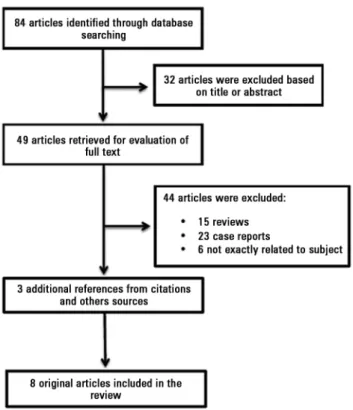
We performed a search of the PubMed/MedLine database for articles written from January 2000 until February 2013 with the following terms: antiphospholipid antibodies, ‘lupus anticoagulant’, ‘anticardiolipin antibody’, ‘anti beta 2 glycoprotein I’, ‘critical illness’, ‘ICU’, ‘sepsis’ and ‘multiple organ failure’. We also reviewed the references of available studies for other potentially eligible studies, and additional published reports were identiied through a manual search of citations from the retrieved articles (Figure 1).
RESULTS
he research resulted in 49 potentially relevant references, most of which consisted of case reports of catastrophic thrombotic events associated with circulating aPL and review articles not speciically about this issue. Of these studies, in addition to three other studies from additional search sources, only eight original studies involving critically ill patients were found. he main characteristics of these studies are summarized in table 1.
In critical illness, we can observe three main types of clinical situations involving aPL. he most frequently cited presentation in the literature is the catastrophic APS, a type of APS that can cause multiple organ dysfunction and therefore requires life sustaining therapies and critical care. However, there are two other important scenarios in clinical practice. Antiphospholipid antibody positive patients with or without APS may require intensive care outside of the context of catastrophic illness.
Additionally, it is reasonable to assume that critical patients are at risk of developing aPL and that such antibodies can contribute to the development of thrombosis and organ dysfunction, afecting the course and outcome of these patients.
Catastrophic antiphospholipid syndrome
his variant of the APS was irst described in 1992 by Asherson(18) and received the eponym “Asherson Syndrome” in 2003. It accounts for <1% of APS cases; however, its severe nature brings attention to the current topic.
In 2000, an international registry of catastrophic antiphospholipid syndrome - CAPS (the CAPS registry: http://www.med.ub.es/MIMMUN/FORUM/ CAPS.HTM) was created by the European Forum on Antiphospholipid Antibodies. Currently, 280 cases have been reported worldwide. Because APS occurs infrequently and possibly because it is under diagnosed, there are no large multicenter studies in the literature, and knowledge on the subject comes mainly from observational studies. Table 2 summarizes the main features of these patients.
he syndrome is characterized by rapid thrombotic involvement (at least one week) of three or more organs associated with the presence of aPL (Table 3).(19) Unlike the classic APS, thrombotic manifestations mainly afect small vessels, and microthromboses are found in 89% of autopsies.(20)
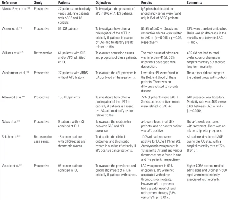
Table 1 - Main study characteristics
Reference Study Patients Objectives Results Comments
Maneta-Peyret et al.(10) Prospective 27 patients mechanically
ventilated, nine patients with ARDS and 18 controls
To investigate the presence of aPL in BAL of ARDS patients.
IgG phosphatidic acid and phosphatidylserine were found only in BAL of ARDS patients.
Wenzel et al.(11) Prospective 51 ICU patients To investigate how often a
prolongation of the aPTT in critically ill patients is caused by LAC and to identify events related to this.
52.9% of LAC +. Sepsis and vasoactive amines were related to LAC + (p=0.006 e p=0.03, respectively).
63% were transient antibodies. There was no difference in the mortality rate between LAC + and -.
Williams et al.(12) Retrospective 61 patients with SLE
and/or APS admitted at ICU
To evaluate admission causes and prognosis of these patients.
The main cause of admission was infection (41%). 58% of patients developed renal dysfunction.
APS did not lead to renal dysfunction or changes in hospital mortality but reduced long-term mortality.
Wiedermann et al.(13) Prospective 27 patients with ARDS
without APS history
To evaluate the aPL presence in BAL or blood of these patients.
Low titles aPL were found in the BAL and blood of these patients. There was no difference related to severity disease.
The authors did not compare the patient group with controls
Aldawood et al.(14) Prospective 155 ICU patients To investigate how often a
prolongation of the aPTT in critically ill patients is caused by LAC and to identify events related to this.
77% of patients were LAC +. Sepsis and vasoactive amines were related to LAC +.
LAC presence was transitory. Mortality rate was 46% versus 5.6% between LAC + and - (p=0.0004)
Nakos et al.(15) Prospective 9 patients with GBS
admitted at ICU
To evaluate the relationship between GBS and aPL presence.
aPL were found in all GBS patients, and no control patient was aPL positive.
The aPL levels decreased with treatment. There was no relationship with prognosis.
Salluh et al.(16) Retrospective
case series
18 cancer patients with SIRS/sepsis and thrombotic events
To describe the clinical outcomes and thrombotic events in a series of critically ill aPL positive cancer patients.
100% of patients were positive for LAC e 11% for aCL. Acrocyanosis was present in 18 patients. Arterial and venous thromboses were found in nine and five patients, respectively.
All patients developed MOF during the ICU stay, with a hospital mortality rate of 72% (13/18).
Vassalo et al.(17) Prospective 95 cancer patients
admitted in ICU
To evaluate the prevalence and prognostic impact of aPL in critically ill patients with cancer.
LAC was present in 61% of patients. aPL were not associated with either thrombosis or mortality. However, aPL + patients had a greater need of renal replacement therapy (33% versus 8%, p=0.017).
Higher SOFA scores, medical admissions and D-dimer >500 ng/dl were independently associated with mortality.
ARDS - acute respiratory distress syndrome; aPL - antiphospholipid antibodies; BAL - bronchoalveolar lavage; IgG - immunoglobulin G; ICU - intensive care unit; aPTT - activated partial thromboplastin time; LAC - lupus anticoagulant; SLE - systemic lupus erythematous; APS - antiphospholipid syndrome; GBS - Guillain-Barré syndrome; MOF - multiple organ failure; SIRS - systemic inflammatory response syndrome; aCL- anticardiolipin; SOFA - Sequential Organ Failure Assessment.
Patients with CAPS develop multiple organ failure and the progression of clinical manifestations depends on which organs were afected by the thrombotic process.(6) In 1998, Kitchens et al. used the term “thrombotic storm”, postulating that a series of changes in the coagulation and ibrinolysis pathway would be responsible for continued thrombosis.(21)
In up to 60% of cases, one or more triggers for thrombosis can be identiied; the most common are
infections and trauma.(6,7,22) Approximately 25% of
patients initially exhibit the clinical manifestations of
pulmonary dysfunction and acute respiratory distress syndrome (ARDS) as the main clinical presentation. During the course of disease, renal dysfunction occurs in up to 71% of patients, followed by neurological, cardiac and skin involvement. Interestingly, thrombosis of large arterial or venous vessels are observed in only one third of patients.(7,8,22)
he most common laboratory abnormalities are
thrombocytopenia and hemolytic anemia.(7) Features
Table 2 - Main characteristics of catastrophic antiphospholipid syndrome patients
Characteristics %
Age (mean) 37
Female 72
Primary APS 46
SLE 40
First thrombotic event 46
Clinical manifestations
Renal involvement 71
Pulmonary involvement 64
Neurological involvement 62
Cardiac involvement 51
Cutaneous involvement 50
Peripheral venous thrombosis 23 Peripheral artery thrombosis 11
Laboratory features
Thrombocytopenia 46
Hemolytic anemia 35
DIC 15
aCL IgG 83
aCL IgM 38
Lupus anticoagulant 82
Precipitating factors
Infection 22
Surgery 10
Anticoagulation withdrawal 8
Obstetric complications 7
Neoplasia 5
Mortality 44
Causes of death
Infection 14
Stroke 13
Multiorgan failure 12
Cardiac failure 12
ARDS 5
Liver failure 3
Pulmonary embolism 1
APS - antiphospholipid syndrome; SLE - systemic lupus erythematous; DIC - disseminated intravascular coagulation; aCL - anticardiolipin; IgG - immunoglobulin G; IgM - immunoglobulin M; ARDS - acute respiratory distress syndrome.
thrombocytopenia (100% versus 59%, p<0.01) was found to be signiicantly diferent between groups of patients with or without DIC.(23)
Despite the decreases in mortality over the past decade, most likely due to treatment regimens combining anticoagulation, corticosteroids and plasmapheresis, the mortality rate is still above 40%.(7,20)
Table 3 - Criteria for the classification of catastrophic antiphospholipid syndrome
Criteria
1. Evidence of the involvement of three or more organs, systems and/or tissuesa
2. Development of manifestations simultaneously or in less than one week
3. Confirmation by histopathology of small vessel occlusion in at least one organ or tissueb
4. Laboratory confirmation of the presence of antiphospholipid antibodies (lupus anticoagulant and/or anticardiolipin)c
Definite catastrophic APS
All four criteria
Probable catastrophic APS
• All four criteria, except for two organs, systems and/or tissues involvement
• All four criteria, except for the absence of laboratory confirmation at least six weeks apart due to the early death of a patient never tested for aPL before the catastrophic APS
• 1, 2 and 4
• 1, 3 and 4 and the development of a third event in more than one week but less than one month despite anticoagulation
a Usually, clinical evidence of vessel occlusions is confirmed by imaging techniques when appropriate. Renal involvement is defined by a 50% rise in serum creatinine, severe systemic hypertension (>180/100mmHg) and/or proteinuria (>500mg/24 hours). b For histopathological confirmation, significant evidence of thrombosis must be present, although vasculitis may coexist occasionally. c If the patient had not been previously diagnosed as having APS, the laboratory confirmation requires that presence of antiphospholipid antibodies must be detected on two or more occasions at least six weeks apart (not necessarily at the time of the event). APS - antiphospholipid syndrome; aPL - antiphospholipid antibodies.
In addition to its severity and association with an intense inlammatory response as well as the frequent triggering factors that contribute to the spectrum of clinical manifestations for 46% of patients this is the irst manifestation of APS, and there was no previous history of positive aPL, thrombosis or fetal loss. hus, its diagnosis becomes a challenge and depends on clinical reasoning because it often becomes indistinguishable from other clinical conditions, such as sepsis, DIC or microangiopathic hemolytic anemia, which may cause delay in treatment and a disastrous course.
Additionally, the diagnostic criteria are complex and diicult to fulill, principally due to the requirement of a biopsy and two tests showing the persistence of antibody positivity. In most cases, due to the severity of illness and early mortality, these patients are not reassessed for aPL status.
herefore, establishing the role of these permanent or transitory antibodies in critically ill patients and the identiication of groups of patients at risk for catastrophic events related to these antibodies is of paramount importance.
Antiphospholipid antibody positive patients admitted to the intensive care unit
and how the presence of these antibodies inluences the clinical course and prognosis in the intensive care unit (ICU) is not well understood.
Data in the literature on critically ill patients and autoimmune diseases are limited and compared to patients with aPL are practically nonexistent. However, the data point to a lower long-term survival of patients with a previous diagnosis of APS that were hospitalized in the ICU compared with other autoimmune diseases.
Two recent studies evaluating the prognosis and predictors of short-term mortality of patients with autoimmune diseases in the ICU included a total of 31 patients who were aPL positive; however, the authors did not analyze this subgroup.(24,25) In multivariate analysis, Faguer et al. identiied hospitalization for bacterial pneumonia or exacerbation of systemic rheumatic disease, need for vasoactive amine during ICU stay and dermatomyositis as underlying disease as predictors of mortality at 30 days.(25)
A small retrospective study in 2002 aimed to evaluate the causes of hospitalization and prognosis of patients with systemic lupus erythematous (SLE) and/or APS admitted to the ICU. Of 61 patients included, 37 had APS, although only one was considered as primary. he main cause of hospitalization was infection (41%). Approximately half of those patients presented with renal dysfunction at admission. During the ICU stay, 61% were mechanically ventilated, 67% required amine support and 63% required hemodialysis. here was no diference between patients with or without aPL. Nevertheless, after adjustment for relevant variables, the presence of APS showed a trend toward an increase in ICU mortality and reduced long-term survival.(12)
The role of antiphospholipid antibodies in critical illness
It is now known that aPL antibodies react with the endothelium and circulating monocytes and stimulate the cells to produce and release interleukin 6 (IL-6)
and tumor necrosis factor (TNF).(26-28) hese cytokines
play an important role in immune and inlammatory responses. However, the same response, in turn, induces the expression of tissue factor (TF), the major trigger of clotting, which can increase the production of activated factor X (FXa) and thrombin.(29) hese last factors act to produce ibrin clots and also have the ability to activate speciic receptors on cells (protease activate receptors or PARs) that generate responses, such as the activation of platelets and endothelium, stimulation of inlammation,
immune response and accumulation of luid in the third space (often found in critical patients), among others.(30)
he intersection among aPL, the immune system and coagulation can occur to regulate self-responses, and thrombotic phenomena as well as the worsening of inlammatory patterns may be a consequence of an imbalance in this regulation.
Maneta-Peyret et al. compared bronchoalveolar lavage (BAL) luid of patients with or without ARDS and identiied the presence of aPL exclusively in patients with ARDS compared to patients mechanically ventilated
for other reasons.(10) Subsequently, Wiedermann et al.
evaluated the presence of aPL in BAL or blood in 27 patients with no previous history of APS who required mechanical ventilation due to ARDS. he authors found low titers of aPL in BAL and the serum of these patients, although the titer was not associated with the severity of lung injury or mortality.(13)
he detection of aPL in BAL of patients with ARDS may suggest an involvement of autoimmune mechanisms in the pathogenesis of the syndrome, but there is no evidence to support the local production of autoantibodies. It is believed that their presence in BAL is due to increased lung permeability. However, it remains unclear whether
aPL hematogeneous production somehow contributes to
the development or severity of ARDS.
Wenzel et al. prospectively evaluated 51 adult patients (>18 years) admitted to the general ICU with increased activated partial thromboplastin time (aPTT) levels. LAC was observed in 52.9% of the patients. No patient
was positive for anticardiolipin or aβ2GLP I. Sepsis
and vasopressor dependence were associated with the development of antibodies, which subsequently became
negative after an average of4 weeks. Among inlammatory
parameters, C-reactive protein (CRP) was higher among patients positive for lupus anticoagulant (14.7 [11.4 to 20.8] versus 6.0 [1.4 to 21.1], p=0.01). here were no clinically detectable thrombotic events.(11)
Likewise, Aldawood et al. prospectively included 155 patients admitted to the general ICU to investigate the incidence of aPTT prolongation caused by lupus anticoagulant and events related to the presence of the antibody. LAC was positive in 77% of the tested patients. Sepsis and vasopressors were also associated with aPL positivity. here were no increased thromboembolic events or bleeding. However, the presence of positive antibodies was associated with a mortality rate of 46% compared to 5.6% in the negative group.(14)
More recently, in a retrospective analysis of a case series
organ dysfunction and thrombotic manifestations, lupus anticoagulant was found in 100% of cases. It was observed that the ICU and hospital stay and 90 day mortality were 61%, 72% and 83%, respectively, a higher rate than reported in the contemporaneous literature for critically ill patients with cancer.(16)
he same group prospectively evaluated the prevalence and the impact of aPL in 95 critically ill patients with cancer. Seventy percent of all patients were positive for at least one aPL, and the most common were LAC (61%) and anti-β2 Glycoprotein I (32%). Vascular complications occurred in 18% of all patients and were comparable between aPL positive and aPL negative patients. In addition, they were associated with severe sepsis or septic shock at admission (40% versus 20%, p=0.047) and with an increased need of renal replacement therapy during the ICU stay (33% versus 8%, p=0.017). Higher SOFA (Sequential Organ Failure Assessment) scores (each point) [HR=2.83 (1.59-5.00)], medical admissions [HR=2.66 (1.34-5.27)] and D-dimer >500ng/dL [HR=1.89 (1.04-3.44)] were independently associated with mortality. After adjusting for these covariates, aPL status was not associated with
outcomes [HR=1.22(0.60-2.47)].(17)
In addition to case reports showing the presence of aPL in patients with severe multiple organ dysfunction, only a few short studies have been published in an attempt to evaluate its pathogenic role in the development of organ dysfunction or its association with mortality and prognosis in these patients.
CONCLUSION
Antiphospholipid antibodies are responsible for a wide spectrum of clinical manifestations, including venous, arterial and microvascular thrombosis and severe catastrophic cases that account for a large morbidly/mortality. hrough the connection among the immune, inlammatory and hemostatic systems, it is possible that these antibodies contribute to the development of organ dysfunction and are associated with a worse short- and long-term prognosis in critically ill patients. According to existing data, the development of antiphospholipid antibodies in critically ill patients seems to be common but transient in most patients and seems to not be associated with thrombotic events or with medium-term survival. However, more studies are necessary to better clarify the role of the antiphospholipid antibodies in critically ill patients.
Os anticorpos antifosfolipídeos são responsáveis por um amplo espectro de manifestações clínicas. A trombose venosa, arterial e microvascular, e casos graves e catastróicos são responsáveis por importante morbidade/mortalidade. Por meio da conexão dos sistemas imune, inlamatório e hemostático, é possível que esses anticorpos contribuam para o desenvolvimento de disfunções orgânicas e sejam associados com um pior prognóstico, tanto em curto quanto em longo prazos, em pacientes gravemente enfermos. Realizamos uma pesquisa do período entre janeiro de 2000 e fevereiro de 2013, utilizando
a base de dados PubMed/MedLine, para avaliar a frequência de anticorpos antifosfolipídeos em pacientes gravemente enfermos e seu impacto nos desfechos desses pacientes. Encontramos apenas oito estudos originais envolvendo pacientes gravemente enfermos. Contudo, o desenvolvimento de anticorpos antifosfolipídeos parece ser frequente em pacientes gravemente enfermos, sendo porém necessários mais estudos para esclarecer seu papel patogênico e suas implicações na prática clínica.
RESUMO
Descritores: Anticorpos antifosfolipídeos; Prognóstico; Es-tado terminal; Doença catastróica; Insuiciência de múltiplos órgãos; Síndrome antifosfolipídica; Unidades de terapia intensiva
REFERENCES
1. Levine JS, Branch DW, Rauch J. The antiphospholipid syndrome. N Engl J Med. 2002;346(10):752-63. Review.
2. Miyakis S, Lockshin MD, Atsumi T, Branch DW, Brey RL, Cervera R, et al. International consensus statement on an update of the classification criteria for definite antiphospholipid syndrome (APS). J Thromb Haemost. 2006;4(2):295-306.
3. de Groot PG, Urbanus RT, Derksen RH. Pathophysiology of thrombotic APS: where do we stand? Lupus. 2012;21(7):704-7.
4. Giannakopoulos B, Krilis SA. The pathogenesis of the antiphospholipid syndrome. N Engl J Med. 2013;368(11):1033-44.
5. Willis R, Harris EN, Pierangeli SS. Pathogenesis of the antiphospholipid syndrome. Semin Thromb Hemost. 2012;38(4):305-21. Review. 6. Asherson RA. The catastrophic antiphospholipid (Asherson’s) syndrome.
Autoimmun Rev. 2006;6(2):64-7.
7. Cervera R, Bucciarelli S, Plasín MA, Gómez-Puerta JA, Plaza J, Pons-Estel G, Shoenfeld Y, Ingelmo M, Espinos G; Catastrophic Antiphospholipid Syndrome (CAPS) Registry Project Group (European Forum On Antiphospholipid Antibodies). Catastrophic antiphospholipid syndrome (CAPS): descriptive analysis of a series of 280 patients from the “CAPS Registry”. J Autoimmun. 2009;32(3-4):240-5.
9. Marshall JC. Inflammation, coagulopathy, and the pathogenesis of multiple organ dysfunction syndrome. Crit Care Med. 2001;29(7 Suppl):S99-106. 10. Maneta-Peyret L, Kitsiouli E, Lekka M, Nakos G, Cassagne C. Autoantibodies
to lipids in bronchoalveolar lavage fluid of patients with acute respiratory distress syndrome. Crit Care Med. 2001;29(10):1950-4.
11. Wenzel C, Stoiser B, Locker GJ, Laczika K, Quehenberger P, Kapiotis S, et al. Frequent development of lupus anticoagulants in critically ill patients treated under intensive care conditions. Crit Care Med. 2002;30(4):763-70. 12. Williams FM, Chinn S, Hughes GR, Leach RM. Critical illness in systemic
lupus erythematosus and the antiphospholipid syndrome. Ann Rheum Dis. 2002;61(5):414-21.
13. Wiedermann FJ, Lederer W, Mayr AJ, Sepp N, Herold M, Schobersberger W. Prospective observational study of antiphospholipid antibodies in acute lung injury and acute respiratory distress syndrome: comparison with catastrophic antiphospholipid syndrome. Lupus. 2003;12(6):462-7. 14. Aldawood AS, Crowther M, Jaeschke R, Dabbagh O, Alkhairy K, Baharoon S,
et al. The incidence and impact of lupus anticoagulants among patients in the intensive care unit. Saudi Med J. 2005;26(12):1994-5.
15. Nakos G, Tziakou E, Maneta-Peyret L, Nassis C, Lekka ME. Anti-phospholipid antibodies in serum from patients with Guillain-Barré syndrome. Intensive Care Med. 2005;31(10):1401-8.
16. Salluh JI, Soares M, De Meis E. Antiphospholipid antibodies and multiple organ failure in critically ill cancer patients. Clinics (São Paulo). 2009;64(2):79-82.
17. Vassalo J, Spector N, de Meis E, Rabello LS, Rosolem MM, do Brasil PE, et al. Antiphospholipid antibodies in critically ill patients with cancer: A prospective cohort study. J Crit Care. 2014 Feb 14. [Epub ahead of print] 18. Asherson RA. The catastrophic antiphospholipid syndrome. J Rheumatol.
1992;19(4):508-12. Review.
19. Asherson A, Cervera R, de Groot PG, Erkan D, Boffa MC, Piette JC, Khamashta MA, Shoenfeld Y; Catastrophic Antiphospholipid Syndrome Registry Project Group. Catastrophic antiphospholipid syndrome: international consensus statement on classification criteria and treatment guidelines. Lupus. 2003;12(7):530-4. Review.
20. Bucciarelli S, Cervera R, Espinosa G, Gómez-Puerta JA, Ramos-Casals M, Font J. Mortality in the catastrophic antiphospholipid syndrome: causes of death and prognostic factors. Autoimmun Rev. 2006;6(2):72-5. Review. 21. Kitchens CS. Thrombotic storm: when thrombosis begets thrombosis. Am
J Med. 1998;104(4):381-5.
22. Cervera R, Espinosa G. Update on the catastrophic antiphospholipid syndrome and the “CAPS Registry”. Semin Thromb Hemost. 2012;38(4):333-8.
23. Asherson RA, Espinosa G, Cervera R, Gómez-Puerta JA, Musuruana J, Bucciarelli S, et al. Disseminated intravascular coagulation in catastrophic antiphospholipid syndrome: clinical and haematological characteristics of 23 patients. Ann Rheum Dis. 2005;64(6):943-6.
24. Antón JM, Castro P, Espinosa G, Marcos M, Gandía M, Merchán R, et al. Mortality and long term survival prognostic factors of patients with systemic autoimmune diseases admitted to an intensive care unit: a retrospective study. Clin Exp Rheumatol. 2012;30(3):338-44.
25. Faguer S, Ciroldi M, Mariotte E, Galicier L, Rybojad M, Canet E, et al. Prognostic contributions of the underlying inflammatory disease and acute organ dysfunction in critically ill patients with systemic rheumatic diseases. Eur J Intern Med. 2013;24(3):e40-4.
26. Satta N, Kruithof EK, Fickentscher C, Dunoyer-Geindre S, Boehien F, Reber G, et al. Toll-like receptor 2 mediates the activation of human monocytes and endothelial cells by antiphospholipid antibodies. Blood. 2011;117(20):5523-31.
27. Zhou H, Sheng L, Wang H, Xie H, Mu Y, Wang T, et al. Anti-β2GPI/β2GPI stimulates activation of THP-1 cells through TLR4/MD-2/MyD88 and NF-kB signaling pathways. Thromb Res. 2013;132(6):742-9.
28. Allen KL, Fonseca FV, Betapudi V, Willard B, Zang J, McCrae KR. A novel pathway for human endothelial cell activation by antiphospholipid/anti-β2 glycoprotein I antibodies. Blood. 2012;119(3):884-93.
29. Ruf W, Disse J, Carneiro-Lobo TC, Yokota N, Schaffner F. Tissue factor and cell signalling in cancer progression and thrombosis. J Thromb Haemost. 2011;9 Suppl 1:306-15