Retrospective karyotype study in mentally retarded patients
W
ELLCYG
ONÇALVEST
EIXEIRA1, F
ABIANAK
ALINAM
ARQUES2*, M
AÍRAC
RISTINAM
ENEZESF
REIRE31MSc in General and Applied Biology – Laboratory Specialist at Instituto Hermes Pardini, Belo Horizonte, MG, Brazil 2MSc in Genetics – Researcher at Instituto Hermes Pardini, Belo Horizonte, MG, Brazil
3PhD in Genetics – Researcher at Instituto Hermes Pardini, Belo Horizonte, MG, Brazil
S
UMMARY
Study conducted at Instituto Hermes Pardini – Pesquisa & Desenvolvimento,
Belo Horizonte, MG, Brazil
Article received: 10/24/2014
Accepted for publication: 11/4/2014
*Correspondence:
Address: Avenida das Nações, 2448, Distrito Industrial Vespasiano, MG – Brazil Postal code: 33200-000 [email protected]
http://dx.doi.org/10.1590/1806-9282.62.03.262
Objective:
To describe the chromosomal alterations in patients with mental
re-tardation (MR) using G-banding karyotype analysis.
Method:
A retrospective study of the results G-banding karyotype analysis of 369
patients investigated for MR was performed. Based on the structural
rearrange-ments found, the authors searched all chromosomal regions related with
break-points, and these were compared with the literature on MR and databases.
Results:
338 (91.6%) normal cases, and 31 (8.4%) with some type of
chromosom-al abnormchromosom-ality were identiied. Among the chromosom-altered cases, 21 patients (67.8%) were
identiied with structural chromosomal alterations, nine (29%) with numerical
alterations, and one (3.2%) with numerical and structural alterations.
Conclusion:
Structural chromosomal abnormalities were observed more
frequent-ly in this study. G-banding karyotyping contributes to the investigation of the
causes of MR, showing that this technique can be useful for initial screening of
pa-tients. However, higher resolution techniques such as array based comparative
ge-nomic hybridization (aCGH) and multiplex ligation-dependent probe
ampliica-tion (MPLA) can detect submicroscopic alteraampliica-tions commonly associated with MR.
Keywords:
karyotype, intellectual disability, chromosome aberrations.
I
NTRODUCTION
According to the deinition of classical cytogenetics, the
term aneuploidy corresponds to changes in number of
chromosomes, including the presence of an extra copy of
a particular chromosome (trisomy) or the absence of one
chromosome (monosomy) leading to abnormal
karyo-type. However, with the advent of new high-resolution
molecular technologies, new aneuploidy syndromes have
been identiied, including deletions and duplications of
chromosomal regions.
1Changes in gene dosage
result-ing from such deletions and duplications are commonly
related to cases of mental retardation. It is estimated that
over 15% of cases of severe mental retardation are due to
microscopic cytogenetic abnormalities.
2Mental retardation (MR), ethically accepted as
intellec-tual retardation or cognitive delay, is characterized by
sig-niicant limitations in intellectual functions and adaptive
behavior of an individual, with onset of symptoms before
the age of 18 years. Adaptive behavior refers to adaptive
con-ceptual, social and practical skills, while intellectual
func-tions are often measured using intelligence test instruments
that generate an intelligence quotient (IQ).
2,3In some cases, MR can be categorized as syndromic,
i.e. the child has associated dysmorphic features
identi-fying a genetic syndrome. However, intellectual
disabili-ty is extremely heterogeneous, a consequence of the large
number of different syndromes related to MR, which
ren-ders accurate diagnosis impossible based only on the
clin-ical picture.
4Accurate diagnosis is essential to provide a
speciic treatment, to establish a clinical predictor of
qual-ity of life, to educate parents on the characteristics and
progression of the syndrome, and inally, to deine a
re-productive prognosis for the family.
Knowing the importance of accurate diagnosis of MR,
and being aware of the extensive clinical use of
G-band-ing as an important method of diagnosis, this study aimed
to carry out a retrospective analysis of all the results of
G-banding karyotyping of patients with MR, performed
in 2009 at Instituto Hermes Pardini, Sector of Human
Cytogenetics, in Belo Horizonte, Minas Gerais, Brazil.
M
ETHOD
The present study followed the guidelines of the
Brazil-ian National Health Council – CNS Resolution 196/96,
and the identity of all patients was kept conidential.
Chro-mosome study was conducted at the Laboratory of
Cyto-genetics of Instituto Hermes Pardini using G-banding
karyotype analysis.
For each patient, peripheral blood was collected in
heparin and from these samples cell cultures were
pre-pared. The cell cultures were treated with colchicine and
subjected to hypotonic shock. Then, they were ixed on
slides for subsequent staining of G-bands with trypsin
and Giemsa.
Slides evaluation was performed using a microscope
(NIKON
®, model E 400) coupled to the karyotype
analy-sis software (Applied Spectral Imaging
®, version 6.0).
Karyotypes were described according to the standards
present in the 2013 International System for Human
Cy-togenetic Nomenclature (ISCN).
5A survey of karyotyping results was carried out,
in-cluding types of rearrangements, chromosomal region,
the possible genes involved, and MR syndromes
associ-ated. Based on the structural rearrangements found, we
searched all chromosomal regions related with
break-points, which were then compared with the literature on
MR. For this comparison, renowned databases available
for public consultation on the international computer
network were used, such as the Mapviewer-National
Cen-ter for Biotechonology Information.
72Then, all the
spe-ciic chromosomal regions were researched in the
scien-tiic literature as to their association with MR.
R
ESULTS
During 2009, we evaluated the karyotyping results of 369
patients with MR, of which 143 (38.8%) were female, and
226 (61.2%) were male. For most of the patients, 338
(91.6%), karyotypes were compatible with normality, while
31 (8.4%) cases had some type of chromosomal
abnor-mality. Of this total of 31 altered cases, 21 patients (67.8%)
were identiied with structural alterations, 9 (29%) with
numerical abnormalities, and 1 (3.2%) with numerical
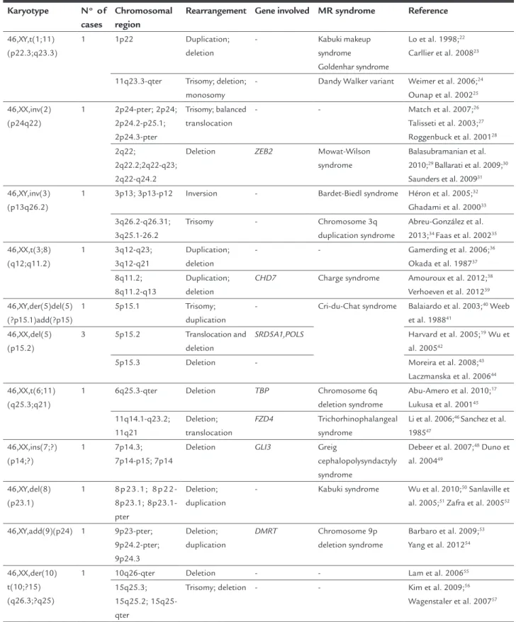
and structural alterations (Figure 1).
Among the patients with identiied numerical
chro-mosomal abnormalities, ive cases had aneuploidy of sex
chromosomes: four cases of 47,XXY karyotype, and one
case of X monosomy (45,X karyotype). Regarding
auto-somal chromosomes, three cases of free trisomy 21
(47,XX,+21 karyotype) were detected. The case of
numer-ical and structural alteration corresponded to a
47,XXY,dup(18)(p12.2 p12.3) karyotype.
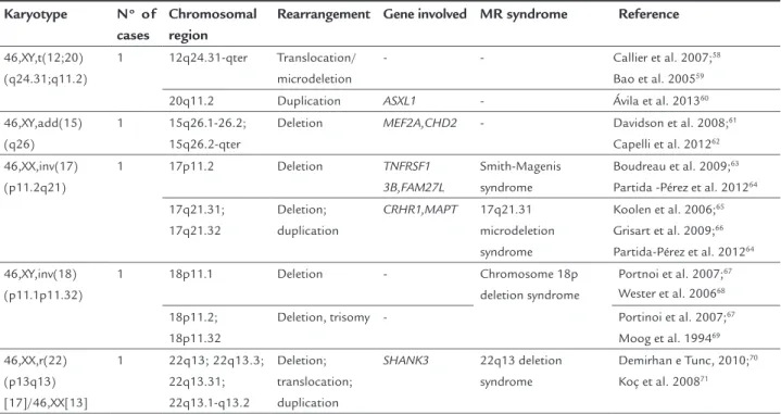
As for structural chromosomal abnormalities,
recip-rocal translocations, inversions, deletions, additional
chro-mosomal segments, and insertions were observed. Based
on the comparison of results of karyotypes with
structur-al structur-alterations and their speciic chromosomstructur-al regions, we
were able to identify breakpoints involving 1p22.3; 2p24;
2q22; 3p13; 3q26.2; 3q12; 5p15.1; 5p15.2; 5p15.3; 6q25.3;
7p14; 8p23.1; 8q11.2; 9p24; 10q26.3; 11q23.3; 11q21;
12q24.31; 15q25; 15q26; 17p11.2; 17q21; 18p11.1; 18p11.32;
20q11.2; 22q13. The respective rearrangements as well as
FIGURE 1
Distribution of results of 369 patients who underwent G-banding karyotyping for investigation of mental retardation in 2009 at
Instituto Hermes Pardini.
Normal
Structural alteration
Numerical alteration
the correlation between the genes involved and syndromes
related with MR are shown in Table 1. With the exception
of the deletion on the short arm of chromosome 5
[46,XX,del(5)(p11.2) karyotype], found in three cases, all
other changes occurred once in this study.
Considering the structural alterations, three cases
re-vealed additional marker chromosomes of unknown
or-igin, two of which were mosaic marker chromosomes. The
cases of mosaic presented 47,XX,+mar[13]/46,XX[17]
karyotype (mosaicism for one lineage with presence of an
additional marker chromosome, and another lineage with
normal complement) and 47,XY,+r[8]/46,XY[22]
karyo-type (mosaicism for one lineage with an additional
mark-er chromosome ring, and anothmark-er lineage with normal
complement). In one case, the additional marker
chro-mosome was seen in all cells analyzed (47,XX,+mar).
D
ISCUSSION
This study presents a retrospective analysis of the results
of G-banding karyotyping of individuals with MR
as-sessed at Instituto Hermes Pardini in 2009, and the
com-parison of these results with literature data. Based on
this evaluation, we were able to identify chromosomal
alterations in 8.4% of patients only. Although this
meth-od is important either to explain or rule out causes of
MR, it has limitations, especially in relation to the
iden-tiication of submicroscopic alterations. Thus, other
tech-niques such as luorescent
in situ
hybridization (FISH),
6comparative genomic hybridization (CGH), array based
comparative genomic hybridization (aCGH),
7and
mul-tiplex ligation-dependent probe ampliication (MLPA),
8are more suitable to detect submicroscopic
rearrange-ments responsible for most cases of MR, especially in
cases of idiopathic MR.
Among the numerical chromosomal abnormalities
identiied in this study, the 47,XXY karyotype was most
frequent. This abnormality causes Klinefelter syndrome,
the most common numerical chromosomal disorder
among men. The estimated frequency of this disorder is
between 1:500 and 1:1,000 live births. Men with
Klinefel-ter syndrome usually have delayed auditory processing,
language dysfunction and, in rare cases, severe
intellec-tual retardation.
2Turner syndrome is less common than other sex
chro-mosome aneuploidies, and its most important
chromo-some constitution is 45,X, found in one case in this study.
Some patients, in addition to the unique physical
char-acteristics of this syndrome, have a non-verbal IQ
signif-icantly lower than their verbal IQ; many require
educa-tional intervention, especially in mathematics.
9,10The only numerical alteration of autosomal
chromo-some found in this study was trisomy 21 (47,XX,+21).
This chromosome constitution is observed in patients
with Down syndrome, an example of neurogenetic
disor-der related to aneuploidy,
1and corresponds to the most
common genetic cause of moderate mental retardation
11,12and intellectual disability.
13In this study we also identiied three cases of
addition-al marker chromosomes, two with mosaic karyotype
(47,XX,+mar[13]/46,XX[17] and 47,XY,+r[8]/46,XY[22]).
Supernumerary marker chromosomes are a heterogeneous
group of structurally abnormal chromosomes derived from
any of the 24 chromosomes, but they cannot be identiied
using conventional cytogenetic techniques.
14They are found
in clinically normal individuals, but are often reported in
patients with mental retardation and infertility.
15Mosa-icism associated with marker chromosomes is a well-known
inding, observed in approximately 50% of cases. Different
types of mosaicism can be found, the most common
be-ing 47,XX,+mar/46,XX.
14The analysis of chromosomal abnormalities and
com-parison with literature data revealed that the alterations
identiied in this study occur in regions determined by
breakpoints also described in previous studies with
pa-tients with MR. In the 46,XX,inv(2)(p24q22) inversion,
for example, there is the q22 breakpoint located close to
the
ZEB2
gene. Deletions involving this gene are
respon-sible for Mowat-Wilson syndrome, and MR is one of its
main clinical manifestations.
16The 6q25.3 point of
46,XX,t(6;11)(q25.3;q21) translocation is associated with
a deletion syndrome with moderate MR.
17The present study found three patients with
termi-nal deletion in the short arm of chromosome 5 and p15.2
breakpoint [46,XX,del(5)(p15.2)]. Most deletions in the
short arm of chromosome 5 are related to Cri-du-Chat
syndrome, which is a genetic disease caused by deletion
of variable size, with breakpoints ranging from p13 to
p15.2.
18A distinctive cry (similar to that of a meowing
kitten), dysmorphisms, psychomotor delay, and MR are
some of the clinical manifestations of the syndrome.
Stud-ies conirm the critical region of the syndrome, mapped
in 5p15, with genes related to Cri-du-Chat in p15.3, and
dysmorphisms and MR in p15.2.
18,19With the aid of websites GenScript
73and ClinVar,
74devel-TABLE 1
List of breakpoints found in the detected structural alterations and comparison with literature data on mental
retardation.
Karyotype
N
oof
cases
Chromosomal
region
Rearrangement Gene involved MR syndrome
Reference
46,XY,t(1;11)
(p22.3;q23.3)
1
1p22
Duplication;
deletion
-
Kabuki makeup
syndrome
Goldenhar syndrome
Lo et al. 1998;
22Carllier et al. 2008
2311q23.3-qter
Trisomy; deletion;
monosomy
-
Dandy Walker variant
Weimer et al. 2006;
24Ounap et al. 2002
2546,XX,inv(2)
(p24q22)
1
2p24-pter; 2p24;
2p24.2-p25.1;
2p24.3-pter
Trisomy; balanced
translocation
-
-
Match et al. 2007;
26Talisseti et al. 2003;
27Roggenbuck et al. 2001
282q22;
2q22.2;2q22-q23;
2q22-q24.2
Deletion
ZEB2
Mowat-Wilson
syndrome
Balasubramanian et al.
2010;
29Ballarati et al. 2009;
30Saunders et al. 2009
3146,XY,inv(3)
(p13q26.2)
1
3p13; 3p13-p12
Inversion
-
Bardet-Biedl syndrome
Héron et al. 2005;
32Ghadami et al. 2000
333q26.2-q26.31;
3q25.1-26.2
Trisomy
-
Chromosome 3q
duplication syndrome
Abreu-González et al.
2013;
34Faas et al. 2002
3546,XX,t(3;8)
(q12;q11.2)
1
3q12-q23;
3q12-q21
Duplication;
deletion
-
-
Gamerding et al. 2006;
36Okada et al. 1987
378q11.2;
8q11.2-q13
Duplication;
deletion
CHD7
Charge syndrome
Amouroux et al. 2012;
38Verhoeven et al. 2012
3946,XY,der(5)del(5)
(?p15.1)add(?p15)
1
5p15.1
Trisomy;
duplication
-
Cri-du-Chat syndrome
Balaiardo et al. 2003;
40Weeb
et al. 1988
4146,XX,del(5)
(p15.2)
3
5p15.2
Translocation and
deletion
SRD5A1,POLS
Harvard et al. 2005;
19Wu et
al. 2005
425p15.3
Deletion
-
Moreira et al. 2008;
43Laczmanska et al. 2006
4446,XX,t(6;11)
(q25.3;q21)
1
6q25.3-qter
Deletion
TBP
Chromosome 6q
deletion syndrome
Abu-Amero et al. 2010;
17Lukusa et al. 2001
4511q14.1-q23.2;
11q21
Deletion;
translocation
FZD4
Trichorhinophalangeal
syndrome
Li et al. 2006;
46Sanchez et al.
1985
4746,XX,ins(7;?)
(p14;?)
1
7p14.3;
7p14-p15; 7p14
Deletion
GLI3
Greig
cephalopolysyndactyly
syndrome
Debeer et al. 2007;
48Duno et
al. 2004
4946,XY,del(8)
(p23.1)
1
8p23.1;
8p22-8p23.1;
8p23.1-pter
Deletion;
duplication
-
Kabuki syndrome
Wu et al. 2010;
50Sanlaville et
al. 2005;
51Zafra et al. 2005
5246,XY,add(9)(p24) 1
9p23-pter;
9p24.2-pter;
9p24.3
Deletion;
duplication
DMRT
Chromosome 9p
deletion syndrome
Barbaro et al. 2009;
53Yang et al. 2012
5446,XX,der(10)
t(10;?15)
(q26.3;?q25)
1
10q26-qter
Deletion
-
-
Lam et al. 2006
5515q25.3;
15q25.2;
15q25-qter
Trisomy; deletion -
-
Kim et al. 2009;
56Wagenstaler et al. 2007
57opmental delay; or signiicant speech delay; or growth
de-lay; or intellectual delay.
20,21C
ONCLUSION
Genetics is one of the areas of expertise that has most
contributed to the elucidation of the causes of MR, with
various possibilities for diagnosis. This study illustrates
the contribution of G-banding karyotype analysis for
in-vestigation of the causes of MR, since our results are in
line with those reported in the literature. Structural
chro-mosomal abnormalities were observed more frequently
in this study. Knowing the critical regions on
chromo-somes is very important to correlate genotype and
phe-notype, and karyotype studies may help determine such
regions. Therefore, although G-banding karyotype
anal-ysis may not be the most suitable test for syndromes
re-lated to mental retardation, since a large number of these
syndromes are due to submicroscopic deletions and
du-plications, this technique proves to be useful for initial
screening of affected individuals, and negative results
should be evaluated using more accurate techniques such
as aCGH and MLPA.
R
ESUMO
Estudo retrospectivo de cariótipo em pacientes com
re-tardo mental
Objetivo:
descrever as alterações cromossômicas em
pa-cientes com retardo mental (RM) pela análise do
carióti-po com bandas G.
Método:
foi realizado um estudo retrospectivo dos
resul-tados de cariótipo com bandas G de 369 pacientes em
in-vestigação de RM. A partir dos rearranjos estruturais
encon-trados, foram levantadas todas as regiões cromossômicas
envolvidas nos pontos de quebra e elas foram comparadas
com a literatura para RM e bancos de dados.
Resultados:
foram identiicados 338 (91,6%) casos normais
e 31 (8,4%) com algum tipo de alteração cromossômica.
Dentre os casos alterados, 21 pacientes (67,8%) foram
iden-tiicados com alterações cromossômicas estruturais, 9 (29%)
com alterações numéricas e 1 (3,2%) com alteração
numé-rica e estrutural.
Conclusão:
as alterações cromossômicas estruturais
fo-ram aquelas observadas com maior frequência. O
carióti-po com bandas G contribui para a investigação das
cau-sas de RM, mostrando que essa técnica pode ser útil como
uma primeira triagem dos pacientes. No entanto, técnicas
mais resolutivas como o
array based comparative genomic
hi-bridization
(aCGH) e o
multiplex ligation dependent probe
am-pliication
(MLPA) permitem detectar alterações
submicros-cópicas comumente associadas ao RM.
Palavras-chave:
cariótipo, deiciência intelectual,
aber-rações cromossômicas.
TABLE 1
(Cont.) List of breakpoints found in the detected structural alterations and comparison with literature data on
mental retardation.
Karyotype
N
oof
cases
Chromosomal
region
Rearrangement Gene involved MR syndrome
Reference
46,XY,t(12;20)
(q24.31;q11.2)
1
12q24.31-qter
Translocation/
microdeletion
-
-
Callier et al. 2007;
58Bao et al. 2005
5920q11.2
Duplication
ASXL1
-
Ávila et al. 2013
6046,XY,add(15)
(q26)
1
15q26.1-26.2;
15q26.2-qter
Deletion
MEF2A,CHD2
-
Davidson et al. 2008;
61Capelli et al. 2012
6246,XX,inv(17)
(p11.2q21)
1
17p11.2
Deletion
TNFRSF1
3B,FAM27L
Smith-Magenis
syndrome
Boudreau et al. 2009;
63Partida -Pérez et al. 2012
6417q21.31;
17q21.32
Deletion;
duplication
CRHR1,MAPT
17q21.31
microdeletion
syndrome
Koolen et al. 2006;
65Grisart et al. 2009;
66Partida-Pérez et al. 2012
6446,XY,inv(18)
(p11.1p11.32)
1
18p11.1
Deletion
-
Chromosome 18p
deletion syndrome
Portnoi et al. 2007;
67Wester et al. 2006
6818p11.2;
18p11.32
Deletion, trisomy -
Portinoi et al. 2007;
67Moog et al. 1994
6946,XX,r(22)
(p13q13)
[17]/46,XX[13]
1
22q13; 22q13.3;
22q13.31;
22q13.1-q13.2
Deletion;
translocation;
duplication
SHANK3
22q13 deletion
syndrome
Demirhan e Tunc, 2010;
70Koç et al. 2008
71R
EFERENCES
1. Dierssen M, Yann H, Estivill X. Aneuploidy: from a physiological mechanism of variance to Down syndrome. Physiol Rev. 2009; 89(3):887-920. 2. Speicher M, Antonarakis SE, Motulski AG. Vogel and Motulsky’s human
genetics: problems and approaches. 4.ed. Berlin: Springer Science & Business Media, 2010.
3. Vasconcelos MM. Retardo mental. J Pediatr (Rio J.). 2004; 80(2):71-82. 4. Stevenson RE: Splitting and lumping in the nosology of XLMR. Am J Med
Genet. 2000; 97(3):174-82.
5. Shaffer LG, Slovak ML, Campbell LJ. ISCN – An International System for Human Cytogenetic Nomenclature. Basel: Karger AG, 2013.
6. Shevell M, Ashwal S, Donley D, Flint J, Gingold M, Hirtz D, et al. Practice parameter: evaluation of the child with global developmental delay: report of the Quality Standards Subcommittee of the American Academy of Neurology and The Practice Committee of the Child Neurology Society. Neurology. 2003; 60(3):367-80.
7. Shaffer LG, Bejjani BA. Using microarray-based molecular cytogenetic methods to identify chromosome abnormalities. Pediatr Ann. 2009; 38(8):440-7. 8. Jehee FS, Takamori JT, Medeiros PF, Pordeus AC, Latini FR, Bertola DR,
et al. Using a combination of MLPA kits to detect chromosomal imbalances in patients with multiple congenital anomalies and mental retardation is a valuable choice for developing countries. Eur J Med Genet. 2011; 54(4):425-32.
9. Nussbaum RL, McInnes RR, Willard HF. Thompson & Thompson genética médica. 6.ed. Rio de Janeiro: Guanabara Koogan, 2002. p.151-4. 10. Murphy MM, Mazzocco MM, Gerner G, Henry AE. Mathematics learning
disability in girls with Turner syndrome or fragile X syndrome. Brain Cogn. 2006; 61(2):195-210.
11. Ahuja AS, Thapar A, Owen MJ. Genetics of mental retardation. Indian J Med Sci. 2005; 59(9):407-17.
12. Rauch A, Hoyer J, Guth S, Zweier C, Kraus C, Becker C, et al. Diagnostic yield of various genetic approaches in patients with unexplained developmental delay or mental retardation. Am J Med Genet. 2006; 140(19):2063-74.
13. Anderson JS, Nielsen JA, Ferguson MA, Burback MC, Cox ET, Dai L, et al. Abnormal brain synchrony in Down syndrome. NeuroImage Clin. 2013; 2:703-15.
14. Liehr T, Karamysheva T, Merkas M, Brecevic L, Hamid AB, Ewers E, et al. Somatic mosaicism in cases with small supernumerary marker chromosomes. Curr Genomics. 2010; 11(6):432-9.
15. Liehr T, Weise A. Frequency of small supernumerary marker chromosomes in prenatal, newborn, developmentally retarded and infertility diagnostics. Int J Mol Med. 2007; 19(5):719-31.
16. Mowat DR, Croaker GD, Cass DT, Kerr BA, Chaitow J, Adès LC, et al. Hirschsprung disease, microcephaly, mental retardation, and characteristic facial features: delineation of a new syndrome and identiication of a locus at chromosome 2q22-q23. J Med Genet. 1998; 35(8):617-23.
17. Abu-Amero KK, Hellani A, Salih MA, Al Hussain A, al Obailan M, Zidan G, et al. Ophthalmologic abnormalities in a de novo terminal 6q deletion. Ophthalmic Genet. 2010; 31(1):1-11.
18. Mainardi PC, Perfumo C, Cali A, Coucourde G, Pastore G, Cavani S, et al. Clinical and molecular characterisation of 80 patients with 5p deletion: genotype-phenotype correlation. J Med Genet. 2001; 38(3):151-8. 19. Harvard C, Malefant P, Koochek M, Creighton S, Mickelson EC, Holden JJ,
et al. A variant Cri du Chat phenotype and autism spectrum disorder in a subject with de novo cryptic microdeletions involving 5p15.2 and 3p24.3-25 detected using whole genomic array CGH. Clin Genet. 2005; 67(4):341-51. 20. Kaminsky EB, Kaul V, Paschall J, Church DM, Bunke B, Kunig D, et al. An
evidence-based signiicance of CNVs in intellectual and developmental disabilities. Genet Med. 2011; 13(9):777-84.
21. Miller DT, Adam MP, Aradhya S, Biesecker LG, Brothman AR, Carter NP, et al. Consensus statement: chromosomal microarray is a irst-tier clinical diagnostic test for individuals with developmental disabilities or congenital anomalies. Am J Hum Genet. 2010; 86(5):749-64.
22. Lo IF, Cheung LY, Ng AY, Lam ST. Interstitial Dup(1p) with indings of Kabuki make-up syndrome. Am J Med Genet. 1998; 78(1):55-7.
23. Callier P, Faivre L, Thauvin-Robinet C, Marle N, Mosca AL, D’Athis P, e tal. Array-CGH in a series of 30 patients with mental retardation, dysmorphic features, and congenital malformations detected an interstitial 1p22.2-p31.1
deletion in a patient with features overlapping the Goldenhar syndrome. Am J Med Genet A. 2008; 146A(16):2109-15.
24. Ounap K, Bartsch O, Uibo O, Laidre P. Girl with combined cellular immunodeiciency, pancytopenia, malformations, deletion 11q23.3 --> qter, and trisomy 8q24.3 --> qter. Am J Med Genet. 2002; 108(4):322-6. 25. Weimer J, Cohen M, Wiedemann U, Heinrich U, Jonat W, Arnold N. Proof
of partial imbalances 6q and 11q due to maternal complex balanced translocation analyzed by microdissection of multicolor labeled chromosomes (FISH-MD) in a patient with Dandy-Walker variant. Cytogenet Genome Res. 2006; 114(3-4):235-9.
26. Mach M, Windpassinger C, Wagner K, Kroisel PM, Petek E. Distal monosomy 16p13.3/distal trisomy 2p24.2-pter: molecular-cytogenetic characterisation and phenotype. Genet Couns. 2007; 18(1):9-16.
27. Talisetti A, Forrester SR, Gregory D, Johnson L, Schneider MC, Kimonis VE. Temtamy-like syndrome associated with translocation of 2p24 and 9q32. Clin Dysmorphol. 2003; 12(3):175-7.
28. Roggenbuck JA, Fink JM, Mendelsohn NJ. Unique case of trisomy 2p24.3-pter with no associated monosomy. Am J Med Genet. 2001; 101(1):50-4. 29. Balasubramaniam S, Keng WT, Ngu LH, Goossens MJ, Giurgea I.
Mowat-Wilson syndrome: the irst two Malaysian cases. Singapore Med J. 2010; 51(3):e54-7.
30. Ballarati L, Recalcati MP, Bedeschi MF, Lalatta F, Valtorta C, Bellini M, et al. Cytogenetic, FISH and array-CGH characterization of a complex chromosomal rearrangement carried by a mentally and language impaired patient. Eur J Med Genet. 2009; 52(4):218-23.
31. Saunders CJ, Zhao W, Ardinger HH. Comprehensive ZEB2 gene analysis for Mowat-Wilson syndrome in a North American cohort: a suggested approach to molecular diagnostics. Am J Med Genet A. 2009; 149A(11):2527-31. 32. Héon E, Westall C, Carmi R, Elbedour K, Panton C, Mackeen L, et al. Ocular
phenotypes of three genetic variants of Bardet-Biedl syndrome. Am J Med Genet A. 2005; 132A(3):283-7.
33. Ghadami M, Tomita HA, Najai MT, Damavandi E, Farahvash MS, Yamada K, et al. Bardet-Biedl syndrome type 3 in an Iranian family: clinical study and conirmation of disease localization. Am J Med Genet. 2000; 94(5):433-7.
34. Abreu-González M, García-Delgado C, Cervantes A, Aparicio-Onofre A, Guevara-Yáñez R, Sánchez-Urbina R, et al. Clinical, cytogenetic, and biochemical analysis of a family with a t(3;13)(q26.2;p11.2): further delineation of 3q duplication syndrome. Case Rep Genet. 2013; 2013:895259. 35. Faas BH, De Vries BB, Van Es-Van Gaal J, Merkx G, Draaisma JM, Smeets DF. A new case of dup(3q) syndrome due to a pure duplication of 3qter. Clin Genet. 2002; 62(4):315-20.
36. Gamerdinger U, Bosse K, Eggermann T, Kalscheuer V, Schwanitz G, Engels H. First report of a partial trisomy 3q12-q23 de novo--FISH breakpoint determination and phenotypic characterization. Eur J Med Genet. 2006; 49(3):225-34.
37. Okada N, Hasegawa T, Osawa M, Fukuyama Y. A case of de novo interstitial deletion 3q. J Med Genet. 1987; 24(5):305-8.
38. Amouroux C, Vincent M, Blanchet P, Puechberty J, Schneider A, Chaze AM, et al. Duplication 8q12: conirmation of a novel recognizable phenotype with duane retraction syndrome and developmental delay. Eur J Hum Genet. 2012; 20(5):580-3.
39. Verhoeven WM, Egger JI, Feenstra I, de Leeuw N. A de novo 3.57 Mb microdeletion in 8q12.3q13.2 in a patient with mild intellectual disability and epilepsy. Eur J Med Genet. 2012; 55(5):358-61.
40. Baialardo EM, Torrado Mdel V, Barreiro CZ, Gallego MS. Partial distal 5p trisomy resulting from paternal translocation (5;15)(p15.1;p13) in a boy with no mental retardation. Clin Dysmorphol. 2003; 12(4):257-9. 41. Webb GC, Voullaire LE, Rogers JG. Duplication of a small segment of 5p
due to maternal recombination within a paracentric shift. Am J Med Genet. 1988; 30(4):875-81.
42. Wu Q, Niebuhr E, Yang H, Hansen L. Determination of the ‘critical region’ for cat-like cry of Cri-du-chat syndrome and analysis of candidate genes by quantitative PCR. Eur J Hum Genet. 2005; 13(4):475-85.
43. Moreira LMA, Carvalho AFL, Borja ALVF, Pinto PSP, Silveira A, de Freitas LM, et al. Mosaic cri-du-chat syndrome in a girl with a mild phenotype. J Appl Genet. 2008; 49(4):415-20.
45. Lukusa T, Willekens D, Lukusa N, De Cock F, Fryns JP. Terminal 6q25.3 deletion and abnormal behaviour. Genet Couns. 2001; 12(3):213-21. 46. Li P, Zhang HZ, Huff S, Nimmakayalu M, Qumsiyeh M, Yu J, et al.
Karyotype-phenotype insights from 11q14.1-q23.2 interstitial deletions: FZD4 haploinsuficiency and exudative vitreoretinopathy in a patient with a complex chromosome rearrangement. Am J Med Genet A. 2006; 140(24):2721-9.
47. Sánchez LM, Labarta JD, De Negrotti TC, Migliorini AM. Complex translocation in a boy with trichorhinophalangeal syndrome. J Med Genet. 1985; 22(4):314-6.
48. Debeer P, Devriendt K, De Smet L, deRavel T, Gonzalez-Meneses A, Grzeschik KH, et al. The spectrum of hand and foot malformations in patients with Greig cephalopolysyndactyly. J Child Orthop. 2007; 1(2):1143-50. 49. Dunø M, Hove H, Kirchhoff M, Devriendt K, Schwartz M. Mapping genomic
deletions down to the base: a quantitative copy number scanning approach used to characterise and clone the breakpoints of a recurrent 7p14.2p15.3 deletion. Hum Genet. 2004; 115(6):459-67.
50. Wu Y, Ji T, Wang J, Xiao J, Wang H, Li J, et al. Submicroscopic subtelomeric aberrations in Chinese patients with unexplained developmental delay/ mental retardation. BMC Med Genet. 2010; 11:72.
51. Sanlaville D, Genevieve DD, Bernardin C, Amiel J, Baumann C, Blois MC, et al. Failure to detect an 8p22-8p23.1 duplication in patients with Kabuki (Niikawa-Kuroki) syndrome. Eur J Hum Genet. 2005; 13(5):690-93. 52. Zafra de la Rosa G, Venegas-Vega CA, Monroy N, Contreras-Bucio G, Friedrich
U, Houman M, et al. Trisomy 3q25.1-qter and monosomy 8p23.1-pter in a patient: cytogenetic and molecular analysis with delineation of the phenotype. Am J Med Genet A. 2005; 136(3):259-64.
53. Barbaro M, Balsamo A, Anderlid BM, Myhre AG, Gennari M, Nicoletti A, et al. Characterization of deletions at 9p affecting the candidate regions for sex reversal and deletion 9p syndrome by MLPA. Eur J Hum Genet. 2009; 17(11):1439-47.
54. Yang Y, Wang C, Wang F, Zhu L, Liu H, He X. Novel chromosomal translocation t(11;9)(p15;p23) involving deletion and duplication of 9p in a girl associated with autism and mental retardation. Gene. 2012; 502(2):154-8.
55. Lam AC, Lam ST, Lai KK, Tong TM, Chau TC. High rate of detection of subtelomeric aberration by using combined MLPA and subtelomeric FISH approach in patients with moderate to severe mental retardation. Clin Biochem. 2006; 39(3):196-202.
56. Kim JH, Lee WM, Ryoo NH, Ha JS, Jeon DS, Kim JR, et al. [A case of partial trisomy 15q25.3-qter]. Korean J Lab Med. 2009; 29(1):66-70.
57. Wagenstaller J, Spranger S, Lorenz-Depiereux B, Kazmierczak B, Nathrath M, Wahl D, et al. Copy-number variations measured by single-nucleotide-polymorphism oligonucleotide arrays in patients with mental retardation. Am J Hum Genet. 2007; 81(4):768-79.
58. Callier P, Faivre L, Marle N, Thauvin-Robinet C, Mosca AL, Masurel-Paulet A, et al. Untreated growth hormone deiciency with extremely short stature, bone dysplasia, cleft lip-palate and severe mental retardation in a
26-year-old man with a de novo unbalanced translocation t(1;12)(q24;q24). Eur J Med Genet. 2007; 50(6):455-64.
59. Bao L, Schorry EK. A girl with partial trisomy 12q24.31 inherited from her father and a possible novel syndrome transmitted from her mother. Am J Med Genet A. 2005; 138(4):361-4.
60. Ávila M, Kirchhoff M, Marle N, Hode HD, Chouchane M, Thauvin-Robinet C, et al. Delineation of a new chromosome 20q11.2 duplication syndrome including the ASXL1 gene. Am J Med Genet A. 2013; 161A(7):1594-8. 61. Davidsson J, Collin A, Björkhem G, Soller M. Array based characterization
of a terminal deletion involving chromosome subband 15q26.2: an emerging syndrome associated with growth retardation, cardiac defects and developmental delay. BMC Med Genet. 2008; 9:2.
62. Capelli LP, Krepischi AC, Gurgel-Guianetti J, Mendes MF, Rodrigues T, Varela MC, et al. Deletion of the RMGA and CHD2 genes in a child with epilepsy and mental deiciency. Eur J Med Genet. 2012; 55(2):132-4. 63. Boudreau EA, Johnson KP, Jackman AR, Blancato J, Huizing M, Bendavid
C, et al. Review of disrupted sleep patterns in Smith-Magenis syndrome and normal melatonin secretion in a patient with an atypical interstitial 17p11.2 deletion. Am J Med Genet A. 2009; 149A(7):1382-91.
64. Partida-Pérez M, Dominguez MG, Neira VA, Figuera LS, Rivera H. De novo inv(17)(p11.2q21.3) in an intellectually disabled girl: appraisal of 21 inv(17) constitutional instances. J Genet. 2012; 91(2):241-4.
65. Koolen DA, Vissers LE, Pfundt R, de Leeuw N, Knight SJ, Regan R, et al. A new chromosome 17q21.31 microdeletion syndrome associated with a common inversion polymorphism. Nat Genet. 2006; 38(9):999-1001. 66. Grisart B, Willat L, Destrée A, Fryns JP, Rack K, de Ravel T, et al. 17q21.31
microduplication patients are characterized by behavioural problems and poor social interaction. J Med Genet. 2009; 46(8):524-30.
67. Portnoï MF, Gruchy N, Marlin S, Finkel L, Denoyelle F, Dubourg C, et al. Midline defects in deletion 18p syndrome: clinical and molecular characterization of three patients. Clin Dysmorphol. 2007; 16(4):247-52. 68. Wester U, Bondenson ML, Edeby C, Annerén G. Clinical and molecular
characterization of individuals with 18p deletion: a genotype-phenotype correlation. Am J Med Genet A. 2006; 140(11):1164-71.
69. Moog U, Engelen JJ, de Die-Smulders CE, Albrechts JC, Loneus WH, Haagen AA, et al. Partial trisomy of the short arm of chromosome 18 due to inversion duplication and direct duplication. Clin Genet. 1994; 46(6):423-9. 70. Dermirhan O, Tunç E. Phenotypic correlations in a patient with ring
chromosome 22. Indian J Hum Genet. 2010; 16(2):97-9.
71. Koç A, Karaer K, Ergün MA, Yirmibeş-Karaoğuz M, Kan D, Cansu A, et al. A case with a ring chromosome 22. Turk J Pedriatr. 2008; 50(2):193-6. 72. MapViewer – National Center for Biotechnology Information. [cited 2014
Sep 3]. Available from: http://www.ncbi.nlm.nih.gov/projects/mapview/. 73. GenScript. [cited 2014 Sep 10]. Available from: http://www.genscript.com/
index.html/.