BLADDEREXSTROPHY
REV ASSOC MED BRAS 2016; 62(3):197-198 197
IMAGE IN MEDICINE
Bladder exstrophy
GUSTAVO GOMES MENDES¹*, JOEL RODRIGO BEAL LUSA¹
1Degree of Specialist in Radiology and Diagnostic Imaging from Colégio Brasileiro de Radiologia e Diagnóstico por Imagem (CBR)/Associação Médica Brasileira (AMB). Full Member of the Imaging Department at Hospital A.C. Camargo Cancer Center, São Paulo, SP, Brazil
S
UMMARYStudy conducted at Hospital A.C. Camargo Cancer Center, São Paulo, SP, Brazil
Article received: 4/6/2015
Accepted for publication: 5/4/2015
*Correspondence:
Address: Rua Professor Antônio Prudente, 211,
Liberdade São Paulo, SP – Brazil Postal code: 01509-010 Phone: +55 11 2189-5000 [email protected]
http://dx.doi.org/10.1590/1806-9282.62.03.197
Bladder exstrophy is a rare congenital anomaly resulting from failure of fusion of the middle of the pelvis line tissues during embryogenesis. It is characterized by malformation of the lower abdominal wall involving the genitourinary tract and the musculoskeletal system. Its incidence is estimated at 1:30,000 to 1:50,000 live births, and it is 2 or 3 times more frequent in males. The child’s age is im-portant and the best results are obtained when treatment is performed shortly after birth.
Keyword: bladder exstrophy.
C
ASEMale patient, aged eight months, referred with clinical di-agnosis of bladder exstrophy for assessment of any associ-ated anorectal and skeletal anomalies. According to the caregiver, this was a term birth with prenatal examinations performed uneventfully, and diagnosis made based on mor-phological routine ultrasound (US) during pregnancy.
D
ISCUSSIONBladder exstrophy is a rare congenital anomaly resulting from failure of fusion of the middle of the pelvis line tis-sues during embryogenesis. It is characterized by malfor-mation of the lower abdominal wall involving the geni-tourinary tract and the musculoskeletal system.
Its incidence is estimated at 1:30,000 to 1:50,000 live births, and it is two or three times more frequent in males. In the classic bladder exstrophy, the anterior wall of the back of the bladder is exposed, and changes such as epi-spadias, dysplasia of the pelvic loor muscles, short penis or clitoris bifurcated are part of the clinical picture.
The child’s age is important and the best results are obtained when treatment is performed shortly after birth. Most pathological changes can be prevented by early clo-sure of bladder exstrophy.
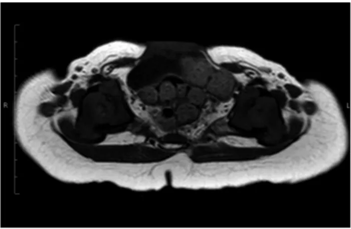
Pubic diastasis (Figures 1 and 2) is the stigma of ex-strophy-epispadias complex malformations; it is narrow-er in epispadias and widnarrow-er in the bladdnarrow-er and cloacal ex-strophy and is always associated with lateral rotation of the femur and acetabulum. The defect of the abdominal wall that remains after closure of the bladder is
triangu-FIGURE 1 Axial T1 magnetic resonance imaging (MRI). Diastasis
of pubic bones associated with a defect of the anterior abdominal wall and insinuation of the anterior wall into the ventral muscles.
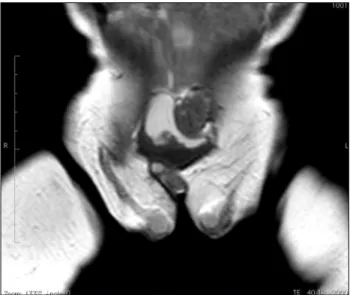
FIGURE 2 Axial T1 MRI. Insinuation of the anterior wall of the bladder
MENDESAND LUSA
198 REV ASSOC MED BRAS 2016; 62(3):197-198
lar, and is limited laterally by the rectus abdominis mus-cle and inferiorly by the inter-symphyseal band, where the external urethral sphincter is inserted. The perineal loor is compromised by the malformation, which ex-plains the recurring rectal prolapse. The anus is more an-terior compared to its original position in both sexes.
Boys present epispadias, short phallus, wide at its base and with upward (dorsal) curvature. The urethra is represented by exposed dorsal mucosa. Penis size is vari-able, but is often small, imposing serious dificulties in obtaining adequate phallus even after reconstruction. The scrotum is split and the testicles are usually palpa-ble (Figure 3). Cryptorchidism is rarely associated anom-aly, and most commonly the testicles are located in the external inguinal ring and can be descended into the tes-ticular pouch. Inguinal hernia, probably related to the fragility of the side of the abdomen, is common.
Typically, girls have biid clitoris; the internal genita-lia is normal, and vaginal and uterine abnormalities may occur. The urethra is also epispadic and extremely short. Differential diagnosis includes diseases that occur with defect of the anterior abdominal wall, such as om-phalocele, gastroschisis and cloacal exstrophy.
R
ESUMOExtroia vesical
A extroia de bexiga é uma anomalia congênita rara decor-rente de falha da fusão dos tecidos da linha média da pelve durante a embriogênese e caracteriza-se por má-formação da região inferior da parede abdominal, envolvendo o trato geniturinário e o sistema musculoesquelético. Apresenta in-cidência estimada de 1:30.000 a 1:50.000 nascidos vivos, sen-do 2 a 3 vezes mais frequente no sexo masculino. A idade da criança é importante e os melhores resultados são obtidos quando o tratamento é realizado logo após o nascimento.
Palavra-chave: extroia vesical.
R
EFERENCES1. Beaty JH. Congenital and developmental anomalies of hip and pelvis In: Canale TS. Campbell’s operative orthopaedics. 10.ed. St. Louis: Mosby, 2003. p.1118-9. 2. Ramírez E, Calderón M. Osteotomía ilíaca en extroia vesical. Rev Mex Ortop
Traumatol. 1997; 11(5):293-6.
3. Segev E, Ezra E, Binyamini Y, Wientroub S, Ben-Chaim J. A combined vertical and horizontal pelvic osteotomy approach for repair of bladder exstrophy: the Dana experience. Isr Med Assoc J. 2004; 6(12):749-52.
4. Frey P. Bilateral anterior pubic osteotomy in bladder exstrophy closure. J Urol. 1996; 156(2 Pt 2):812-5.
5. Satsuma S, Kobayashi D, Yoshiya S, Kurosaka M. Comparison of posterior and anterior pelvic osteotomy for bladder exstrophy complex. J Pediatr Orthop B. 2006; 15(2):141-6.
6. Martín C, Darnell A, Durán C, Bermúdez P, Mellado F, Rigol S. Magnetic resonance imaging of the intrauterine fetal genitourinary tract: normal anatomy and pathology. Abdom Imaging. 2004; 29(3):286-302.