Breast cancer features in women under the age of 40 years
DEISE SANTIAGO GIRÃO EUGÊNIO1*, JULIANA A. SOUZA1, RUBENS CHOJNIAK1, ALMIR G. V. BITENCOURT1,LUCIANA GRAZIANO1, ELVIRA F. SOUZA1
1Department of Diagnostic Imaging, A.C.Camargo Cancer Center, Fundação Antonio Prudente, São Paulo, SP, Brazil
S
UMMARYStudy conducted at the Department of Diagnostic Imaging, A.C.Camargo Cancer Center, Fundação Antonio Prudente, São Paulo, SP, Brazil
Article received: 11/23/2015
Accepted for publication: 3/16/2016
*Correspondence:
Address: R. Professor Antônio Prudente, 211 São Paulo, SP – Brazil Postal code: 01509-010 [email protected]
http://dx.doi.org/10.1590/1806-9282.62.08.755
Objective: To describe the clinical features, imaging indings and pathological aspects of breast cancer diagnosed in women under the age of 40 years.
Method: A retrospective, descriptive study was performed through analysis of medical records between November 2008 and August 2012. One hundred and twenty (120) patients were included, of whom 112 underwent mammography, 113 underwent ultrasonography, and 105 underwent magnetic resonance imag-ing (MRI). The histopathological data was obtained in most cases from post-surgical analysis, which was available for 113 patients.
Results: The mean age at diagnosis of primary breast cancer was 34 years. Only 11 patients (9.0%) had a family history of breast or ovarian cancer in irst-degree relative. Ninety-two (92) patients sought medical attention after showing breast symptoms, and the presence of a palpable nodule was the main complaint. One hundred and twenty-two (122) primary tumors were diagnosed, of which 112 were invasive (95%). The most common histological type was invasive ductal carcinoma (73.8%). Luminal B was the predominant molecular subtype (42.6%). Ultrasonography was positive in 94.5% of the cases and the most common inding were nodules (94.8%). At mammography, the malignancy was observed in 92.8% and the presence of suggestive calciications was the dominant feature. The MRI was positive in 98% of patients, and mass lesions were the most common.
Conclusion: Most cases of breast cancer diagnosed in patients under the age of 40 years, in our population, had symptoms at diagnosis and tumor with more aggressive biological behavior. Despite the ultrasound has been the most wide-ly used method, we found improved characterization of breast lesions when also used mammography and MRI.
Keywords: breast neoplasms, mammography, mammary ultrasonography, magnetic resonance imaging.
I
NTRODUCTIONBreast cancer is the second most frequent neoplasm world-wide and the most common among women. The disease is mainly found in postmenopausal women, given that about 75% of cases are diagnosed in women over 50 years of age.1,2 Despite being a relatively uncommon condition,
current statistics indicate an increase in the incidence of such tumors in young women.1-4
The increased incidence of breast tumors in young patients may be related to behavioral factors such as changes in diet, exposure to exogenous and endogenous hormones and late age of irst pregnancy.5 A positive
family history of breast cancer is also an important risk factor associated with the development of breast cancer in young women, as it can be related to the presence of a familial syndrome.6 However, certain studies suggest that
many of the young patients who develop breast tumors do not present a signiicant family history, and these tumors are classiied as sporadic.7,8
In women under 40 years of age, breast cancer may present more aggressive behavior and a worse prognosis.9,10
seeking medical care, lack of screening programs in this age group, and fast tumor growth and dense pattern of breast parenchyma, which can hinder the identiication of lesions both on clinical examination and on certain imaging methods.11 Therefore, imaging methods are of
fundamental importance in the diagnosis and monitoring of mammary lesions, being ultrasound, mammography (MMG), and magnetic resonance imaging (MRI) the most widely used methods.
Knowledge of the clinical and imaging forms of breast cancer in young women in association with the anatomo-pathological aspects of these tumors is important for improving the detection of mammary lesions in this group. The aim of this study was to describe the clinical proile, image indings and pathological aspects of breast cancer diagnosed in a group of women under the age of 40 years.
M
ETHODA retrospective, descriptive study was conducted based on an analysis of medical records and data collection from a group of patients that underwent diagnostic tests at the institution’s Imaging Department. These patients had a diagnosis of breast cancer, and were under 40 years of age. The study was carried out between November 2008 and August 2012, after approval by the institution’s Research Ethics Committee.
During the study period, 370 women under the age of 40 that received a diagnosis of breast cancer underwent imaging exams at the institution. The sample included those undergoing diagnostic or staging tests in the period indicated, who had not undergone any therapeutic mo-dality prior to such. At least one of these diagnostic tests (MMG, ultrasound or MRI) was carried out within the research institution, totaling 120 patients.
Among the 120 patients studied, 112 underwent the MMG examination (93.3%), with 70 patients (62.5%) doing so within the institution, using digital devices, while 42 patients (37.5%) underwent MMG at other in-stitutions. All patients evaluated by MMG underwent the standard positions for craniocaudal and mediolat-eral oblique views, with additional views as necessary. Ultrasound examinations were conducted on 113 patients, with 59 examinations (52.2%) carried out at the institu-tion and 54 (47.8%) at other diagnostic centers. All ul-trasounds were performed with linear transducers at a frequency between 7.5 and 12 MHz. MRI examination was performed on 105 patients (87.5%), with 96 (91.4%) at the institution and 9 (8.6%) at other services. The MRI examinations were conducted using high-ield devices (1.5 Tesla) with a coil dedicated to studying the breast.
The radiological information collected was in accordance with the standard adopted by the American College of Radiology (ACR-BI-RADS®).
Conirmation of malignancy in most of the lesions was undertaken using an ultrasound-guided, percutane-ous core-needle biopsy (72.9%). A stereotactic-guided, vacuum-assisted biopsy was conducted in 16 cases (13.1%). A smaller percentage of lesions was biopsied using other methods, including ultrasound-guided, vacuum-assisted biopsy (n=3), ultrasound-guided, ine needle aspiration (FNA) (n=3), and surgical biopsy (n=3). Histopathological data collected, in most cases originating from post-sur-gical information, were available for 113 patients (94.2%). The histological types were reported according to the WHO Classiication of Tumors12 and the inal Nottingham
histological grade, according to the ELSTON-ELLIS modiication (1991)13 of the SBR
(Scarff-Bloom-Richard-son) grading system.
The statistical analysis was descriptive, in which each variable of interest was described using the main sum-mary measures or through their frequency distributions. The calculations were carried out with the aid of the free statistical software R, version 2.15.2 (www.r-project.org).
R
ESULTSClinical data
The age upon diagnosis of primary breast neoplasm in the group under study ranged from 24 to 39 years, with a mean of 34 years, and was comprised of 22.1% in the age group between 24 and 29 years, 43.3% between 30 and 34 years, and 34.2% between 35 and 39 years. The use of oral con-traceptives was reported by 88 (73.3%) patients, with aver-age use for 8.7 years (ranging from 2.0 months to 20 years). In relation to family history of breast or ovarian cancer, 73 patients (60.7%) denied any positive family history, while 11 (9.0%) presented a positive family history for breast/ ovarian cancer in irst-degree relatives. Three patients (2.5%) reported a past history of cancer: one patient was diagnosed with retinoblastoma and melanoma, another presented a diagnosis of Li-Fraumeni syndrome, and one had been treated for lymphoma during adolescence, including tho-racic irradiation during treatment.
asymptom-atic upon diagnosis, with the majority of the lesions diagnosed after conducting examinations for early detec-tion of breast cancer. The most common examinadetec-tion was ultrasound (n=17; 60%), followed by MMG (n=15; 52%) and MRI (n=1; 3.5%). The asymptomatic patient diagnosed via MRI undertook this examination for as-sessment of breast implants.
At the time of the clinical examination carried out at the reference institution, 107 patients (89.2%) showed signs of the disease, and the clinical examination of the breast was negative in 19 patients (15.8%). The most relevant ind-ing in the physical examination performed by the physician was the presence of a palpable nodule, which was found in 63 patients (52.5%). A nodule associated with other clinical signs was found in 25 patients (20.8%), with papillary re-traction, thickening of the skin and edema as the main associations, which in some cases suggested the presence of lesions at more advanced stages. The average size of the palpable lesions upon physical examination was 44 mm, varying from around 10 to 200 mm. Palpable lymph nodes were found in the ipsilateral axillary chain in 62 patients (51.6%), as well as in the supraclavicular fossa in one case. In relation to clinical staging, 3.3% of patients were classi-ied as stage 0 (carcinoma in situ), 14.2% as stage I, 40.8% as
stage II, 33.3% as stage III and 6.7% as stage IV. Locally advanced tumors were diagnosed in 44 patients (39.3%). Metastatic disease was found in eight cases (6.6%).
Histopathological results
Two of the 120 patients studied presented synchronous malignant lesions in the contralateral breast, with 122 primary tumors diagnosed. One hundred and twelve (112) of these were invasive tumors (95%), while the remainder was represented by four microinvasive ductal carcinomas, ive pure ductal carcinoma in situ (DCIS), and one case of
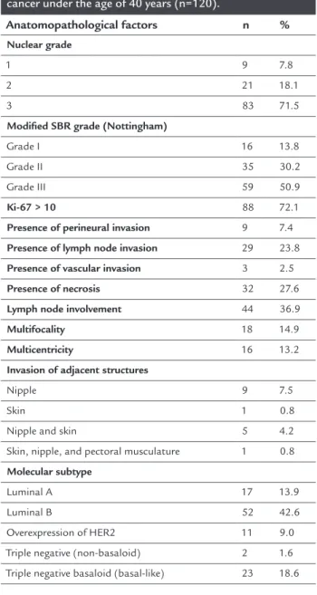
Paget’s disease of the nipple. The most common histo-logical type was invasive ductal carcinoma – not otherwise speciied (IDC-NOS), representing 90 lesions (73.8%). Other special types of invasive tumors corresponded to 18% of the cases. The average size of the invasive lesion in the histopathological evaluation was 31 mm. The histo-logical characteristics of the breast tumors are listed in Table 1. In relation to immunohistochemical classiication, the luminal B subtype was predominant and found in 52 cases (42.6%), followed by the basaloid triple-negative subtype in 23 cases (18.6%).
Diagnostic imaging
Ultrasound was the most requested imaging method for the initial assessment of the breast in young patients, and
was used as a single method in 48% of cases (Table 2). The ultrasound was performed on 113 of the 120 patients included in the study. In four patients (3.3%), the ultra-sound examination was negative or presented benign indings, while in 109 patients (96.5%) it was positive, showing evidence of a malignant lesion. The analysis of the ultrasound examinations showed that most malignant lesions were classiied with nodules (n=109, 94.8%). Most of the nodules presented an irregular shape (54.1%) and indistinct margins (34.8%).
One hundred and twelve (112) of the patients studied underwent MMG, with 65.2% presenting heterogeneously dense or extremely dense breasts. Malignant changes were detected by MMG in 104 patients, while the indings
TABLE 1 Pathological findings of patients with breast cancer under the age of 40 years (n=120).
Anatomopathological factors n %
Nuclear grade
1 9 7.8
2 21 18.1
3 83 71.5
Modified SBR grade (Nottingham)
Grade I 16 13.8
Grade II 35 30.2 Grade III 59 50.9
Ki-67 > 10 88 72.1
Presence of perineural invasion 9 7.4
Presence of lymph node invasion 29 23.8
Presence of vascular invasion 3 2.5
Presence of necrosis 32 27.6
Lymph node involvement 44 36.9
Multifocality 18 14.9
Multicentricity 16 13.2
Invasion of adjacent structures
Nipple 9 7.5
Skin 1 0.8
Nipple and skin 5 4.2 Skin, nipple, and pectoral musculature 1 0.8
Molecular subtype
were negative in eight of them. The presence of suggestive calciications as the only change or associated with other indings was found in 52 lesions, representing the domi-nant mammographic pattern and accounting for 45.6% of the indings. Calciications associated with the nodule accounted for 21 cases (18.4%) and suggestive calciica-tions, viewed as the only change, corresponded to 18 cases (15.8%). A predominance of high-density pleomor-phic calciications (n=21, 40.4%) was found, and grouped lesions prevailed in relation to the distribution (n=25, 48.1%). Nodule-type lesions were the second most com-mon form, accounting for 44.7% of the indings (n=51). An isolated nodule was found in 27 cases (23.7%) and associated with other indings such as calciications and distortion, in 21 (18.4%) and three (2.1%) cases, respec-tively.Nodular lesions with an irregular shape (n=23, 44.2%) and spiculated margins (n=15, 29.4) were the most frequently encountered.
The MRI examination was conducted on 105 patients, with positive indings for 103 (98%). In two cases, the ex-amination’s indings were negative and corresponded to groups of calciications diagnosed solely on MMG. In relation to the morphological characteristics, mass-type lesions were the most frequently encountered (n=67, 62.6%), followed by non-mass lesions (n=17, 15.9%). There was an association between the two patterns in 19.6% of cases (n=21). The irregular (46.6%) and lobular (43.2%) forms were the most prevalent mass-type lesions. Lesions with irregular margins (61.4%) and spiculated lesions (20.4%) were the most frequent. Heterogeneous enhancement (57.9%) was predominant in the assessment of the internal echo pattern. Type III enhancement kinetic curve was the most frequently found (39.8%), followed by the type II or
plateau (23.8%). In relation to non-mass lesions, the most common morphology was that of ductal distribution (28.9%) and, in relation to the internal enhancement pat-tern, the heterogeneous type was the most common (52.6%), followed by the homogeneous pattern (18.4%).
Table 3 describes the BI-RADS® classiication adopt-ed for MMG, ultrasound, and MRI examinations. All of the eight lesions not viewed on MMG (categories 1 and 2) corresponded to invasive tumors. Nineteen (19) invasive tumors did not show openly suggestive features on MMG and were classiied as categories 0 and 3 according to the BI-RADS®. On ultrasound, 13 malignant invasive lesions
were classiied as probably benign indings (category 3), while 3 of the 4 examinations with negative indings rep-resented carcinomas in situ. On MRI, all of the invasive
lesions were characterized as suggestive and the two lesions not viewed corresponded to lesions in situ.
High-risk patients
Thirteen (10.8%) of the 120 patients studied presented high risk for breast cancer. Eleven of these patients had a positive family history for breast or ovarian cancer in irst-degree relatives, one had undergone thoracic irra-diation during adolescence for treatment of lymphoma, and the last had a conirmed diagnosis of genetic muta-tion (Li-Fraumeni syndrome). In relamuta-tion to this group of 13 patients with a high risk for cancer, seven (53.8%) participated in screening programs for the early detection of breast cancer. Four (57%) of the seven patients under-going screening were diagnosed in the symptomatic phase of the disease and three (42%) presented locally advanced disease. The imaging examinations carried out the most among high-risk patients for the prevention
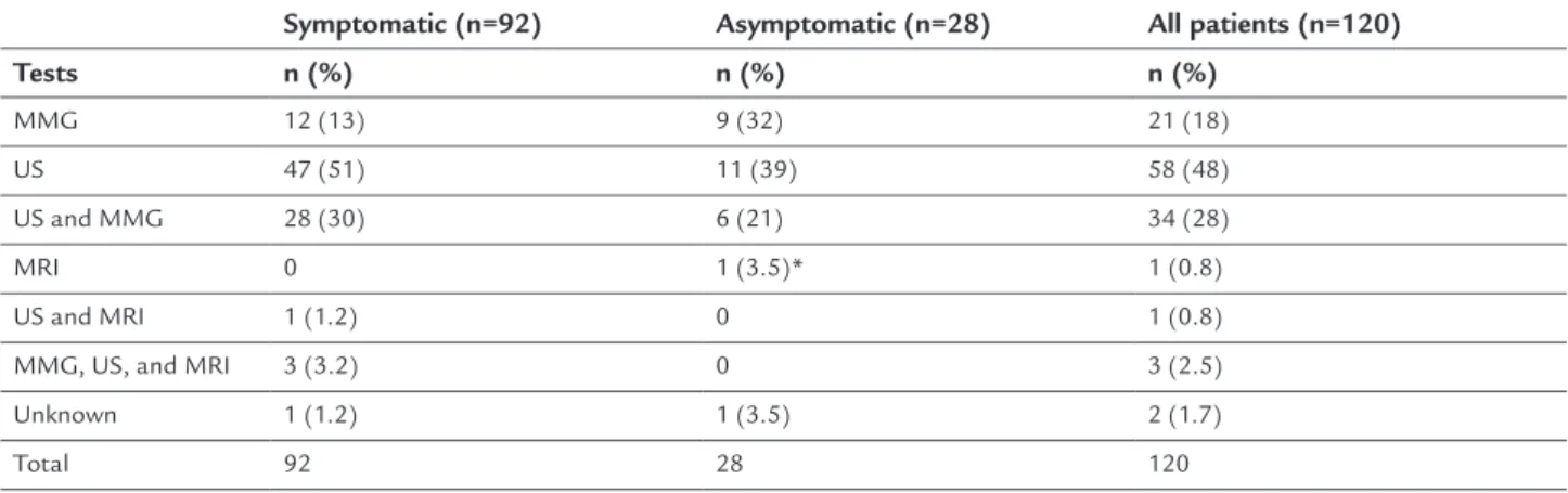
TABLE 2 Imaging methods initially requested for the study of patients with breast cancer under the age of 40 years (n=120).
Symptomatic (n=92) Asymptomatic (n=28) All patients (n=120)
Tests n (%) n (%) n (%)
MMG 12 (13) 9 (32) 21 (18)
US 47 (51) 11 (39) 58 (48)
US and MMG 28 (30) 6 (21) 34 (28)
MRI 0 1 (3.5)* 1 (0.8)
US and MRI 1 (1.2) 0 1 (0.8) MMG, US, and MRI 3 (3.2) 0 3 (2.5) Unknown 1 (1.2) 1 (3.5) 2 (1.7)
Total 92 28 120
of breast cancer were the combination of ultrasound and MMG. Only one patient performed an MRI during screen-ing for high risk.
D
ISCUSSIONAt times, the diagnosis of malignant neoplasm of the breast in women under 40 years of age is more dificult than in women at an older age. Malignant lesions are less common in this group, more dificult to detect and can be more easily interpreted as benign lesions, which are predominant in this age group.
The clinical proile of patients under 40 years of age diagnosed with primary breast neoplasm was represented by women with an average age of 34 years, 73% of whom were users of oral contraceptives (for 8.7 years on average) and 61% of whom had a negative family history of breast or ovarian cancer. In relation to the patients studied, 75.6% reported breast symptoms at diagnosis (64% represented by the presence of a palpable nodule), which is consistent with other studies in the literature.7,14-16 In our case series,
the average time between the onset of the complaint and conirmation of the disease was 6 months.
The histopathological and immunohistochemical aspects of the lesions studied corroborated the data in the literature, with a greater number of lesions with more ag-gressive biological behavior. According to the literature, the anatomopathological presentation of mammary car-cinomas in young women is related to a worse prognosis.17-19
The tumors are often poorly differentiated and may present higher rates of vascular and lymphatic embolization, as well as a higher locoregional recurrence rate.9,20 This was
also veriied in the current study, where most of the tumors showed a high nuclear grade and high proliferative rates, associated with important lymph node involvement.
Although the luminal molecular type is still most fre-quent among young women, the portion of non-hormone
sensitive tumors, both HER2 and triple-negative, is great-er. In a study published in Brazil, a percentage of 27.1% was found for the triple-negative proile in patients aged up to 35 years. This is 17.6% in patients over 60 years of age.21
Ultrasound was the method used the most in the initial assessment of mammary lesions in young patients and was positive in the detection of malignant lesions in 96.5% of the cases. In our study, the sensitivity of the ultrasound for detecting changes to the breasts of young women was slightly higher (96.5%) than that of the MMG (92.2%). Zadelis and Houssami (2003) reported 84% sen-sitivity in the detection of lesions using ultrasound,
com-pared to 76% found using MMG.22 However, Di Nubila
et al. (2006) also found slightly higher sensitivity for ultrasound in relation to MMG (88.7% vs. 84.9%).15 Despite
these indings, the use of ultrasound as an isolated initial method in the assessment of the breasts of young patients should be undertaken with caution, especially among those presenting palpable changes or other symptoms, in which case an attempt should be made to correlate the indings with MMG.
The current study highlighted the fact that MMG can provide essential information in the diagnosis of breast cancer in women less than 40 years of age. Despite the MMG traditionally presenting lower sensitivity in young patients,16 recent technical advances related to the
use of MMG with digital techniques have enabled con-siderable improvement in the pattern of the mammo-graphic image, especially in dense breasts, meaning that its value in the study of younger women has been reas-sessed, especially in symptomatic patients. In our study, positive indings were detected in 92% of the MMG ex-aminations. The dominant pattern was the presence of microcalciications as a single presentation or associated with other indings such as nodules, architectural distor-tion or asymmetry. The change detected on MMG indings
TABLE 3 BI-RADS® classification of the mammary lesions identified on mammogram (MMG), ultrasound (US), and
magnetic resonance imaging (MRI) of patients with breast cancer under the age of 40 years (n=120).
MMG (n=112) US (n=115) MRI (n=105)
BI-RADS® n (%) n (%) n (%)
Category 0 15 13.1 - - -
-Categories 1 and 2 8 7.0 4 3.5 2 1.9
Category 3 4 3.5 13 11.3 -
was characterized as suggestive or highly suggestive in 56.2% of the examinations.
In general, MMG is the irst imaging technique recom-mended to assess most clinical changes in the breasts of women over 40 years old, but its routine use as the irst examination method in younger patients does not repre-sent a consensus. The current study corroborates data from the literature and conirms the importance of MMG in symptomatic patients or those with relevant ultrasound indings, as this method is especially able to detect the presence of suggestive calciications that can sometimes be associated with nodules with benign morphology, which could have a delayed diagnosis if diagnosed by ultrasound alone. In addition, lesions viewed solely using MMG, such as microcalciications, may still represent the only sugges-tive inding, meaning that the lesion may not be diagnosed if an MMG is not performed.
MRI was used especially as a complementary method in the staging of lesions with a conirmed diagnosis for malignancy, and showed an important role in the assess-ment of locoregional disease. The examination was posi-tive in the detection of 98% of tumors and the dominant pattern found was the presence of mass-type lesions with an irregular shape, with the type III kinetic curve found the most. Despite the limitations relating to cost, MRI has advantages compared to other examination, such as lack of exposure to radiation compared to MMG, thereby decreasing the carcinogenic effect. It has excellent perfor-mance in the characterization of other suggestive indings of malignancy not detected using ultrasounds or MMG because it is a highly sensitive method. Furthermore, it can demonstrate the functional behavior of lesions and provide greater clarity in determining their extent, thus enabling a more targeted treatment.23-25
For women under 40 years old there are no recom-mendations for performing breast imaging examinations for early detection of breast cancer, except in an indi-vidualized manner among those at high risk or symp-tomatic individuals.26 In this study, half of the patients
with important risk factors did not undergo screening before diagnosis, while others reported having undergone imaging examinations for breast cancer prevention even without any formal recommendation, although not regularly. This reality relects the shortcomings in the implementation of the screening recommendations for this age group. Additional efforts are needed to identify relevant primary and secondary preventive approaches, including not only advanced research seeking to iden-tify the predictors of early risk and biomarkers, but also strengthening the healthcare practices with effective
measures for warning the younger population about the importance of the disease.
The usefulness of ultrasounds as a supplementary screening method in asymptomatic patients with negative MMG has already been conirmed in the literature, with an increase of up to 42% demonstrated in the detection of breast cancer in patients with dense breasts.27 The use
of ultrasounds can also be indicated as a diagnostic al-ternative for screening of breast cancer in high-risk wom-en who have no access to MRI, which is a common situ-ation in our country. MRI has been underused for screening purposes in high-risk patients despite its use being recommended and widely advocated in the inter-national literature.28-30
This study has shown that, in our country, most cases of breast cancer diagnosed in patients less than 40 years of age presented symptoms at diagnosis and tumors with more aggressive biological behavior. Despite ultra-sounds being the most widely used method in the diag-nosis of mammary lesions in this group of patients, we noted more precise characterization of mammary lesions in young patients when MMG and MRI are used in com-bination with the ultrasound examination. The discussion of these indings is essential for identifying preventive approaches to warn the younger population about the importance of the disease, as well as developing effective early diagnostic measures in this population.
R
ESUMOPeril do câncer de mama em mulheres com idade inferior a 40 anos
Objetivo: descrever o peril clínico, os achados de imagem e os aspectos anatomopatológicos do câncer de mama em mulheres com idade inferior a 40 anos.
Método: estudo retrospectivo, descritivo, com análise de prontuários de novembro de 2008 a agosto de 2012. Foram estudadas 120 pacientes, das quais 112 realizaram ma-mograia, 113 ultrassonograia e 105 ressonância magné-tica (RM). A coleta dos dados histopatológicos foi reali-zada com informações pós-cirúrgicas, disponíveis para 113 pacientes.
tu-mores primários, dos quais 112 eram invasivos (95%). O tipo histológico mais encontrado foi o carcinoma ductal invasivo (73,8%). Em relação ao subtipo molecular, o lu-minal B foi predominante (42,6%). A ultrassonograia foi positiva em 94,5% dos casos e o achado mais comum foi nódulo (94,8%). Na mamograia, a lesão maligna foi evi-denciada em 92,8% e a presença de calciicações suspeitas foi o padrão dominante. O exame de RM foi positivo em 98% dos pacientes, sendo lesões tipo massa as mais comuns.
Conclusão: a maioria dos casos de câncer de mama em pacientes com idade inferior a 40 anos apresentavam sintomas ao diagnóstico e tumores de comportamento biológico mais agressivo. Apesar de a ultrassonograia ter sido o método mais utilizado, observamos uma melhora da caracterização das lesões mamárias quando utilizadas também a mamograia e a RM.
Palavras-chave: neoplasias da mama, mamograia, ultras-sonograia mamária, imagem por ressonância magnética.
R
EFERENCES1. Mintzer D, Glassburn J, Mason BA, Sataloff D. Breast cancer in the very young patient: a multidisciplinary case presentation. Oncologist. 2002; 7(6):547-54. 2. Kothari AS, Beechey-Newman N, D’Arrigo C, Hanby AM, Ryder K, Hamed
H, et al. Breast carcinoma in women age 25 years or less. Cancer. 2002; 94(3):606-14.
3. Agarwal G, Pradeep P V, Aggarwal V, Yip CH, Cheung PS. Spectrum of breast cancer in Asian women. World J Surg. 2007; 31(5):1031-40.
4. Villarreal-Garza C, Aguila C, Magallanes-Hoyos MC, Mohar A, Bargalló E, Meneses A, et al. Breast cancer in young women in Latin America: an unmet, growing burden. Oncologist. 2013; 18(12):1298-306.
5. Fancher TT, Palesty JA, Paszkowiak JJ, Kiran RP, Malkan AD, Dudrick SJ. Can breast self-examination continue to be touted justiiably as an optional practice? Int J Surg Oncol. 2011; 2011:965464.
6. Lalloo F, Varley J, Moran A, Ellis D, O’Dair L, Pharoah P, et al. BRCA1, BRCA2 and TP53 mutations in very early-onset breast cancer with associated risks to relatives. Eur J Cancer. 2006; 42(8):1143-50.
7. Samphao S, Wheeler AJ, Rafferty E, Michaelson JS, Specht MC, Gadd MA, et al. Diagnosis of breast cancer in women age 40 and younger: delays in diagnosis result from underuse of genetic testing and breast imaging. Am J Surg. 2009;198(4):538-43.
8. Eccles BK, Copson ER, Cutress RI, Maishman T, Altman DG, Simmonds P, et al.; POSH Study Steering Group. Family history and outcome of young patients with breast cancer in the UK (POSH study). Br J Surg. 2015; 102(8):924-35. 9. Anders CK, Hsu DS, Broadwater G, Acharya CR, Foekens JA, Zhang Y, et al.
Young age at diagnosis correlates with worse prognosis and deines a subset of breast cancers with shared patterns of gene expression. J Clin Oncol. 2008; 26(20):3324-30.
10. Gewefel H, Salhia B. Breast cancer in adolescent and young adult women. Clin Breast Cancer. 2014; 14(6):390-5.
11. Lee H-B, Han W. Unique features of young age breast cancer and its management. J Breast Cancer. 2014; 17(4):301-7.
12. Tavassoli F, Devilee P. Pathology and genetics of tumours of the breast and female genital organs. World Health Organization Classiication of Tumours. 5. ed. Lyon: IARC Press; 2003.
13. Elston CW, Ellis IO. Pathological prognostic factors in breast cancer. I. The value of histological grade in breast cancer: experience from a large study with long-term follow-up. Histopathology. 1991; 19(5):403-10.
14. Ruddy KJ, Gelber S, Tamimi RM, Schapira L, Come SE, Meyer ME, et al. Breast cancer presentation and diagnostic delays in young women. Cancer. 2014; 120(1):20-5.
15. Di Nubila B, Cassano E, Urban LABD, Fedele P, Abbate F, Maisonneuve P, et al. Radiological features and pathological-biological correlations in 348 women with breast cancer under 35 years old. Breast. 2006; 15(6):744-53. 16. Foxcroft LM, Evans EB, Porter AJ. The diagnosis of breast cancer in women
younger than 40. Breast. 2004; 13(4):297-306.
17. El Saghir NS, Seoud M, Khalil MK, Charafeddine M, Salem ZK, Geara FB, et al. Effects of young age at presentation on survival in breast cancer. BMC Cancer. 2006; 6:194.
18. Weigelt B, Horlings HM, Kreike B, Hayes MM, Hauptmann M, Wessels LFA, et al. Reinement of breast cancer classiication by molecular characterization of histological special types. J Pathol. 2008; 216(2):141-50.
19. Yao Y, Cao M, Fang H, Xie J. Breast cancer in 30-year-old or younger patients: clinicopathologic characteristics and prognosis. World J Surg Oncol. 2015; 13(1):38. 20. Rudra S, Yu DS, Yu ES, Switchenko JM, Mister D, Torres MA. Locoregional and distant recurrence patterns in young versus elderly women treated for breast cancer. Int J Breast Cancer. 2015; 2015:213123.
21. Bacchi LM, Corpa M, Bacchi CE Carvalho FM. Caracterização anatomopatológica e imunofenotípica de carcinomas de mama em mulheres jovens. Rev Bras Mastol. 2009; 19(2):42-6.
22. Zadelis S, Houssami N. Mammographic features of breast cancer in young symptomatic women. Australas Radiol. 2003; 47(4):404-8.
23. Kuhl C, Weigel S, Schrading S, Arand B, Bieling H, König R, et al. Prospective multicenter cohort study to reine management recommendations for women at elevated familial risk of breast cancer: the EVA trial. J Clin Oncol. 2010; 28(9):1450-7.
24. Sardanelli F, Giuseppetti GM, Panizza P, Bazzocchi M, Fausto A, Simonetti G, et al. Sensitivity of MRI versus mammography for detecting foci of multifocal, multicentric breast cancer in fatty and dense breasts using the whole-breast pathologic examination as a gold standard. AJR Am J Roentgenol. 2004; 183(4):1149-57.
25. Salem DS, Kamal RM, Mansour SM, Salah LA, Wessam R. Breast imaging in the young: the role of magnetic resonance imaging in breast cancer screening, diagnosis and follow-up. J Thorac Dis. 2013; 5(Suppl.1):S9-18. 26. Aguiar RMB, Bitencourt AGV, Chojniak R, Valadão ACR, Maciel M do S,
Marques EF. Multimodality imaging in the optimization of breast cancer screening : literature review. Appl Cancer Res. 2013; 33(4):194-8. 27. Kolb TM, Lichy J, Newhouse JH. Comparison of the performance of screening
mammography, physical examination, and breast US and evaluation of factors that inluence them: an analysis of 27,825 patient evaluations. Radiology. 2002; 225(1):165-75.
28. Kuhl CK, Schrading S, Leutner CC, Morakkabati-Spitz N, Wardelmann E, Fimmers R, et al. Mammography, breast ultrasound, and magnetic resonance imaging for surveillance of women at high familial risk for breast cancer. J Clin Oncol. 2005; 23(33):8469-76.
29. Morris EA. Diagnostic breast MR imaging: current status and future directions. Radiol Clin North Am. 2007; 45(5):863-80.