(1) Universidade Veiga de Almeida, UVA, Rio de Janeiro, RJ, Brasil.
(2) Universidade Federal do Rio de Janeiro, UFRJ, Rio de Janeiro, RJ, Brasil.
Conlict of interest: non-existent
Brainstem auditory evoked potentials in smokers
Potenciais evocados auditivos de tronco encefálico em fumantes
Denise Miranda Terra Martins(1)
Cristiane Fregonesi Dutra Garcia(2)
Heidi Elisabeth Baeck(1)
Silvana Frota(2)
Received on: April 19, 2015 Accepted on: December 07, 2015
Mailing address:
Denise Miranda Terra Martins Rua Oswaldo Cruz 28/1001 – Icaraí Niterói – RJ – Brasil
CEP: 24230-210
E-mail: denisemterra@hotmail.com
doi: 10.1590/1982-0216201618113915
ABSTRACT
Purpose: to perform a comparative study of brainstem evoked auditory potentials between smokers and non-smokers.
Methods: the group studied was composed of 40 individuals, being 20 non-smokers and 20 smokers within the range of 20 to 59 years of age. All participants had to present responses to tonal thresholds within normal range and tympanometry type A, with the presence of ipsilateral and contralateral acoustic relexes. Both groups underwent brain stem auditory evoked potential (BAEP). The parameters used to compare the two groups were the absolute latencies of waves I, III and V, the inter-latency waves I-III, IV and III-V in both ears, the difference between the IV peak latency between the two ears and the inter--aural difference of wave V absolute latency between the two ears.
Results: in our results, it was ascertained that the group of smokers showed latency I in the RE (p= 0.036), latency V in the RE (p= 0.007), latency V in the LE (p=0.014), inter-latency III-V in the RE (p=0.015) and LE (p= 0.016) signiicantly higher than the non-smokers. There was no signiicant diffe-rence in wave V latency between the two ears.
Conclusion: the results of the study led to the conclusion that tobacco is a risk factor for the central auditory nervous system, interfering with latencies and with BAEP inter-wave latencies in the group of smokers when compared to the group of non-smokers.
Keywords: Smoking; Evoked Potentials, Auditory, Brain Stem; Hearing
RESUMO
Objetivo: comparar os resultados dos exames de potenciais evocados auditivos de tronco encefálico em indivíduos não tabagistas e tabagistas.
Métodos: foram estudados 40 indivíduos, sendo 20 não tabagistas e 20 tabagistas, com idades entre 20 e 59 anos. Todos os participantes incluídos na pesquisa deveriam apresentar respostas de limiares tonais dentro dos padrões da normalidade e timpanometria tipo A com presença de relexos acústicos contralaterais e ipsilaterais. Em ambos os grupos foram realizados os potenciais evocados auditivos de tronco encefálico (PEATE), por meio de cliques. Os parâmetros que foram utilizados na comparação dos dois grupos foram as latências absolutas das ondas I, III e V; as interlatências das ondas I-III, I-V e III-V em ambas as orelhas; a diferença da latência interpico I-V entre as duas orelhas e a diferença interaural da latência absoluta da onda V entre as duas orelhas.
Resultados: os resultados encontrados mostraram que o grupo de tabagistas apresentou latência I da Orelha Direita (p=0,036), latência V da Orelha Direita (p=0,007), latência V da Orelha Esquerda (p=0,014), interlatência III-V da Orelha Direita (p=0,015) e Orelha Esquerda (p=0,016) signiicante-mente maior que o grupo de não tabagistas. Não houve diferença signiicante na latência da onda V entre as duas orelhas.
Conclusão: os resultados da pesquisa levaram à conclusão de que o tabaco é um fator de risco para o sistema nervoso auditivo central, que pode interferir nas latências e interlatências das ondas do PEATE no grupo de tabagistas quando comparado com o grupo de não tabagistas.
Descritores: Tabagismo; Potenciais Evocados Auditivos de Tronco Encefálico; Audição Original articles
INTRODUCTION
According to the World Health Organization (WHO, 2003)1,2, the cigarette, in a general way, affects the
health of the individual and it is responsible for cancer in the lungs, larynx, cervix (in female smokers), pancreas, bladder, esophagus, stomach and kidneys, besides cardiovascular and respiratory diseases, among others. These are fatal diseases, and the amount of cigarettes smoked per day is proportional to the risk of contracting them.
The cigarette can also be harmful for the hearing because of the antioxidant mechanism effect or because of the vascular suppression of the auditory system, as this may cause conductive, mixed, senso-rineural or central hearing loss. The brainstem auditory evoked potentials (ABR) test allows one to check the electrophysiological activity of the auditory system at brainstem level, to map the synapses of the auditory pathways from the cochlear nerve, cochlear nuclei, superior olivary complex (bridge) to the inferior colliculus (midbrain)3.
The most commonly performed and used analysis for the ABR test are waves I, III and V latency values and their interpeaks I-III, III-V and I-V, since these three waves have higher amplitude and stability. Absolute wave latency V, the interaural difference of wave V latency and interpeaks I-III, I-V, and III-V are good parameters for diagnostic purposes4. One of the
qualities of the ABR test is the ability to evaluate the neurophysiological integrity of the brain stem auditory pathways. You can compare the speed of progress of the stimulus (latencies) in both ears5.
For over 100 years it´s been suggested that there is a relation between smoking and hearing loss6 , and
recent research carried out by Belgian researchers offer convincing evidence on the subject. These researchers
stated that smoking signiicantly increased
high-frequency hearing loss and the dose dependent effect. According to Erik Fransen, one of the researchers of
the beating rate of hair cells lashes in the lining of the middle ear, reducing the frequency of its beating rate and leading to persistence of middle ear infections10.
A study on the effects of smoking on brainstem auditory evoked potentials (ABR) in twelve smokers found that the latency and amplitude of wave peaks I, III and V were evaluated and analyzed, and there was
no signiicant effect of peaks I and III. A signiicant effect
for peak V with tobacco, resulting in higher latencies was observed11.
The information above motivated this research, and its goal is to carry out a comparative study of the brainstem auditory evoked potentials in non-smokers and in smokers, observing absolute latencies of waves I, III and V; interpeak latencies of waves I-III, III-V, I-V in both ears; the difference in interpeak latency I-V between the two ears and the interaural difference of wave V latency between the two ears.
METHODS
This research was approved by the Research Ethics Committee (CEP) of the University Veiga de Almeida (UVA), under number 302/11. It is a cross-sectional, observational study, of descriptive and exploratory type.
The literature review was performed using the Regional Medical Library (BIREME), and we used the databases from LILACS and SCIELO. Articles and
scientiic publications were also reviewed.
The study was conducted in the Audiology Clinic of the University Hospital, Department of Audiology.
The study was divulged by the Program of Study and Treatment of Smoking - PROJETA, a project linked to the INCA (National Institute of Cancer). This project was developed in the Hospital, which recruits volun-teers for the study.
To select the sample, we initially carried out anamnesis, otoscopy, pure-tone audiometry threshold and immitanciometry exams.
The anamnesis was composed of questions in order to gather information on personal background, such as audiological history, general health and exposure to occupational noise. We excluded individuals who presented otalgia, otitis, ear surgery history, neuro-logical disorders and exposure to occupational noise/ acoustic trauma.
An ENT doctor performed the otoscopy, in order to detect any alterations that could affect the implemen-tation of the remaining stages of the research. When the presence of earwax was detected, the participant was referred to earwax removal and advised to return to the study after that procedure. In cases where the otorhinolaryngology evaluation detected other ear abnormalities, the participant was excluded from the research.
In the acoustic cabin, patients were previously
instructed on the dynamics of the exam. We veriied
pure tone hearing thresholds in the frequencies of 0.25 kHz, 0.5 kHz, 1 kHz, 2 kHz, 3 kHz, 4 kHz, 6 kHz and 8 kHz for air conduction, and 0.5 kHz, 1 kHz, 2 kHz, 3 kHz and 4 kHz for bone conduction. We excluded from the research subjects with alterations in the audiometric thresholds, i.e., those who obtained thresholds worse than 25 dBNA12.
The individuals underwent tympanometry and
research of acoustic relexes in frequencies 0.5 kHz,
1 kHz, 2 kHz and 4 kHz. We also excluded individuals who presented type B or C curve and absence of
contralateral acoustic relex in two or more frequencies,
among frequencies 0.5 kHz, 1 kHz, 2 kHz and 4 kHz. All participants selected for the research in both groups had to present responses to tonal thresholds within the standards of normality and type A tympa-nometry, with the presence of contralateral and
ipsilateral acoustic relexes.
The register of the ABR test was obtained with the patient lying awake, in a quiet environment, with the use of insert earphones (Eartone), surface electrodes
placed in the vertex and bilateral mastoid region, ixed
with electrolytic paste. The sound stimulus used, issued on intensity of 85 dB SPL, consisted of 100 µs long clicks, alternating polarity at rate 27.7 Hz and prome-diation of 1024 stimuli, and the collected signal was
iltered between 100 and 5000 Hz. The electric stimulus
generated in the computer was turned into acoustic stimulus and transmitted through the auditory system to generate the tone-evoked potential13.
Two records were performed, for each ear, in order to check the reproducibility of the tracing and
conirm the presence or absence of the waves. For this
research, between the two records we chose the one with the clearest morphology.
In the electrophysiological evaluation, we analyzed the latency values of waves I, III and V, as well as the interpeak intervals IIII, IV and IIIV, for both ears.
We studied interpeak latency I-III, which represents the activity between the auditory nerve and the lower
brainstem, interpeak latency III-V, which relects the
activity of the higher brainstem and interpeak latency I-V, which is the most important one, because it repre-sents all the activity from the auditory nerve to the nuclei and tracts of the brainstem13.
We also studied the comparison of interpeak latency I-V between the two ears, granted that the inter-aural difference should not exceed 0.3 ms in normal individuals. In the absence of wave I, the interaural difference was calculated between absolute latencies of waves V and it should not exceed 0.3 ms in normal individuals either.
Statistical analysis was done using the following methods: We applied the Mann-Whitney Test to
ascertain if there was signiicant difference between
the smoking and the non-smoking groups in latency and inter-latency measurements (at 85 dB NHL). We applied the Fisher’s Exact Test to check the rate of change of these measurements. In order to check
if there was signiicant variation in latency and
inter-latency measurements from the right to the left ear we used the Wilcoxon signed-rank test.
Clinical variables were analyzed using Fisher’s exact Test (sex) and Mann-Whitney Test.
Nonparametric tests were applied because latency and inter-latency measurements did not present Gaussian distribution, due to the rejection of the hypothesis of normality according the
Kolmogorov-Smirnov test. The criteria for determining signiicance
level was 5%. The Statistical analysis was processed by the software SAS 6.11 (SAS Institute Inc., Cary, NC).
RESULTS
Using the descriptive level of the Mann-Whitney test it was ascertained that the smoking group showed latency I for the RE (p = 0.036), latency V for the RE
(p = 0.007) and latency V for the LE (p = 0.014) signii
inter-latencies III-V for the LE (p = 0.016) signiicantly
higher than those of the non-smoking group (Table 1).
There was no signiicant difference, at the 5% level, in
inter-latencies between the two groups.
In order to check if there was a signiicant difference
in the change in the of latency measure between non-smoking groups (G1) and smoking groups (G2), table 2 provides the frequency (n) and percentage (%) of change in latency measurements of right and left ear. Table 2 also provides the corresponding descriptive level (p-value) of Fisher´s exact test.
It was ascertained that there was no signiicant
difference, at 5% level, in the rate of change of latency measurements between the two groups. Table 2 also provides the interaural difference analysis (IA) between the groups.
In table 3, we can observe if there is signiicant
difference in latency and inter-latency measurements (at 85 dB NHL) from the right to the left ear. Table 3 also provides the average, standard deviation (SD) and the median latencies (at 85 dB NHL) according to the ear (right and left) and the corresponding descriptive level (p-value) of the Wilcoxon signed-rank test for the total sample and per group (smokers and non-smokers). It
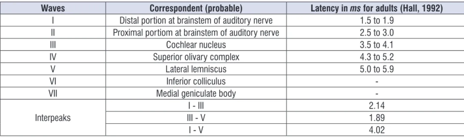
Waves Correspondent (probable) Latency in ms for adults (Hall, 1992)
I Distal portion at brainstem of auditory nerve 1.5 to 1.9 II Proximal portiom at brainstem of auditory nerve 2.5 to 3.0 III Cochlear nucleus 3.5 to 4.1 IV Superior olivary complex 4.3 to 5.2
V Lateral lemniscus 5.0 to 5.9
VI Inferior colliculus
-VII Medial geniculate body
-Interpeaks III - VI - III 2.141.89
I - V 4.02
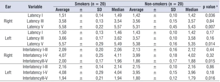
Table 1. Latency values of wavesI, III e V and inter-latencies of I-III, I-V e III-V, right and left ears, for the group of smokers and for the group of non-smokers at 85dB NHL
Ear Variable Smokers (n = 20) Non-smokers (n = 20) p value a
Average ± SD Median Average ± SD Median
Right Latency I 1,51
± 0,14 1,49 1,42 ± 0,10 1,42 0,036
Latency III 3,58 ± 0,13 3,54 3,56 ± 0,15 3,57 0,84
Latency V 5,58 ± 0,22 5,57 5,31 ± 0,45 5,43 0,007
Left Latency I 1,50
± 0,13 1,46 1,43 ± 0,10 1,42 0,17
Latency III 3,66 ± 0,17 3,62 3,57 ± 0,10 3,58 0,16
Latency V 5,57 ± 0,29 5,49 5,38 ± 0,16 5,35 0,014
Right Interlatency I-lll 2,09
± 0,20 2,06 2,13 ± 0,16 2,12 0,44
Interlatency I-V 4,07 ± 0,25 4,11 3,99 ± 0,18 4,02 0,32
Interlatencylll-V 2,00 ± 0,17 1,96 1,86 ± 0,17 1,88 0,015
Left Interlatency I-lll 2,16
± 0,14 2,14 2,15 ± 0,10 2,16 0,86
Interlatency I-V 4,08 ± 0,29 4,04 3,95 ± 0,15 3,96 0,18
Interlatencylll-V 1,94 ± 0,21 1,94 1,80 ± 0,12 1,79 0,016
SD: Standard deviation
a descriptive level of Mann-Whitney test.
Table 2. Alteredlatency values, in the group of smokers,compared to the unaltered values, in the group of non-smokers, for waves I, III, V and for inter-latencies I-III,I-V, III-V
Ear Altered
Value
Smokers (n = 20) Non- smokers (n = 20)
p value a
n % n %
Right Latency I 0 0,0 0 0,0
NA
Latency III 0 0,0 0 0,0 NA
Latency V 2 10,0 0 0,0 0,24
Left Latency I 0 0,0 0 0,0
NA
Latency III 0 0,0 0 0,0 NA
Latency V 1 5,0 0 0,0 0,50
Right Inter-latency I-lllInter-latency I-V 12 10,05,0 00 0,00,0 0,500,24 Inter-latency lll-V 1 5,0 0 0,0 0,50
Left Inter-latency I-lll 0 0,0 0 0,0
NA
Inter-latency I-V 2 10,0 0 0,0 0,24 Inter-latency lll-V 1 5,0 0 0,0 0,50 Difference IA < 0,3ms 20 100,0 20 100,0 NA
When studying latencies I, III and V (table 1), better latency measurements value I were found in the right ear (RE) and left ear (LE) best for the non-smoking
group (G1). However, statistically signiicant difference
was found only in the right ear (RE).
In latency III, no signiicant change between the two groups was observed. However, signiicantly higher
change in latency V in the RE (p = 0.007) and LE (p = 0.014) was found for the smoking group (G2) when compared to the non-smoking group (G1). These results are consistent with a study on the effects of
smoking on the latencies of the ABR where a signiicant
DISCUSSION
The ABR test stands out for being an effective method in measuring the brainstem
electrophysi-ological proile, considering the ascending auditory
pathways, which occupy the segment of this structure in the central nervous system. The brainstem runs various functions of the human organism, from the simplest,
such as primitive relexes, to integrated relexes, like the relexes responsible for heart rate, breathing and blood
pressure control13-17.
The deterioration of the nervous system function, for
Table 3. Latency values for waves I, III, V and inter-latencies of I-III, I-V, III-V, according to right and left ears, in all the sample per groups – smokers and non-smokers, at 85 dB NHL
Sample Variable Right Ear Left Ear p value a
Average ± SD Median Average ± SD Median
To
tal
(n
= 40)
Latency I 1,47 ± 0,13 1,45 1,46 ± 0,12 1,42 0,58
Latency III 3,57 ± 0,14 3,56 3,62 ± 0,15 3,59 0,010
Latency V 5,45 ± 0,38 5,51 5,47 ± 0,25 5,41 0,81
Inter-latency I-lll 2,11 ± 0,18 2,10 2,16 ± 0,12 2,15 0,10
Inter-latency I-V 4,03 ± 0,22 4,05 4,01 ± 0,24 3,99 0,97
Inter-latency lll-V 1,93 ± 0,19 1,92 1,87 ± 0,18 1,84 0,063
Gro
up
o
f s
m
ok
ers
(n
= 20)
Latency I 1,51 ± 0,14 1,49 1,50 ± 0,13 1,46 0,52
Latency III 3,58 ± 0,13 3,54 3,66 ± 0,17 3,62 0,010
Latency V 5,58 ± 0,22 5,57 5,57 ± 0,29 5,49 0,90
Inter-latency I-lll 2,09 ± 0,20 2,06 2,16 ± 0,14 2,14 0,22
Inter-latency I-V 4,07 ± 0,25 4,11 4,08 ± 0,29 4,04 0,54
Inter-latency
lll-V 2,00 ± 0,17 1,96 1,94 ± 0,21 1,94 0,23
Gro
up
o
f n
on
-s
m
ok
ers
(n
= 20)
Latency I 1,42 ± 0,10 1,42 1,43 ± 0,10 1,42 0,93
Latency III 3,56 ± 0,15 3,57 3,57 ± 0,10 3,58 0,33
Latency V 5,31 ± 0,45 5,43 5,38 ± 0,16 5,35 0,66
Inter-latency I-lll 2,13 ± 0,16 2,12 2,15 ± 0,10 2,16 0,47
Inter-latency I-V 3,99 ± 0,18 4,02 3,95 ± 0,15 3,96 0,42
Inter-latency
lll-V 1,86 ± 0,17 1,88 1,80 ± 0,12 1,79 0,13
SD: Standard Deviation
For the group of non-smokers (G1) we found that 100% of the sample did not present any alterations in the items studied (latency, inter-latency and interpeak).
Thus, in the total of the group of smokers (G2), three individuals were found (15%) with alterations in one or more of the items studied (latency, inter-latency and interpeak).
In the analysis of interaural difference, it was
observed that there was no signiicant difference at the
level of 5% in wave V latency between the two ears. Given the importance of the study of the interaural difference of wave V latency for diagnostic purposes13,
we decided to conduct this study according to the groups, but no alterations were found.
CONCLUSION
There are signiicant differences in the absolute
latencies of wave I, electric impulse transmission as far as the auditory nerve, in the right ear, and of wave V, electrical impulse to the lateral lemniscus, in both ears, for the groups of smokers when compared with the group of non-smokers.
There is an increase in inter-latencies III-V, which may indicate impairment of the higher brainstem, in both ears, for the group of smokers.
There is no interaural difference of wave V between the ears, in both groups.
The nicotine found in tobacco, which interferes with the neural transmission of auditory information, is a risk factor for the central auditory nervous system. It can affect the latencies and inter-latencies of the auditory brainstem response test in the group of smokers, when compared to the group of non-smokers, when we consider that this test is able to evaluate the neurophys-iological integrity of the brainstem auditory pathways.
REFERENCES
1. Ministério da Saúde (Brasil), Instituto Nacional de Câncer. Programa Nacional de controle do tabagismo e outros fatores de risco de câncer: modelo lógico e avaliação. Brasília: Ministério da Saúde, 2003.
2. Ministério da Saúde (Brasil), Instituto Nacional de Câncer. Relatório da OMS sobre a epidemia global de tabagismo. Brasília: Ministério da Saúde, 2008. 3. Angrisani RMG, Matas CG, Furtado JRB. Análise
dos potenciais evocados auditivos em fumantes. Acta ORL. 2008;26(3):146-50.
compared to the group of non-smokers, and this
difference is signiicant. There was no similarity found in studies that presented statistically signiicant values
only in the right ear (RE); perhaps, this fact can be
justiied because of the small size of the sample.
According to Table 1, in the analysis of inter-latencies I-III and I-V, it was observed that there was
no statistically signiicant difference between the two
groups, at 5% level. It was observed that the group of smokers (G2) presented inter-latencies III-V in the RE (p = 0.015) and inter-latencies III-V in the LE (p =
0.016) signiicantly higher than those of the group of
non-smokers (G1). This increase in III-V inter-latencies found in the group of smokers (G2) may represent an abnormality of neural transmission, possibly located between the cochlear nuclei (wave III) and lateral lemniscus (wave V) 15. Due to the size of the sample, it was not possible to carry out a study considering the time of smoking, the minimum amount of cigarettes smoked per day and inter-latency III-V. However, we suggest studies with a larger population, with a view
to the identiication of the inluence of the quantity of
tobacco in neural transmission of auditory information,
relected in the latencies and inter-latencies of the ABR.
To analyze the criterion of normality in table 2, we adopted the values suggested by Hall (1992)13.
In studies of the latencies according to groups, in agreement with the proposal of normality, individuals with altered latencies were found only in the group of smokers (G2), where the latencies of wave V were altered in two individuals, granted that one individual had it in both ears (RE and LE) and the other in his left
ear (LE). In consulted literature, we did not ind other
studies using individual values of latency, inter-latency and interpeak and these criteria of normality, which
makes it impossible to compare the indings with other
studies.
Still based on the normality criteria of Hall (1992)13,
an analysis of individuals of inter-latencies I-V was
presented in table 2. It was veriied that there were more
alterations in the group of smokers (G2.) We observed three individuals with altered values, considering that the participant had alterations in both ears (RE and LE), one participant (RE) and another participant (LE). However, statistical analysis only pointed difference in inter-latencies III-V.
diagnóstico de morte encefálica. Pró-Fono R Atual. Cient. 2008;20(2):123-8.
17. Sousa LCA, Rodrigues LS, Pizza MRT, Ferreira DR, Ruiz DB. Achado ocasional de doenças neurológicas durante a pesquisa da surdez infantil através do BERA. Rev Bras Otorrinolaringol. 2007;73(3):424-8.
18. Harkrider AW, Champlin CA, McFadden D. Acute effect of nicotine on non-Smokers: OAEs and ABRs. Hear Res. 2001;(160):73-88.
19. Weis W. How Smoking affects hearing. Medical Times. 1970;98(11):84-8.
20. Knott VJ, Harr A, Mhoney C. Smoking history and aging-associated cognitive decline: an event-related potential study. Neuropsychobiology. 1999;40:95-106.
4. Lima MAMT. Potencial evocado auditivo:
eletrococleograia e audiometria de tronco
encefálico. In: Frota S. Fundamentos em fonoaudiologia: audiologia. 2.ed. Rio de Janeiro: Guanabara Koogan; 2003. p.157-72.
5. Musiek FE, Borestein SP, Hall III JW, Schwaber MK. Audiometria de tronco encefálico (ABR): neurodiagnóstico e aplicações intra-operatórias. In: Katz J (org). Tratado de Audiologia Clínica. 4.ed. São Paulo: Manole; 1999. p.349-71.
6. Domino EF. Effects of tobacco smoking on electroencephalographic auditory evoked and event related potentials. Brain and Cognition. 2003;53:66-74.
7. Fransen E, Topsakal V, Hendrickx JJ, Van Laer L, Huyghe JR, Van Eyken E et al. Occupational noise, smoking, and a high body mass index are risk factors for age-related hearing impairment and moderate alcohol consumption is protective: a European population-based multicenter study. J Assoc Res Otolaryngol. 2008;9(3):264-76.
8. Jess D. Lifestyle Choises can affect hearing. Disponível em www.advanceweb.com 2008. Acesso em; 24mai 2010.
9. Paschoal CP, Azevedo MF. O cigarro como fator de risco para alterações auditivas. Braz J Otorhinolaryngol 2009;75(6):893-902.
10. Rodrigues J, Malatesta R. Células ciliadas da mucosa da orelha média. Acta Médica Misericordia. 1998;1(1):26-8.
11. Knott VJ. Acute effects of tobacco on human brain stem evoked potentials. Addictive Behaviors Journal.1987;12(4):375-9.
12. Frota S. Avaliação básica da audição. In: Frota S. Fundamentos em fonoaudiologia: audiologia. 2.ed. Rio de Janeiro: Guanabara Koogan; 2003. p. 41-56. 13. Hall III, JW. Anatomy and physiology. In: Hall III,