Kinetics of TNF-alpha and IFN-gamma
mRNA expression in islets and spleen
of NOD mice
1Disciplina e Laboratório de Imunologia Clínica, Departamento de Clínica Médica, and 2Departamento de Medicina Preventiva e Social, Faculdade de Ciências Médicas, Universidade Estadual de Campinas, Campinas, SP, Brasil
D. Ventura-Oliveira1, C.A. Vilella1, M.E. Zanin1, G.M. Castro1, D.C. Moreira Filho2 and R.L. Zollner1
Abstract
Insulin-dependent diabetes mellitus is caused by autoimmune destruc-tion of pancreatic ß cells. Non-obese diabetic (NOD) mice spontane-ously develop diabetes similar to the human disease. Cytokines pro-duced by islet-infiltrating mononuclear cells may be directly cytotoxic and can be involved in islet destruction coordinated by CD4+ and CD8+ cells. We utilized a semiquantitative RT-PCR assay to analyze
in vitro the mRNA expression of TNF-a and IFN-g cytokine genes in
isolated islets (N = 100) and spleen cells (5 x 10⁵
cells) from female NOD mice during the development of diabetes and from female CBA-j mice as a related control strain that does not develop diabetes. Cy-tokine mRNAs were measured at 2, 4, 8, 14 and 28 weeks of age from the onset of insulitis to the development of overt diabetes. An increase in IFN-g expression in islets was observed for females aged 28 weeks
(149 ± 29 arbitrary units (AU), P<0.05, Student t-test) with advanced
destructive insulitis when compared with CBA-j mice, while TNF-a
was expressed in both NOD and CBA-j female islets at the same level at all ages studied. In contrast, TNF-a in spleen was expressed at
higher levels in NOD females at 14 weeks (99 ± 8 AU, P<0.05) and 28 weeks (144 ± 17 AU, P<0.05) of age when compared to CBA-j mice. The data suggest that IFN-g and TNF-a expression in pancreatic islets
of female NOD mice is associated with ß cell destruction and overt diabetes.
Correspondence
R.L. Zollner
Disciplina e Laboratório de Imunologia Clínica
Departamento de Clínica Médica FCM, UNICAMP
Caixa Postal 6111 13081-970 Campinas, SP Brasil
Fax: +55-19-3289-3709 E-mail: [email protected]
Research supported by FAPESP (No. 99/02039-2). D. Ventura-Oliveira was the recipient of a FAPESP fellowship.
Received January 18, 2002 Accepted July 30, 2002
Key words
·Diabetes ·NOD mice ·Cytokines ·RT-PCR ·Pancreatic islets ·Time course
Introduction
Type I diabetes mellitus is a T cell-dependent autoimmune disease resulting in selective destruction of ß cells of the islet of Langerhans (1). Non-obese diabetic (NOD) mice spontaneously develop type I diabetes mellitus and serve as an animal model for human type I diabetes mellitus (2-4). The
occurrence of a mixed lymphocytic popula-tion in pancreatic ß-islets (insulitis) can re-sult in progressive ß cell destruction, insulin deficiency and hyperglycemia. Cells such as macrophages and dendritic cells appear early, followed by CD4+ and CD8+ T cells (5-7).
destruction and usually begins at 4 weeks of age in both female and male NOD mice. The second stage, that occurs between 12 and 25 weeks of age (median at 15 weeks of life), is characterized by intra-islet infiltration and an aggressive attack upon the insulin-producing cells, leading to a loss of ß cell mass and overt diabetes. Interestingly, in most NOD mouse colonies (8) a clear sexual dimorphism is present showing high inci-dence of diabetes among females (60-90%) and low incidence among males (10-20%). Both CD4+ and CD8+ T cell subsets are required for development of diabetes in NOD mice (9-12). However, the precise mechan-isms by which T cells destroy ß cells are unclear. Studies on the correlation between cytokines expressed in islets and autoim-mune diabetes in NOD mice have demon-strated that ß cell destructive insulitis is as-sociated with increased levels of proinflam-matory cytokines (IL-1, TNF-a and IFN-g) (13-20).It has now been well documented in vitro that certain cytokines are cytotoxic to
pancreatic islets. IL-1, TNF-a and IFN-g impair insulin secretion and when they are added to synergic cytokines their phlogistic effect is amplified, leading to ß cell destruc-tion in vitro (14,20), although in vivo the direct cytotoxicity of cytokines to ß cells remains to be demonstrated. On the other hand, the cytokines can be cytotoxic to ß cells by inducing nitric oxide and oxygen free radicals (21-23).
Expression of cytokine mRNA in islet lesion has been characterized by the reverse transcriptase-polymerase chain reaction (RT-PCR) assay. In NOD mice, the numbers of infiltrating cells increase with age and there is elevated expression of IL-2 and IFN-g after 13 weeks of age in NOD females (24). Furthermore, there is evidence suggesting that IFN-g may be directly toxic to ß cells, decreasing the secretion of insulin in vitro (14,25). TNF-a mRNA expression is de-tected in islets of NOD mice; however, the direct effect of TNF-a on ß cells upon the
progression to diabetes remains unclear; it is possible that TNF-a can lead to insulitis but not to diabetes (21,26). Nevertheless, taken as a whole, the observations suggest that the presence of these cytokines, IFN-g and TNF-a, may be playing an important role in ß cell destruction and development of diabe-tes (27). However, the relation between the expression of these cytokines in islets and in spleen cells and the progression of autoim-mune diabetes in NOD mice aged 2 to 28 weeks remains to be studied. The purpose of the present study was to analyze the mRNA expression of IFN-g and TNF-a in islets and spleen of female NOD mice at five times (2, 4, 8, 14 and 28 weeks) from the onset of insulitis to the development of overt diabe-tes, using a semiquantitative RT-PCR assay. Furthermore, we compared the degree of insulitis severity detected by morphological studies, and IFN-g/TNF-a expression at the same time points.
Material and Methods
Animals and experimental groups
Ltda., São Paulo, SP, Brazil) twice a week. The glucose concentration in blood obtained from a tail vein was measured using Prestige LX Smart System Test-strips (Home Diag-nostic, Inc., Fort Lauderdale, FL, USA). Consecutive readings of blood glucose lev-els ³300 mg/dl (12 mmol/l) accompanied by glycosuria on two consecutive days were considered to be diagnostic of diabetes on-set.
In the first set of experiments, 42 female NOD mice divided into six groups of 7 ani-mals each at 2, 4, 8, 14 and 28 weeks of age, 10 diabetic mice (aged 17 to 28 weeks), and 25 CBA-j mice, as a non-correlated strain, were divided into five groups of 5 animals each at same age as NOD mice. The mice were killed by a sodium pentobarbital over-dose and the pancreas was removed for mor-phological classification of islet inflamma-tion stages (insulitis). In the second set of experiments, 35 female NOD mice and 35 female CBA-j mice divided into five groups of 7 animals each at 2, 4, 8, 14 and 28 weeks of age were used. The pancreas was re-moved at each time and islets were isolated and separately processed. Total RNA was extracted from a pool of 100 islets obtained from 7 animals for semiquantitative study of cytokine mRNA expression by RT-PCR.
Histological analysis
To evaluate the severity of insulitis in non-diabetic females and diabetic female NOD mice the pancreas from each animal was snap-frozen in Tissue-Tek OTC embed-ding compound (Miles Laboratories Inc., Clifton, NJ, USA) on liquid nitrogen. Cryo-stat sections were prepared with a cryoCryo-stat model CM1850 (Leica Instruments, Nußluch, Germany) as follows: nine consecutive 5-µm sections were cut and placed on differ-ent slides coated with g -methacryloxypropyl-methoxysilane (Sigma, St. Louis, MO, USA) with a cryostatic distance of 300 µm be-tween sections. Eighteen slides with three
sections were obtained for each specimen. The histological sections were stained with hematoxylin and eosin and examined by light microscopy (Zeiss, Axioscop, Jena, Ger-many). The severity of insulitis was classi-fied by the magnitude of mononuclear cell infiltration: grade 0, intact islet, with no infiltration by mononuclear cells; 1, peri-and islet infiltration of <25%; 2, intra-islet infiltration of 25-80%; 3, invasive insulitis of >80%; 4, destructive insulitis, with total islet invasion by mononuclear cells. A mean insulitis score was calculated. The number of total islets was determined and the mean was estimated considering the num-ber of sections examined and animals studied.
Islet isolation
Pancreatic islets were isolated from mice by stationary collagenase digestion of the pancreas using previously described meth-ods (29,30) with minor modifications. Mice were anesthetized by intraperitoneal injec-tion of 3% sodium pentobarbital (20 mg/kg). The pancreas was excised, minced and di-gested with collagenase V (Sigma) for 15 min. Islets were isolated and collected under a dissection microscope with a micropipette. The number of islets (50-100/mice) recov-ered depended on age. After isolation, the cell number was counted and viability was checked by Trypan blue exclusion. Only a batch of islets with more than 90% viability was used for RT-PCR procedure.
Spleen cell isolation
Mice were anesthetized by intraperito-neal injection of 3% sodium pentobarbital (20 mg/kg). Spleen cells were excised, passed through a stainless-steel mesh screen and washed three times in Hank’s (Hyclone, CA, USA) balanced salt solution. Isolated spleen cells were diluted to 5 x 10⁶
sodium citrate, pH 7.0, and 0.5% sarkosyl (N-lauroylsarcosine, sodium salt; Sigma), fro-zen in liquid nitrogen and maintained at -80ºC until use.
Total RNA extraction
Total RNA was prepared by the guanidi-nium thiocyanate method (31) from pooled islets isolated from 7 female NOD mice (100 islets/mouse) and CBA-j mice (non-diabetic animal control) at different ages (2, 4, 8, 14 and 28 weeks of age). Pooled islets were immediately transferred to Rnase-free plas-tic tubes containing 4 M guanidinium thio-cyanate, 0.1 M 2-ß-mercaptoethanol, 25 mM sodium citrate, pH 7.0, and 0.5% sarkosyl, frozen in liquid nitrogen and maintained at -80ºC until the time for use. All aliquots were analyzed by spectrophotometry (Spec-tra Max 190, Molecular Devices, Sunnyvale, CA, USA) before use. The ratio of readings at 260/280 nm was determined and only samples presenting a ratio between 1.6 and 1.8 were used.
RT-PCR
cDNA was synthesized from 5 µg of total RNA using 0.5 µl of Oligo d(pt) and ultrapure water to complete 31 µl. Tubes were heated to 65ºC for 10 min and then refrigerated at 4ºC for 5 min when 18 µl of reaction solution (10 µl super RT buffer, 2 µl 0.5 mM dNTP mix, 5 µl 0.1 M DTT and 1 µl RNAsin; GibcoBRL, Life Technologies, Gaithersburg, MD, USA) was added. The temperature was then set at 42ºC for 2 min and 1 µl Super-scriptTM RT (GibcoBRL) was added to each
tube. The reaction was developed at 42ºC for 50 min followed by 15 min at 70ºC and then 4ºC at the end. cDNA samples were stored at -20ºC until the time for use.
PCR was performed in 200-µl tubes con-taining 2 µl cDNA, 100 ng sense primer, 100 ng antisense primer, and 43 µl reaction solu-tion (5 µl PCR buffer, 5 µl 0.5 mM dNTP
mix, 1.5 µl 50 mM MgCl2, and 31.5 µl
ultrapure water). Each sample was overlaid with mineral oil (Sigma) and incubated in a thermocycler (GeneAmpl 9700, Perkin-Elmer, Foster City, CA, USA) using one cycle at 94ºC for 3 min followed by 40 cycles of 94ºC for 60 s, 58ºC for 45 s and 72ºC for 90 s. PCR fragments were visual-ized by agarose gel electrophoresis and ethi-dium bromide staining. Sample contamina-tion by genomic DNA was verified by sub-mitting the RNA sample to PCR amplifica-tion omitting the RT step. Cyclophilin (house-keeping gene) was co-amplified as an inter-nal control. The following primers were used: TNF-a (254 bp), sense 5'-CTTAGACTTTG CGGAGTCCG-3', antisense 5'-CCCTGTCA CTGGACCTGACA-3'; IFN-g (428 bp), sense 5'-CGCTACACACTGCATCTTGG, antisense 3'-GGCTGGATTCCGGGCAA CA; cyclophilin (276 bp), sense 5'-GACA GCAGAAAACTTTCGTGC-3', antisense 5'-GGTTCTGACTCACCGACCT-3'; insulin (257 bp), sense 5'-GCTATAATCAGAGA CATC-3', antisense 5'-GTTGCAGTAGTT CTCCAGCTG-3'.
Semiquantitative analysis of RT-PCR products
PCR products were submitted to electro-phoretic analysis in 1.5% agarose gel with ethidium bromide (2/50 µl). Samples con-tained 8 µl cDNA, 1.5 µl PCR loading buffer and 5.5 µl ultrapure water and migrated in the presence of TBE buffer containing 2 µl/ 100 ml ethidium bromide for 45 min (70 V, 150 mA). PCR products were visualized by excitation of ethidium bromide under ultraviolet light and digitally recorded us-ing the Nucleovision® system (Nucleo
Tech, San Mateo, CA, USA) and their molecular weight and band pixel area were calculated using the Gel Expert®
Software (NucleoTech).
ex-pressed as arbitrary absorbance units (AU): SE (AU) = pixel area of the product to be analyzed/pixel area of cyclophilin x 100.
Statistical analysis
All results are reported as means ± SD. Comparisons of semiquantitative measure-ments of cytokine expression and score se-verity were made using ANOVA or the un-paired Student t-test.
Results
Insulitis kinetics in female NOD mice
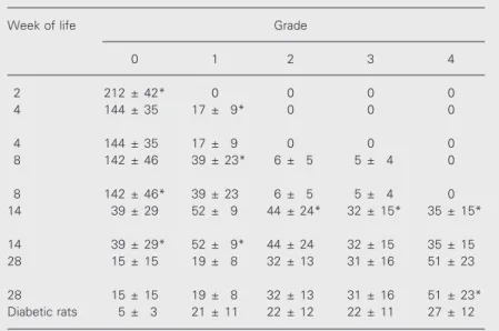
Pancreatic islet infiltration by mono-nuclear cells in female NOD mice was pro-gressive from 4 to 28 weeks of age for both diabetic and non-diabetic animals. These re-sults are described below as mean ± SD insulitis score and are summarized in Table 1. At 8 weeks of life an increase of infiltra-tive cells surrounding the islets (peri-islet, grade 1, 39 ± 23, P<0.05) was observed and the severity score increased at age 14, with most islets being invaded by infiltrating cells (grade 2, 44 ± 24, grade 3, 32 ± 15 and grade 4, 35 ± 15, P<0.01). The severity score at age 14 compared to 28 weeks showed grade 0, 39 ± 30 (P<0.05) and grade 1, 52 ± 9 (P<0.01). The values for grades 2, 3 and 4 were not statistically different (P<0.1).
In 28-week-old CBA-j animals and in diabetic animals, insulitis scores between grades 0 and 3 were the same for both groups. Nevertheless, in 28-week-old animals the grade 4 score (51 ± 23, P<0.02) was signifi-cantly higher than in the diabetic group prob-ably due to the smaller amount of islets than in the non-diabetic animals.
Mean numbers of pancreatic islets per animal did not differ significantly among non-diabetic animals, in contrast to diabetic animals whose total number of islets (96 ± 31, P<0.05) was significantly lower than in the other animals.
Expression of IFN-g and TNF-a mRNA
In order to investigate the expression of TNF-a and IFN-g mRNA in the autoimmune diabetes process, multiple time point analy-ses were performed to identify the associa-tion between cytokine gene expression and the infiltration of immune cells during dis-ease development. All cDNA samples used for the study expressed the product of the housekeeping gene cyclophilin (276 bp) and insulin (257 bp), confirming their quality and the source. The expected products for the genes of TNF-a (254 bp) and IFN-g (428 bp) were expressed in all animals studied.
Semiquantitative analysis of RT-PCR products
A cross-sectional study involving 7 fe-male NOD mice at 2, 4, 8, 14 and 28 weeks of age and CBA-j mice was performed. Two cytokines, TNF-a and IFN-g, were
exam-Table 1. Insulitis scores as a function of age.
Week of life Grade
0 1 2 3 4
2 212 ± 42* 0 0 0 0
4 144 ± 35 17 ± 9* 0 0 0
4 144 ± 35 17 ± 9 0 0 0
8 142 ± 46 39 ± 23* 6 ± 5 5 ± 4 0
8 142 ± 46* 39 ± 23 6 ± 5 5 ± 4 0
14 39 ± 29 52 ± 9 44 ± 24* 32 ± 15* 35 ± 15*
14 39 ± 29* 52 ± 9* 44 ± 24 32 ± 15 35 ± 15
28 15 ± 15 19 ± 8 32 ± 13 31 ± 16 51 ± 23
28 15 ± 15 19 ± 8 32 ± 13 31 ± 16 51 ± 23*
Diabetic rats 5 ± 3 21 ± 11 22 ± 12 22 ± 11 27 ± 12
The pancreases were removed from 7 female NOD mice at the age of 2, 4, 8, 14 and 28 weeks. Three tissue sections from each diabetic animal were analyzed and pancre-atic islet insulitis was graded from 0 to 4 according to severity. The analysis was performed comparing ages 2-4, 4-8, 8-14, 14-28, and 28 weeks and diabetic rats. A mean insulitis score was calculated and the total number of islets was estimated considering the number of sections examined and the animals studied. Data concern-ing grades of insulitis and severity of the islet inflammation process were assessed by the unpaired Student t-test.
ined for these analyses. Total RNA samples were prepared from the islets of NOD mice at different ages, i.e., 2, 4, 8, 14 and 28 weeks, representing different stages of dis-ease. From 2 to 14 weeks there was a nonsig-nificant increase in IFN-g mRNA expression, which then increased significantly (149 ± 29 AU, P<0.05) in female NOD mice at 28 weeks compared to both youngest groups of animals and CBA-j mice at 14 and 28 weeks of life (Figure 1A). However, in the spleen of these animals the level of IFN-g expression was not significantly increased (P<0.1) (Figure 1B).
In contrast, the expression of TNF-a in islets of NOD and CBA-j mice progressively increased from 2 weeks of age, being statis-tically significant at 14 (99 ± 8 AU, P<0.05) and 28 weeks of age (144 ± 17 AU, P<0.05) and being related to the increase of infiltrat-ing cells in pancreatic islets (Figure 2A). In comparison with CBA-j mice, TNF-a mRNA levels in spleens from NOD females started to increase significantly (168 ± 14 AU,
P<0.05) at 8 weeks of age (Figure 2B). Thus, TNF-a mRNA levels in islets and spleens correlated with the inflammatory process and diabetes risk in older NOD mice. In contrast, islets of CBA-j animals expressed low and constant levels at all ages studied.
Discussion
Several studies have correlated cytokine expression by islets with the development of autoimmune diabetes in NOD mice and have demonstrated that islet destruction is associ-ated with increased expression of cytokines such as TNF-a and IFN-g (13-20). However, the present study is the first to focus on mRNA expression and the morphological features of pancreatic islets in NOD mice aged 2 to 28 weeks. Our results are consist-ent with published data concerning the ex-pression of TNF-a and IFN-g as a candidate for the final effector of autoimmune diabetes (20). The expression of a particular group of
Figure 2. Kinetics of TNF-a mRNA expression in islets (A) and spleen (B) assessed by semiquantitative RT-PCR in fe-male NOD and CBA-j mice in a time-course study (2, 4, 8, 14 and 28 weeks of age) represent-ing different stages of the dis-ease process. *P<0.05 com-pared to CBA-j mice of the same age (Student t-test). N = 55 fe-male NOD and 25 fefe-male CBA-j mice.
Arbitrary units
200
Arbitrary units
200
150
100
50
0
150
100
50
0
NOD CBA-j NOD CBA-j
2 4 8 14 28 2 4 8 14 28
Age (weeks) Age (weeks)
*
*
A B * * *
* Figure 1. Kinetics of IFN-g mRNA
expression in islets (A) and spleen (B) assessed by semi-quantitative RT-PCR in female NOD and CBA-j mice in a time-course study (2, 4, 8, 14 and 28 weeks of age) representing dif-ferent stages of the disease pro-cess. *P<0.05 compared to CBA-j mice of the same age (Stu-dent t-test). N = 55 female NOD and 25 female CBA-j mice.
Arbitrary units
200
Arbitrary units
200
150
100
50
0
150
100
50
0
NOD CBA-j NOD CBA-j
2 4 8 14 28 2 4 8 14 28
Age (weeks) Age (weeks)
*
*
cytokines in islets can modulate the inflam-matory process leading to ß cell destruction or suppressing inflammation. In addition, cytokines such as IL-1, TNF-a or IFN-g impair insulin secretion and, when added in combination, are destructive to islets (13,20). In the present study, we examined the rela-tions between TNF-a and IFN-g mRNA ex-pression in pancreatic islets of NOD mice and the progression of insulitis, its severity, islet destruction and overt diabetes. Further-more, the expression of these cytokine genes was examined in spleen isolated at the same time as the islets, as a central lymphoid organ. IFN-g is a product of the Th1 subset of T lymphocytes, cells involved in cell-medi-ated immune responses and relcell-medi-ated to modu-lation of islet ß cell destruction (11,18,32). Indeed, previous studies showed that diabe-tes could be transferred to neonatal NOD mice, suggesting that both CD4+ and CD8+ are required for the initiation of insulitis, contributing to the development of diabetes (9,33). Furthermore, the morphological find-ings of the presence of macrophages in islet infiltrating cells suggest their participation in early events of the inflammatory reaction which lead to the recruitment of T cells (5). In the early stages of insulitis, at 2 and 4 weeks of age, the mild IFN-g mRNA expres-sion in islets and spleen coincides with the low number of activated T cells in pancreatic islets (24,26,34). However, with the increase of infiltrating T cells in islets the level of IFN-g expression increases in older mice after 14 weeks of age, associated with de-structive insulitis (21,24,32). The cytotoxic action of IFN-g against islet ß cells has been described in in vitro experiments (13,20). In transgenic mice, the expression of IFN-g in ß cells can lead to insulitis and diabetes (35-37). Thus, our results concur with studies showing that IFN-g mRNA expression cor-relates with islet destructive insulitis and diabetes development in NOD mice and emphasizes the importance of IFN-g in the pathogenesis of autoimmune diabetes.
How-ever, in the present study, the group of fe-male NOD mice studied at 14 weeks of age (pre-diabetic stages) showed a decreased expression of IFN-g despite the presence of advanced insulitis (grades 3 and 4). In con-trast, the genetic absence of IFN-g did not prevent diabetes in NOD mice, but delayed the onset of diabetes (38). Moreover, a pre-vious study dissociated class I major histo-compatibility complex upward regulation from progression to diabetes due to local IFN-g action, suggesting that ß cells are not the direct targets of IFN-g in autoimmune diabetes (37). This apparent contradiction suggests that the levels of IFN-g expression at this age could determine, together with other factors, the rate of development of overt diabetes. On the other hand, the ex-pression of IFN-g mRNA levels in spleen did not show a correlation with severity of insulitis progression, suggesting a compart-mental IFN-g response.
The levels of TNF-a gene expression progressively increased with age, with over-expression being observed at the 28thweek. In some cases, TNF-a expression did not correlate with the progression of diabetes, suggesting that TNF-a can be more impor-tant for the development and maintenance of insulitis than diabetes (39,40).
References
1. Tisch R & McDevitt HO (1996). Insulin-dependent diabetes mellitus. Cell, 85: 291-297.
2. Kikutani H & Makino S (1992). The murine autoimmune diabetes model: NOD and related strains. Advances in Immunology, 51: 285-322.
3. Castaño L & Eisenbarth GS (1990). Type I diabetes: A chronic autoimmune disease of human, mouse and rat. Annual Review of Immunology, 8: 647-679.
4. Leiter EH, Prochazka M & Coleman DL (1987). Animal model of human disease: The non-obese diabetic (NOD) mouse.
American Journal of Pathology,128: 380-383.
5. Hee-Sook J, Chang-Soon Y, Zbytnuik L, Van Rooijen N & Ji-Woon Y (1999). The role of macrophages in T cell-mediated autoimmune diabetes in non-obese dia-betic mice. Journal of Experimental Medi-cine, 18: 347-358.
6. Lee KU, Amano K & Yoon JW (1988). Evidence for initial involvement of macro-phages in development of insulitis in NOD mice. Diabetes, 37: 989-991.
7. Wong FS & Janeway Jr CA (1997). The role of CD4 and CD8 T cells in type I diabetes in the NOD mouse. Research in Immunology, 148: 327-332.
8. Pozzilii P, Signore A, Williams AJK & Beales PE (1993). NOD mouse colonies around the world - Recent facts and fig-ures. Immunology Today, 14: 193-196. 9. Kay TWH, Chaplin HL, Parker JL,
Stephens LA & Thomas HE (1997). CD4+ and CD8+ T lymphocytes: Clarification of their pathogenic roles in diabetes in the NOD mouse. 70th Forum in Immunology.
Research in Immunology, 148: 320-326. 10. Faulkner-Jones B, Dempsey-Collier M,
Mandel ET & Harrison LC (1996). Both Th1 and Th2 cytokine mRNAs are ex-pressed in the NOD mouse pancreas in vivo. Autoimmunity, 23: 99-110. 11. Bradley LM, Asensio VC, Schioetz LK,
Harbertson J, Krahl T, Patstone G, Woolf N, Campbell IL & Sarvetnick N (1999). Islet-specific Th1, but not Th2, cells se-crete multiple chemokines and promote rapid induction of autoimmune diabetes.
Journal of Immunology, 62: 2511-2520. 12. Hartemann AH, Richard MF & Boitard C
(1999). Absence of significant Th2 re-sponse in diabetes-prone non-obese dia-betic (NOD) mice. Clinical and Experimen-tal Immunology, 116: 225-230.
13. Yang X-D, Tisch R, Singer SM, Cao ZA, Liblau RS, Schreiber RD & McDevitt HO (1994). Effect of tumor necrosis factor a on insulin-dependent diabetes mellitus in NOD mice. I. The early development of autoimmunity and the diabetogenic pro-cess. Journal of Experimental Medicine, 180: 995-1004.
14. Rabinovitch A (1998). An update on cyto-kines in the pathogenesis of insulin-dependent diabetes mellitus. Diabetes/ Metabolism Reviews, 14: 129-151. 15. Pakala SV, Chivetta M, Kelly CB & Katz JD
(1999). In autoimmune diabetes the tran-sition from benign to pernicious insulitis requires an islet cell response to tumor necrosis factor a. Journal of Experimental
Medicine, 189: 1053-1562.
16. Stephens LA, Thomas HE, Ming L, Grell M, Darwiche R, Volodin L & Kay TW (1999). Tumor necrosis factor a-activated cell death pathways in NIT-1 insulinoma cells and primary pancreatic beta cells.
Endocrinology, 140: 3219-3227. 17. Green EA, Eynon EE & Flavell RA (1998).
Local expression of TNF-a in neonatal NOD mice promotes diabetes by enhanc-ing presentation of islet antigens. Immu-nity, 9: 733-743.
18. Green EA, Wong FS, Eshima K, Mora C & Flavell RA (2000). Neonatal tumor necro-sis factor a promotes diabetes in non-obese diabetic mice by CD154-independ-ent antigen presCD154-independ-entation to CD81 T cells.
Journal of Experimental Medicine, 191: 225-238.
19. Green EA & Flavell RA (2000). The tempo-ral importance of TNF-a expression in the development of diabetes. Immunity, 12: 459-469.
20. Suk K, Kim S, Kim Y-H, Kim K-A, Chang I, Yagita H, Shong M & Lee M-S (2001). IFN-g/TNF-a synergism as the final effec-tor in autoimmune diabetes: A key for STAT1/IFN regulatory factor-1 pathway in pancreatic ß cell death. Journal of Immu-nology, 166: 4441-4489.
21. Rabinovitch A, Suarez-Pinzon WL, Soren-sen O & Bleackley RC (1996). Inducible
nitric oxide synthase (iNOs) in pancreatic islet of non-obese diabetic mice: Identifi-cation of iNOs-expressing cells and rela-tionships to cytokines expressed in the islets. Endocrinology, 137: 2093-2099. 22. Gurlo T, Kawamura K & Grafenstein HV
(1999). Role of inflammatory infiltrate in activation and effect function of cloned islet reactive non-obese diabetic CD8+ T cells: involvement of a nitric oxide-dependent pathway. Journal of Immunol-ogy, 163: 5770-5780.
23. Suarez-Pinzon WL, Strynadka K, Schulz R & Rabinovitch A (1994). Mechanisms of cytokine-induced destruction of rat insuli-noma cells: The role of nitric oxide. Endo-crinology, 134: 1006-1010.
24. Rabinovitch A, Suarez-Pinzon WL, Soren-sen O, Bleackley RC & Power PR (1995). IFN-g gene expression in pancreatic islet-infiltration mononuclear cells correlates with autoimmune diabetes in nonobese diabetic mice. Journal of Immunology, 154: 4874-4882.
25. Thomas HE & Kay TWH (2000). Beta cell destruction in development of autoim-mune diabetes in the non-obese diabetic (NOD) mouse. Diabetes/Metabolism Re-search and Reviews, 16: 251-261. 26. Hirai H, Kaiano T, Ito T & Kida K (2000).
Analysis of cytokine mRNA expression in pancreatic islet of nonobese diabetic mice. Journal of Pediatric Endocrinology and Metabolism, 13: 91-98.
27. Rabinovitch A & Suarez-Pinzon WL (1998). Cytokines and their roles in pancreatic is-let ß-cell destruction and insulin-depend-ent diabetes mellitus. Biochemical Phar-macology, 55: 1139-1149.
28. Pavin EJ & Zollner RL (1994). Implantação da linhagem “NOD-mice” (camundongos diabéticos não obesos) no Brasil: Contri-buição deste modelo animal ao estudo do diabetes mellitus insulino-dependente e outras doenças auto-imunes. Arquivos Brasileiros de Endocrinologia e Metabolo-gia, 38: 105-108.
29. Gotoh M, Maki T, Kiyoizumi T, Satomi S & Monaco AP (1985). An improved method for isolation of mouse pancreatic islet.
Transplantation, 40: 437-438.
30. Lacy PE & Kostianovsky M (1967). A method for the isolation of intact islet of insulitis and diabetes (20). Furthermore, our
findings suggest that spleen TNF-a and IFN-g were not correlated with severe insulitis and
Langerhans from the rat pancreas. Diabe-tes, 16: 35-39.
31. Chomczynski P & Sacchi N (1987). Single-step method of RNA isolation by acid gua-nidinium thiocyanate-phenol-chloroform extraction. Analytical Biochemistry, 162: 156-159.
32. Campbell IL, Iscalo A & Harrison LC (1988). IFN-g and tumour necrosis
factor-a: cytotoxicity to murine islets of Langer-hans. Journal of Immunology, 141: 2325-2329.
33. O’Reilly LA, Hutchings PR, Crocker R, Simpson E, Lund T, Kioussis D, Takei F, Baird J & Cooke A (1991). Characteriza-tion of pancreatic islet cell infiltrates in NOD mice: effect of cell transfer and transgene expression. European Journal of Immunology, 21: 1171-1180. 34. Toyoda H, Formby B, Magalong D,
Redford A, Chan E, Takei S & Charles MA (1994). In situ islet cytokine gene expres-sion during development of type I diabe-tes in the non-obese diabetic mouse. Im-munology Letters, 39: 283-288. 35. Stewart TA, Hultgren B, Huang X,
Pitts-Meek S, Hully J & MacLachlan NJ (1993). Induction of type I diabetes by interferon-gamma in transgenic mice. Science, 260: 1942-1946.
36. Sarvetnick N, Shizuru J, Liggitt D, Martin L, McIntyre B, Gregory A, Parslow T & Stewart T (1990). Loss of pancreatic islet tolerance induced by beta-cell expression of interferon-gamma. Nature, 346: 844-847.
37. Thomas HE, Parker JL, Schreiber RD & Kay TW (1998). IFN-g action on pancreatic ß cells causes class I MHC upregulation but not diabetes. Journal of Clinical
Inves-tigation, 102: 1249-1257.
38. Hultgren B, Huang X, Dybdal N & Stewart TA (1996). Genetic absence of g-interferon delays but does not prevent diabetes in NOD mice. Diabetes, 45: 812-817. 39. Higuchi Y, Herrera P, Muniesa P, Huarte
J, Belin D, Ohashi P, Aichele P, Orci L, Vassalli JD & Vassalli P (1992). Expression of a tumor necrosis factor alpha trans-gene in murine pancreatic beta cells re-sults in severe and permanent insulitis without evolution towards diabetes. Jour-nal of Experimental Medicine, 176: 1719-1731.