Cell lineage relationship in the
stomach of normal and genetically
manipulated mice
Department of Anatomy, Faculty of Medicine, Kuwait University, Safat, Kuwait S.M. Karam
Abstract
The oxyntic mucosa of the mouse stomach is lined with a heteroge-neous population of cells that form numerous short pits continuous with long tubular glands. Tritiated thymidine radioautography has made it possible to pinpoint the origin of all cell types and to follow the differentiation/migration of different cell lineages along the pit-gland unit. The proliferating multipotent stem cells functionally anchored in the upper glandular region, the isthmus, give rise to three main lineage precursors: 1) pre-pit cells, which migrate upward to the pit while differentiating into mucus-producing pit cells; 2) pre-neck cells, which migrate downward to the glandular neck while differentiating into mucus-producing neck cells that, by approaching the glandular base, gradually change their phenotype into pepsinogen- and intrinsic fac-tor-producing zymogenic cells; 3) pre-parietal cells, which differenti-ate into acid-producing parietal cells in the isthmus and then undergo bipolar migration towards the pit and the glandular base. Thus, parietal cells are the only cells that complete their differentiation in the isthmus and then migrate to be scattered throughout the pit-gland unit. To determine whether parietal cells play a role in controlling decisions about cell fate within the pit-gland unit, the gastric epithelium has been examined in transgenic mice expressing the H,K-ATPase ß-subunit-1035 to +24/simian virus 40 large T antigen fusion gene. The blockade in parietal cell differentiation in these mice produces an amplification of lineage precursors, a marked depletion of zymogenic cells and an increase in pit cell census. Ablation of parietal cells in another transgenic mouse model expressing the H,K-ATPase ß-sub-unit-1035 to +24/diphtheria toxin fragment A fusion gene also produces amplification of lineage precursors, and similar effects on zymogenic and pit cell census. These findings strongly suggest that parietal cells produce regulatory signals that control the cellular differentiation program of both pit and zymogenic cell lineages, and would hopefully improve our ability to identify the cellular pathways leading to malig-nant transformation.
Correspondence
S.M. Karam
Department of Anatomy Faculty of Medicine Kuwait University P.O. Box 24923 Safat 13110 Kuwait
Fax: 00-965-531-8454 E-mail: [email protected]
Presented at the 5th International Symposium on Radioautography, São Paulo, SP, Brasil, August 24-26, 1997.
Research supported by the Kuwait Foundation for Advancement of Sciences (No. KFAS 95-07-02).
Received August 4, 1997 Accepted August 20, 1997
Key words
•Radioautography
•Gastric epithelium
•Stem cells
•Cell proliferation/ differentiation programs
•Lineage progenitors
Introduction
The mouse stomach consists of three main regions: the fundus which is lined with strati-fied squamous keratinized epithelium, the pyloric antrum which is continuous with the duodenum and lined with one layer of cells that invaginates to form numerous long pits continuous with short mucous glands, and the body (or corpus) which forms the main central region of the stomach and is lined with one layer of cells that forms numerous short pits continuous with long tubular glands. In this review, we will concentrate only on the epithelial cells of the body region of the mouse stomach.
The pit-gland units found in the corpus region of the mouse stomach are not static structures. Cellular proliferation, commit-ment, migration-associated differentiation, and death programs occur perpetually along the pit-gland axis of these units. The exten-sive dynamism of this epithelium is further indicated by its rapid regeneration after dam-age. In addition, interactions amongst the different gastric epithelial cell populations play a major role in determining the structure and function of the pit-gland unit. The main objective of this review is to summarize all of these fundamental features of the pit-gland units in the body region of the mouse stomach from studies in which tritiated thy-midine radioautography, electron micros-copy, multilabeled immunocytochemistry, and genetic manipulation techniques have been utilized.
Normal mice
Morphological identification of the main cell types along the pit-gland unit. Since the 1830s, it has been recognized that the mam-malian gastric mucosa contains numerous glandular structures comprising mucus-pro-ducing glands in the pylorus and pepsino-gen-producing glands in the corpus (1). In the mouse, the glands of the corpus open into
the luminal surface via short pits. These pit-gland units, also called “zymogenic units” (2), consist of a structurally and functionally heterogeneous population of cells that in-clude several cell types. 1) The mucus-se-creting pit cells, or surface mucous cells, are found in the pit region and on the luminal surface and are characterized by a group of dense mucous granules (3-5). The granules are packed in an organelle-free apical area called ectoplasm (2,6). In the mouse, there are 37 pit cells per unit; their Golgi apparatus produces a uniformly fine particulate con-tent packed in prosecretory vesicles which eventually form secretory granules. The di-ameter of the granules varies around 350 nm. The granule contents are homogeneously dense except on the free surface where they may acquire a core (2,6). Two lectins, Ulex europaeus type 1 agglutinin and cholera toxin B subunit, can be used as markers for pit cells in adult and developing mice (7,8). 2) The acid-secreting parietal cells have been extensively investigated by Forte et al. (9), Helander (4) and Ito (5). In the mouse, there are 26 parietal cells per unit; they are scat-tered along the pit and all three glandular regions and are characterized by an intracel-lular canalicular system, cytoplasmic tubu-lovesicular elements, long numerous mi-crovilli lining canalicular/apical membranes and large numerous mitochondria. Antibod-ies against the α- and ß-subunits of the H,K-ATPase (10-12), the cytoskeletal protein ezrin (13) and the Lewisx blood group antigen,
packed at the periphery of the same vesicles. These vesicles form cored granules scattered throughout the cytoplasm in comparison to the apical granules of pit cells. The diameter of neck cell granules varies around 570 nm. The Grifforia simplifolica II lectin can be used as a marker for neck cells (7,8) in both adult and developing mice. 4) The pepsino-gen-secreting zymogenic cells have been extensively studied by Samloff (16) and Hersey (17). The mouse stomach contains 67 zymogenic cells per unit; they are typical serous cells characterized by a basal stack of rough ER cisternae and apical zymogen gran-ules with a homogeneously pale content (2). Antibodies against pepsinogen and intrinsic factor are utilized as markers specific for mouse zymogenic cells (8,18,19). During morphogenesis, while pit, neck and parietal cells appear as early as embryonic day 18, typical zymogenic cells only appear at post-natal day 21 (8). 5) The peptide-secreting entero-endocrine cells have been character-ized and extensively studied by Solcia et al. (20). The subtypes of these cells vary based on the shape of the secretory granules and their peptide content. In the mouse, there are 13 entero-endocrine cells per unit compris-ing several subtypes. They are scattered along the four unit regions but are mainly found in the base (2). 6) The villin-rich caveolated cells have been discovered by Hammond and LaDeur (21) and Nabeyama and Leblond (22); they are characterized by a microvil-lous tuft protruding into the glandular lu-men, and long narrow convoluted caveoli that open between the microvilli. In the mouse, they are very few (1 cell per 2-3 units) but may be found in any of the four gland regions (2). 7) In the upper segment of the base region, there are pre-zymogenic cells (2) which are characterized by a Golgi apparatus producing prosecretory vesicles and secretory granules whose contents ap-pear to be intermediate between those of neck cells and zymogenic cells. In the mouse stomach, there are 5 pre-zymogenic cells per
unit. These cells can be identified by neck cell-specific lectins and pepsinogen-specific antibodies, markers for both neck and zy-mogenic cells (8,19).
for the granule-free cells or the precursors of both pre-pit and pre-neck cells. 4) Pre-pit cells are characterized by a Golgi apparatus producing prosecretory vesicles similar to those of pre-pit cell precursors and pit cells. They also have dense secretory granules simi-lar to those of pit cells, but are fewer and smaller; their diameter varies around 200 nm. 5) Pre-neck cells are characterized by a Golgi apparatus producing prosecretory vesicles similar to those of pre-neck cell precursors and neck cells. They also have cored secretory granules similar to those of neck cells, but are fewer and smaller; their diameter varies around 398 nm. 6) Pre-pari-etal cells are characterized by pariPre-pari-etal cell-like features, i.e., long apical microvilli and an incipient intracellular canaliculus; they include three subtypes: one carrying a few secretory granules similar to those of pre-pit cells, the second with cored granules similar to those of pre-neck cells, and the third de-void of any granules. 7) Pre-entero-crine cells are characterized by a few endo-crine-type secretory granules. 8) Pre-caveolated cells are characterized by few caveoli and microvilli similar to those of caveolated cells.
The stem cell of the mouse gastric epithe-lium. In renewing epithelia, stem cells are defined by their high proliferative capacity to ensure their own persistence while pro-ducing committed cells (27) and their primi-tive embryonic cell-like features (28). Triti-ated thymidine radioautography combined with electron microscopy revealed that the undifferentiated granule-free cells are the most proliferative cell type (30 min labeling index = 32%) and the most primitive among other isthmal cells, and are therefore consid-ered to be the stem cells of the gastric epithe-lium (26).
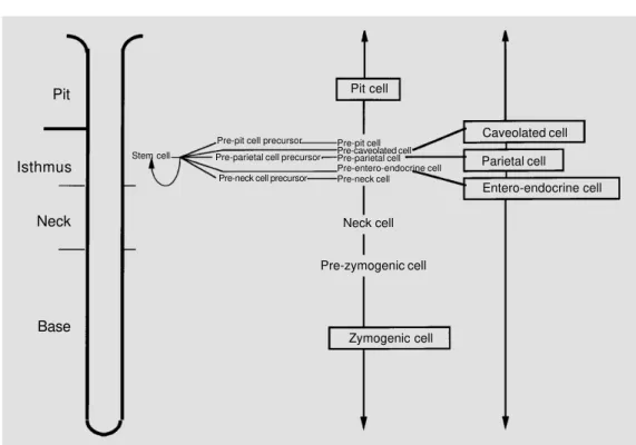
Proliferation, differentiation and migra-tion pathways along the pit-gland unit as revealed by tritiated thymidine radioautog-raphy. Tritiated thymidine has been utilized to label the proliferating cells of the
pit-gland unit and to follow their differentiation/ migration pathways with time. Radioauto-graphs thus represent the source of valuable data on the labeling indices of various cell types at different time intervals. Cell prolif-eration is restricted to the isthmus region where granule-free cells are the most prolif-erative; pre-pit and pre-neck cells and their precursors also have some potential for mi-tosis, whereas pre-parietal cells do not di-vide. The shift in tritiated thymidine labeling that occurs with time from pre-pit to pit cells has confirmed morphological findings and indicated that they constitute one lineage (6) that migrates upwards to the free surface (Figure 1). The shift in the labeling from pre-neck to pre-neck cells has confirmed morpho-logical studies and indicated that they be-long to one lineage. The morphological fea-tures of pre-zymogenic cells and the fact that they acquire thymidine labeling after neck cells and before the zymogenic cells indicate that they represent a transition during the transformation of neck cells into zymogenic cells (29). Thus, pre-neck, neck, prezymo-genic and zymoprezymo-genic cells all constitute one lineage that migrates towards the bottom of the gland (Figure 1). Also, the shift in label-ing from pre-parietal to parietal cells indi-cates that they constitute a third lineage, but with a bipolar mode of migration towards either the pit orifice or gland bottom (30). Similar to parietal cells, the entero-endo-crine and caveolated cells develop in the isthmus and undergo bipolar migration (31; Figure 1). The turnover time of the different gastric epithelial cell types is determined by continuous infusion of a low dose of tritiated thymidine into mice, which are then sacri-ficed at different time intervals. From the cumulative increase in the labeling indices of each cell type, the rate of cellular turnover and the turnover time can be estimated (Fig-ure 1).
parietal cells appear to be strategically lo-cated among the precursor cells of the isth-mus, and also scattered throughout the unit, they seem to have a role in cellular prolifera-tion and commitment that perpetually occur in the isthmus. Immunohistochemical and in situ hybridization experiments have shown that both the α- and ß-subunits of H,K-ATPase are expressed in the pre-parietal and parietal cells (12,32). However, the synthetic activity of parietal cells is higher in the isth-mus and neck regions than in the base. In addition, functional assays have revealed that parietal cells in the isthmus and neck are more active in acid secretion than basal pari-etal cells. Thus, a possible role for paripari-etal cells other than acid secretion has been pro-posed. Covalent blockade of H,K-ATPase, the major protein of parietal cells, with omeprazole enhances the physiological de-generation of parietal cells. Furthermore, al-teration in isthmal cell proliferation and com-mitment programs occurs, as indicated by the increase in the census of both the prolif-erating precursor cells and the non-prolifer-ating committed pre-parietal cells (12).
To date there is no cell culture system that can reproduce cell lineages of the gastric epithelium for identifying factors that modu-late cellular proliferation and differentiation. An alternative approach is to design a gain-of-function experiment by introducing a bio-logically interesting foreign gene into the mouse genome and generating transgenic mice.
Genetically manipulated mice
Transgenic mice overexpressing trans-forming growth factor α (TGFα). The epi-dermal growth factor (EGF) family of pep-tides includes TGFα which seems to play an important role in the regulation of cellular proliferation and differentiation programs in various tissues. The activity of TGFα is me-diated by the EGF receptor, a tyrosine kinase that phosphorylates both itself and additional substrates upon ligand-specific activation/ dimerization (33). Both TGFα and EGF re-ceptors have been localized to the gastric epithelial cells by using immunohistochem-istry and Northern blot analysis (34,35). It Pit
Isthmus
Neck
Base
Pit cell
Caveolated cell
Parietal cell
Entero-endocrine cell
Neck cell
Pre-zymogenic cell
Zymogenic cell Stem cell
Pre-pit cell precursor
Pre-parietal cell precursor
Pre-neck cell precursor
Pre-pit cell Pre-caveolated cell Pre-parietal cell Pre-entero-endocrine cell Pre-neck cell
has been shown that TGFα stimulates gas-tric mucosal growth (36) and also inhibits gastric acid secretion (37). Merlino and co-workers (38) have used the mouse metallo-thioneine I promoter to generate transgenic mice overexpressing human TGFα in sev-eral tissues. In these mice, TGFα disrupts the normal program of gastric epithelial cell differentiation. While pit cells and isthmal precursor cells are greatly expanded, pari-etal and zymogenic cells are depleted. These results suggest that TGFα plays an important role in the regulation of gastric epithelial cell differentiation.
A more powerful approach to the study of gastric epithelial biology is to utilize cis-acting regulatory elements that control tran-scription of a gastric cell lineage-specific gene in expressing any foreign gene in that cell lineage. Lorenz and Gordon (18) first found that nucleotides -1035 to +24 of the H,K-ATPase ß-subunit gene include tran-scriptional regulatory elements able to direct expression of any foreign gene in the parietal cell lineage.
Transgenic mice expressing the H,K-ATPase ß-subunit-1035 to +24/simian virus 40
large T antigen fusion gene. Simian virus 40 large T antigen (SV40 TAg) is an oncoprotein that binds p53 and induces cell proliferation. To test whether re-entry of the committed non-proliferating pre-parietal cells into the cell cycle would affect the cellular differen-tiation program in the gastric epithelium, Li et al. (19) used the regulatory elements of the H,K-ATPase ß-subunit gene to generate transgenic mice expressing SV40 TAg in their parietal cell lineage. In these mice, the proliferation-associated amplification of pre-parietal cells and the blockade of their differ-entiation into parietal cells starting from embryonic day 18 was not unexpected, whereas the accompanying loss of zymogenic cells was surprising. The latter finding indi-cates that members of the parietal cell lin-eage regulate the differentiation of neck cells to zymogenic cells (8,19). Furthermore,
elec-tron microscopic examination of the ampli-fied population of pre-parietal cells in these transgenic mice at embryonic day 18 and postnatal day 1 revealed signs of their early commitment and transformation of undiffer-entiated (stem) cells into pre-parietal cells. Thus, the first member of the parietal cell lineage was identified, i.e., the pre-parietal cell precursor (Figure 1), which is character-ized by loss of apical membrane glycocalyx and gradual elongation of microvilli (8). The amplification of pre-parietal cells and their precursors in these transgenic mice provides an excellent system to study the molecular and biochemical features of the early com-mitted precursors of the acid-secreting pari-etal cell. With age these transgenic mice develop gastric adenocarcinoma (Karam SM, Li Q and Gordon JI, unpublished results).
Transgenic mice expressing the H,K-ATPase ß-subunit-1035 to +24/diphtheria toxin
the migration-associated terminal differen-tiation of both pit and zymogenic cell lin-eages. With age, the diphtheria toxin trans-genic mice develop gastric adenocarcinoma in which the predominant cells are the nor-mally proliferating isthmal cells, granule-free cells and their pre-neck and pre-pit de-scendants (Li Q, Karam SM and Gordon JI, unpublished results).
Transgenic mice expressing the H,K-ATPase ß-subunit promoter/herpes simplex virus 1 thymidine kinase fusion gene. Fur-ther progress in the field of genetic manipu-lation techniques to study gastric epithelial cell biology has been provided by the possi-bility of controlling the expression of a for-eign gene in a specific cell lineage. Canfield et al. (40) have used the thymidine kinase ablation technique which is based on the selective toxicity of the antiherpetic drug ganciclovir to cells expressing a herpes virus thymidine kinase suicide gene. The gener-ated transgenic animals provide a powerful system in which parietal cells are normally produced, but upon treatment with the antiherpetic drug ganciclovir, they are com-pletely ablated. Discontinuation of the drug was associated with reemergence of parietal cells and restoration of the glandular archi-tecture. In support of the findings of Gordon and coworkers (8,19,39), parietal cell abla-tion in these mice is found to be associated with a block in the terminal differentiation program of zymogenic cells (40). Thus, pari-etal cells appear to be the source of trophic factors that are essential for maintaining the differentiation of zymogenic cells.
Inhibin knockout mice. Activins A and B are members of the transforming growth fac-tor ß superfamily which is known to regulate cellular proliferation and differentiation pro-grams in several tissues (41). Matzuk and co-workers (42) have generated mice
ho-mozygous for a null allele of the inhibin α subunit. These inhibin knockout mice de-velop gonadal tumor and overexpress activins. The stomachs of these mice have no parietal cells. Removal of the gonads prior to development of tumors prevents the ablation of parietal cells. Detailed immunohistochemi-cal and electron microscopic analysis of inhibin knockout mice has revealed that over-expression of activins produces a block in the differentiation of pre-parietal to parietal cells, a block in the transformation of neck into zymogenic cells, and a marked increase in pit and pit cells as well as pre-caveolated and pre-caveolated cells (Li Q, Karam SM, Coerver KA, Matzuk MM and Gordon JI, unpublished results). Thus, 1) activins play an important role in modulating the differentiation program of the gastric epithe-lial cell lineages, 2) several lines of evidence strongly suggest that members of the parietal cell lineage are essential for maintaining the normal programs of gastric epithelial cell proliferation and differentiation, and 3) it seems that altering parietal cells by using H2-receptor antagonists or proton pump in-hibitors during treatment of peptic ulcer dis-ease may elucidate the dynamics of the whole epithelium.
References
1. Bensley RR (1932). The gastric glands. In: Cowdry EV (Editor), Special Cytology. PB Hoeber, New York.
2. Karam SM & Leblond CP (1992). Identify-ing and countIdentify-ing all epithelial cell types present in the “corpus” of the mouse stomach. Anatomical Record, 232: 231-246.
3. Stevens CE & Leblond CP (1953). Re-newal of the mucous cells in the gastric mucosa of the rat. Anatomical Record, 115: 231-245.
4. Helander HF (1981). The cells of the gas-tric mucosa. International Review of Cy-tology, 70: 217-289.
5. Ito S (1987). Functional gastric morpholo-gy. In: Johnson LR (Editor), Physiology of the Gastrointestinal Tract. 2nd edn. Raven Press, New York.
6. Karam SM & Leblond CP (1993). Dynam-ics of epithelial cells in the “corpus” of the mouse stomach. II. Outward migra-tion of pit cells. Anatomical Record, 236: 280-296.
7. Falk PG, Roth KA & Gordon JI (1994). Lectins are sensitive tools for defining differentiation programs of mouse gut epithelial cell lineages. American Journal of Physiology, 266 (Gastrointestinal and Liver Physiology, 29): G987-G1003. 8. Karam SM, Li Q & Gordon JI (1997).
Gas-tric epithelial morphogenesis in normal and transgenic mice. American Journal of Physiology, 272 (Gastrointestinal and Liver Physiology, 35): G1209-G1220. 9. Forte JG, Hanzel DK & Urushidani T
(1989). Mechanisms of parietal cell func-tion. In: Garner A & Whittle BJR (Editors), Advances in Drug Therapy of Gastrointes-tinal Ulceration. John Wiley & Sons, Sus-sex.
10. Smolka A, Helander H & Sachs G (1983). Monoclonal antibodies against H+-K+ -ATPase. American Journal of Physiology, 245 (Gastrointestinal and Liver Physiolo-gy, 8): G589-G596.
11. Crothers Jr JM, Chow DC & Forte JG (1993). Omeprazole decreases H+-K+ -ATPase protein and increases permeabil-ity of oxyntic secretory membranes in the rabbit. American Journal of Physiology, 256 (Gastrointestinal and Liver Physiolo-gy, 19): G231-G241.
12. Karam SM & Forte JG (1994). Inhibiting gastric H+-K+-ATPase activity by omepra-zole promotes degeneration and produc-tion of parietal cells. American Journal of Physiology, 266 (Gastrointestinal and Liver Physiology, 29): G745-G758.
13. Hanzel DK, Urushidani T, Usinger WR, Smolka A & Forte JG (1989). Immunologi-cal loImmunologi-calization of an 80-kD phosphopro-tein to the apical membrane of gastric parietal cells. American Journal of Physi-ology, 256 (Gastrointestinal and Liver Physiology, 19): G1082-G1089.
14. Bry LP, Falk PG & Gordon JI (1996). Ge-netic engineering of carbohydrate biosyn-thetic pathways in transgenic mice dem-onstrates cell cycle-associated regulation of glycoconjugate production in small in-testinal epithelial cells. Proceedings of the National Academy of Sciences, USA, 93: 1161-1166.
15. Wattel W & Geuze JJ (1978). The cells of the rat gastric groove and cardia. Cell and Tissue Research, 186: 375-391. 16. Samloff IM (1971). Pepsinogens, pepsin
and pepsin inhibitors. Gastroenterology, 60: 586-604.
17. Hersey S (1994). Gastric secretion of pep-sins. In: Johnson LR (Editor), Physiology of the Gastrointestinal Tract. 3rd edn. Raven Press, New York.
18. Lorenz RG & Gordon JI (1993). Use of transgenic mice to study regulation of gene expression in the parietal cell lin-eage of gastric units. Journal of Biological Chemistry, 268: 26559-26570.
19. Li Q, Karam SM & Gordon JI (1995). Sim-ian virus 40 T antigen-induced amplifica-tion of pre-parietal cells in transgenic mice. Effects on other gastric epithelial cell lineages and evidence for a p53-inde-pendent apoptotic mechanism that oper-ates in a committed progenitor. Journal of Biological Chemistry, 270: 15777-15788. 20. Solcia E, Capella C, Buffa R, Usillini L,
Fiocca R & Sessa F (1987). Endocrine cells of the digestive system. In: Johnson LR (Editor), Physiology of the Gastrointesti-nal Tract. 2nd edn. Raven Press, New York.
21. Hammond JB & LaDeur L (1969). Fibrillo-vesicular cells in the fundic glands of the canine stomach. Evidence for a new cell type. Anatomical Record, 161: 393-412. 22. Nabeyama A & Leblond CP (1974).
“Caveolated cells” characterized by deep surface invaginations and abundant fila-ments in mouse gastro-intestinal epithe-lia. American Journal of Anatomy, 140: 147-166.
23. Messier B & Leblond CP (1960). Cell pro-liferation and migration as revealed by ra-dioautography after injection of thymidine-H3 into male rats and mice. American Journal of Anatomy, 105: 247-285.
24. Corpron RE (1966). The ultrastructure of the gastric mucosa in normal and hypo-physectomized rats. American Journal of Anatomy, 118: 53-90.
25. Kataoka K & Sakano Y (1984). Panoramic observation of the mouse gastric mucosa by superwide-field electron microscopy. Archivum Histologicum Japonicum, 47: 209-221.
26. Karam SM & Leblond CP (1993). Dynam-ics of epithelial cells in the “corpus” of the mouse stomach. I. Identification of proliferative cell types and pinpointing of the stem cell. Anatomical Record, 236: 259-279.
27. Hall PA (1989). What are stem cells and how are they controlled? Journal of Pa-thology, 158: 275-277.
28. Leblond CP (1981). The life history of cells in renewing systems. American Journal of Anatomy, 160: 113-158.
29. Karam SM & Leblond CP (1993). Dynam-ics of epithelial cells in the “corpus” of the mouse stomach. III. Inward migration of neck cells followed by progressive transformation into zymogenic cells. Ana-tomical Record, 236: 297-313.
30. Karam SM (1993). Dynamics of epithelial cells in the “corpus” of the mouse stom-ach. IV. Bidirectional migration of parietal cells ending in their gradual degeneration and loss. Anatomical Record, 236: 314-322.
31. Karam SM & Leblond CP (1993). Dynam-ics of epithelial cells in the “corpus” of the mouse stomach. V. Behavior of entero-endocrine and caveolated cells. General conclusions on cell kinetics in the oxyntic epithelium. Anatomical Record, 236: 333-340.
32. Karam SM, Yao X & Forte JG (1997). Func-tional heterogeneity of parietal cells along the pit-gland axis. American Journal of Physiology, 272 (Gastrointestinal and Liver Physiology, 35): G161-G171. 33. Carpenter G (1987). Receptors for
epider-mal growth factor and other polypeptide mitogens. Annual Review of Biochemis-try, 56: 881-914.
35. Yasui W, Ji ZQ, Kuniyasu H, Ayhan A, Yokozaki H, Ito H & Tahara E (1992). Ex-pression of transforming growth factor al-pha in human tissues: immunohisto-chemical and Northern blot analysis. Virchows Archiv A Pathological Anatomy and Histopathology, 421: 513-519. 36. Chen MC, Lee AT & Soll AH (1991).
Mito-genic response of canine fundic epithelial cells in short-term culture to transforming growth factor α and insulin-like growth factor I. Journal of Clinical Investigation, 87: 1716-1723.
37. Guglietta A, Lesch CA, Romano M, McClure RW & Coffey RJ (1994). Effect of transforming growth factor-α on gas-tric acid secretion in rats and monkeys. Digestive Diseases and Sciences, 39: 177-182.
38. Sharp R, Babyatsky MW, Takagi H, Tagerud S, Wang TC, Bockman DE, Brand SJ & Merlino G (1995). Transforming growth factor α disrupts the normal pro-gram of cellular differentiation in the gas-tric mucosa of transgenic mice. Develop-ment, 121: 149-161.
39. Li Q, Karam SM & Gordon JI (1996). Diph-theria toxin-mediated ablation of parietal cells in the stomach of transgenic mice. Journal of Biological Chemistry, 271: 3671-3676.
40. Canfield V, West AB, Goldenring JR & Levenson R (1996). Genetic ablation of parietal cells in transgenic mice: A new model for analyzing cell lineage relation-ships in the gastric mucosa. Proceedings of the National Academy of Sciences, USA, 93: 2431-2435.
41. Jenkin G, McFarlane J & de Kretser DM (1995). Inhibin and activin in embryonic and fetal development in ruminants. Jour-nal of Reproduction and Fertility, 49 (Suppl): 177-186.