A third generation regimen VACOP-B
with or without adjuvant radiotherapy
for aggressive localized non-Hodgkin’s
lymphoma - Report from the Italian
Non-Hodgkin’s Lymphoma Co-operative
Study Group
1Department of Hematology, San Martino Hospital, Genoa, Italy 2Centro de Hematologia e Transfusão de Sangue,
Universidade Estadual de Campinas, Campinas, SP, Brasil 3Oncology Division, Padua Hospital, Padua, Italy
4Department of Hematology, Cervello Hospital, Palermo, Italy 5Oncology Division, San Carlo Borromeo Hospital, Milan, Italy
Departments of 6Medicine and 7Hematology, Ancona University, Ancona, Italy 8Department of Hematology, SS. Giovanni and Paolo Hospital, Venice, Italy 9Department of Biostatistics, National Cancer Institute and University of Genoa, Genoa, Italy 10Department of Medicine, Civitanova Marche Hospital, Civita, Italy
11Department of Pathology, San Martino Hospital, Genoa, Italy G. Santini1, C. De Souza2,
S. Aversa3, C. Patti4, L. Tedeschi5, M. Candela6, A. Olivieri7, T. Chisesi8, A. Rubagotti9, R. Centurioni10, V. Nardi1, M. Congiu1, M. Gennaro1 and M. Truini11
Abstract
The objective of this multicenter prospective study was to determine the clinical efficacy and toxicity of a polychemotherapeutic third generation regimen, VACOP-B, with or without radiotherapy as front-line therapy in aggressive localized non-Hodgkin’s lymphoma. Ninety-three adult patients (47 males and 46 females, median age 45 years) with aggressive localized non-Hodgkin’s lymphoma, 43 in stage I and 50 in stage II (non-bulky), were included in the study. Stage I patients received VACOP-B for 6 weeks plus involved field radiotherapy and stage II patients received 12 weeks VACOP-B plus involved field radiotherapy on residual masses. Eighty-six (92.5%) achieved com-plete remission and 4 (4.3%) partial remission. Three patients (3.2%) were primarily resistant. Ten-year probability of survival, progres-sion-free survival and disease-free survival were 87.3, 79.9 and 83.9%, respectively. Eighty-four patients are surviving at a median observa-tion time of 57 months (range: 6-126). Statistical analysis showed no difference between stages I and II in terms of response, ten-year probability of survival, progression-free survival or disease-free sur-vival. Side effects and toxicity were negligible and were similar in the two patient groups. The results of this prospective study suggest that 6 weeks of VACOP-B treatment plus radiotherapy may be the therapy of choice in stage I aggressive non-Hodgkin’s lymphoma. Twelve weeks of VACOP-B treatment with or without radiotherapy was shown to be effective and feasible for stage II. These observations need to be confirmed by a phase III study comparing first and third generation protocols in stage I-II aggressive non-Hodgkin’s lymphoma. Correspondence
G. Santini
Dipartimento di Ematologia Ospedale San Martino Largo Rosanna Benzi 10 16132 Genova Italia
Fax: +39-010-555-6854 E-mail:
[email protected] Publication supported by FAPESP.
Received July 15, 2003 Accepted February 26, 2004
Key words
•Localized aggressive non-Hodgkin’s lymphoma •VACOP-B
Introduction
In the 1960’s and 70’s radiotherapy was widely used for the treatment of localized non-Hodgkin’s lymphoma (NHL) but results were unsatisfactory due to the high inci-dence of relapse and progression (1-5). Pa-tients with localized, aggressive, non-bulky stage I and II NHL can be cured using first generation regimens containing doxorubi-cin, alone or in combination with involved field radiotherapy (6-8). In 1998, Miller et al. (7) published a randomized study com-paring 3 cycles of classical first generation polychemotherapy containing doxorubicin (CHOP) plus involved field radiotherapy with 6-8 cycles of CHOP treatment for localized intermediate and high-grade NHL, without considering stage during randomization. The study cited suggested the advantage of com-bined therapy (CHOP plus radiotherapy) in terms of overall and progression-free sur-vival. Further advantages included a decrease in both life-threatening toxic effects and heart failure due to cumulative doxorubicin toxic-ity (7,8). The CHOP regimen seems to repre-sent a good treatment for early stage aggres-sive NHL. However, 6-8 cycles of CHOP treatment resulted in an increase in extra-hematological toxicity (7) without the po-tential advantages of disease eradication or avoiding the complications of radiotherapy (5,7,8). Left ventricular function can be sig-nificantly reduced in patients treated with 8 cycles of CHOP (7). Phase II studies seem to confirm the encouraging results obtained by combining chemotherapy and radiotherapy (9). Three cycles of doxorubicin-based che-motherapy and involved field radiotherapy seem to be a successful approach with re-spect to long-term outcome for most patients with early stage aggressive NHL (10). When compared to standard CHOP therapy, inten-sive second and third generation regimens seem to offer no advantage when used as front-line therapy in advanced diffuse and aggressive NHL (6,11,12). However, there
are few reports of second or third generation regimens alone or combined with radio-therapy as front-line treatment for localized NHL (13,14).
VACOP-B is a third generation regimen consisting of etoposide, doxorubicin, cyclo-phosphamide, vincristine, prednisone, and bleomycin which was effective as front-line therapy for advanced and diffuse NHL (15,16). When compared with the CHOP protocol, toxicity was very low and treat-ment time was shorter in this subset of pa-tients.
Thus, in October 1991, the Italian Non-Hodgkin’s Lymphoma Co-operative Study Group (NHLCSG) began a controlled, pro-spective, non-randomized multicenter study to analyze the role of VACOP-B with or without radiotherapy in improving outcome and reducing chemotherapy-related toxicity in patients with aggressive localized NHL.
Design and Methods
Eligibility criteria and treatment
patients or those with a positive serology for human immunodeficiency virus and hepati-tis B or C virus were excluded. All patients were ambulatory. Patients with a history of congestive heart disease, another cancer, or symptoms or findings compatible with cen-tral nervous system involvement by lym-phoma were excluded. All participants gave written informed consent before treatment. The study protocol was approved by the institutional review board of each participat-ing hospital. All cases were reviewed by a central committee to confirm the diagnosis. After pretreatment evaluation, 93 patients were assigned to receive 6 cycles of VACOP-B followed by involved field radiotherapy (stage I, 43 patients) or 12 cycles of VACOP-B (stage II, 50 patients). Patients in stage II who still presented a residual mass were treated with involved field radiotherapy. The radiotherapy doses ranged from 4000 to 5000 cGy administered in 20 fractions over a pe-riod of 4 weeks. Most patients received 4,500 cGy. VACOP-B was given according to the original protocol (15). VACOP-B is a 12-week third generation protocol consisting of doxorubicin, 50 mg/m2 iv, in the 1st, 3rd, 5th, 7th, 9th, and 11th weeks of treatment; cyclophosphamide, 350 mg/m2iv, in the 1st, 5th and 9th weeks; VP-16, 50 mg/m2iv, on day 1 and 100 mg/m2po on days 2-3 in the 3rd, 7th, and 11th weeks; bleomycin, 10 U/ m2iv, and vincristine, 1.2 mg/m2 iv, in the 2nd, 4th, 6th, 8th, 10th, and 12th weeks, and prednisone, 45 mg/m2 per day po, in the 1st week and on alternate days until the end of treatment. Stage II patients who achieved complete remission after 12 cycles of VACOP-B were followed until the end of the study.
Disease status was defined on comple-tion of treatment (chemotherapy alone or combined chemoradiotherapy). Patients in partial remission, non-responders, or relapsed patients received a second line conventional chemotherapy: 6 cycles of “CEMP” proto-col repeated at 21-day intervals
(cyclophos-phamide, 650 mg/m2 on day 1; VP-16, 150 mg/m2 on day 1; mitoxanthrone, 12 mg/m2 on day 1; methylprednisone, 60 mg daily given orally on day 1 to 5) (20) or 6 cycles of the traditional CHOP protocol (6). Patients who had not obtained complete remission after two conventional chemotherapy series underwent an intensified phase including cyclophosphamide at the dosage of 7 g/m2 to reduce tumor burden and to collect peripher-al blood progenitor cells for subsequent au-tologous stem cell transplant (21).
Staging and response criteria
Staging included routine blood chemis-try tests, complete blood cell counts and differentials, ECG, and chest X-ray. Extent of disease was confirmed by physical exami-nation, bilateral iliac crest bone marrow bi-opsies, and computed tomography (CT) of the chest, abdomen, and pelvis. Magnetic resonance imaging (MRI) and radionuclide scans were performed when required. Lapa-rotomy and/or laparoscopy to confirm clini-cal and instrumental stage were not per-formed.
Complete re-staging was performed at the end of treatment. This included two pos-terior iliac crest biopsies, CT of chest, abdo-men, and pelvis, MRI scan and radionuclide scan when required. Re-staging was per-formed every three months during the first year after completion of therapy, every 6 months in the second year, and annually thereafter. In addition, patients were care-fully followed and all necessary tests were performed when clinically required.
reduction of all measurable lesions. Non-response was defined as a less than 50% reduction in tumor mass, and progressive disease as an increase of at least 25% in the size of disease or the appearance of new lesions.
Patients who received consolidation ra-diotherapy were assessed for response on completion of therapy. The toxicity of con-ventional chemotherapy was evaluated ac-cording to World Health Organization (WHO) criteria.
Statistical analysis
This was an open-label phase II study. The primary end-point was tumor response. Overall survival, progression-free survival (progression-free survival) and disease-free survival (disease-free survival) were assessed as secondary end-points.
It is assumed that the combination thera-py will be of no further interest for patients with stage I-II NHL if the true tumor re-sponse rate is less than 60% (H0). The alter-nate hypothesis (H1) assumes that a true response rate of 75% or more would be of considerable interest in patients with the disease. The study took place in two stages: the first stage included 34 patients. If less than 21 responses had been seen, the trials would have been terminated. Otherwise, ac-crual was to continue up to a total of 95 patients with a 5% alpha-error and a power of 90%. If more than 64 responses were observed we would conclude that the combi-nation therapy was promising for further study.
Patients were enrolled by telephone from the Central Office (University of Genoa, Istituto dei Tumori, Italy). Analysis was based on disease status on December 15th, 2002. Overall survival was measured from the date of enrollment to the date of death or last follow-up evaluation. Progression-free sur-vival was applied to all patients and was calculated from the date of enrollment to the
date of relapse, progression, death or last follow-up evaluation. Disease-free survival was only applied to patients who achieved complete remission. Duration was calculated from the date of complete remission assess-ment to the date of relapse or last confirma-tion of complete remission status. Actuarial curves were estimated according to the Kaplan and Meier (23) method. The statisti-cal significance of the difference between groups was determined by the log-rank test. The relationship between parameters and outcome was examined by univariate and multivariate analysis according to Cox’s haz-ards regression model (24). Survival analy-sis according to a number of prognostic fac-tors included in the step-wise Cox analysis was as follows: PS (0 vs 1), stage (I vs II), and lactate dehydrogenase (LDH) (normal vs
abnormal). We also carried out a retrospec-tive analysis of patients according to the International Non-Hodgkin’s Lymphoma Prognostic Factors Project (International Prognostic Index, IPI) (25) stage modified by Miller et al. (7) and adjusted for age ≤60 years. The adverse factors considered were stage II, increase of LDH level, and a PS > 1. Age over 60 years was removed from the analysis according to the original IPI indica-tion. Statistical tests for comparison of main objectives were regarded as significant if the two-sided P value was less than 0.05. The χ2 test or the Fisher exact test was used to compare toxicity according to group.
Results
Patient characteristics
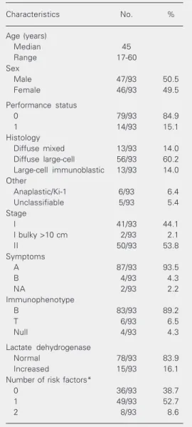
mediastinal bulky disease. Extranodal in-volvement was present in 8 stage I-E patients (sinus, 4; breast, 2; bladder, 1; bone, 1) and in 5 stage II-E patients (sinus, 4; thyroid, 1). All patients received VACOP-B with or with-out radiotherapy according to the protocol. There were no patients with a PS > 1. The distribution of patients according to the IPI stage, modified by Miller et al. (7) and ad-justed for age ≤60 years, only included pa-tients with no more than 2 negative prognos-tic factors. Histology features and other pa-tient characteristics at diagnosis are reported in Table 1.
Response to treatment and survival
All patients were available for response. At the end of therapy, 86 of 93 patients (92.4%) were in complete remission or in unconfirmed complete remission (3 patients), 4 in partial remission (4.3%) and 3 (3.2%) were non-responders.
After 6 cycles of VACOP-B, 37 stage I patients entered complete remission + un-confirmed complete remission (1 patient) (86%), 2 achieved partial remission and 4 were non-responders. After involved field radiotherapy, 41 stage I patients were in complete remission (95%) and 2 were non-responders (5%). These last 2 patients died 7 and 16 months after diagnosis because of progression in spite of a polychemoterapeutic salvage regimen containing cisplatinum and Citarabin (DHAP) (26) in one case and high-dose therapy in the other. Eleven stage I patients (26%) had received treatment fol-lowing a diagnostic biopsy without visible tumor masses. Four patients refused to un-dergo involved field radiotherapy.
After 12 courses of VACOP-B, 40 stage II patients achieved complete remission + unconfirmed complete remission (2 patients) (80%), 8 entered partial remission and 2 patients were non-responders. Ten patients received involved field radiotherapy. At the end of treatment 45 patients were in
com-plete remission (90%), 4 in partial remission and 1 patient was a non-responder. Four patients of this last group showed a stable persistence of minimal residual masses (≤1.5 cm) after radiotherapy, and required exten-sive re-staging including an MRI scan and radionuclide scan. They were judged to be in complete remission three months after ra-diotherapy. Salvage therapy in the 4 partial remission patients and in 1 non-responder
Table 1. Characteristics of the patients before treatment.
Characteristics No. %
Age (years)
Median 45
Range 17-60
Sex
Male 47/93 50.5
Female 46/93 49.5
Performance status
0 79/93 84.9
1 14/93 15.1
Histology
Diffuse mixed 13/93 14.0 Diffuse large-cell 56/93 60.2 Large-cell immunoblastic 13/93 14.0 Other
Anaplastic/Ki-1 6/93 6.4 Unclassifiable 5/93 5.4 Stage
I 41/93 44.1
I bulky >10 cm 2/93 2.1
II 50/93 53.8
Symptoms
A 87/93 93.5
B 4/93 4.3
NA 2/93 2.2
Immunophenotype
B 83/93 89.2
T 6/93 6.5
Null 4/93 4.3
Lactate dehydrogenase
Normal 78/93 83.9 Increased 15/93 16.1 Number of risk factors*
0 36/93 38.7
1 49/93 52.7
2 8/93 8.6
3 and 6 months after complete remission. After conventional therapy both achieved a second complete remission. Eight of 45 (18%) stage II patients relapsed within a median time of 14 months (range: 6-43 months) after complete remission, 4 at a different site and 4 at the initial site of disease (3 of these had previously received involved field radio-therapy). These patients received DHAP (4 patients), CHOP plus radiotherapy (1 pa-tient), and DHAP plus high-dose therapy (3 patients). Four patients achieved a second complete remission, while 4 died from pro-gressive disease within a median time of 9 months after relapse.
On July 7, 2003, 84 of 93 patients were still alive. The median follow-up observa-tion time for these patients was 57 months (range: 6-126 months). The ten-year sur-vival estimate for patients was 87.3% (SEM 4.2%). The ten-year probability of disease-free survival and of progression-disease-free sur-vival was 83.9% (SEM 4.6%) and 79.9% (SEM 4.6%), respectively (Figure 1).
The overall survival rates at 8 years were 94.5% (SEM 3.8%) for stage I patients and 82.8% (SEM 6.1%) for stage II patients (P = 0.2; Figure 2). The probability of disease-free survival and of progression-disease-free sur-vival was similar in both groups of patients with a trend in favor of stage I patients (Figure 2). Univariate analysis for PS and disease-free survival did not show any dif-ference in relation to prognostic factors. Univariate analysis for progression-free sur-vival showed PS as an adverse factor pre-dicting a poor outcome (P = 0.02).
The ten-year estimates of overall sur-vival according to the IPI stage, modified by Miller et al. and adjusted for age ≤60 years, were 96.1% (SEM 3.8%) for patients with zero risk factors (36 patients), 85.3% (SEM 5.8%) for patients with 1 risk factor (49 patients), and 57.1% (SEM 24.9%) for pa-tients with 2 risk factors (8 papa-tients) (P = 0.07). Pairwise comparison showed a sig-nificant difference between patients without Figure 1. Overall survival
(con-tinuous black line), disease-free survival (dotted line) and pro-gression-free survival (broken line) for 93 non-Hodgkin’s lym-phoma patients treated with VACOP-B with or without radio-therapy.
patient consisted of the DHAP regimen for 2 patients and high-dose therapy for 3 pa-tients. Two patients in partial remission achieved complete remission after DHAP and 3 died because of progressive disease 7, 9 and 9 months after diagnosis, respectively. Two of 41 (51%) stage I patients relapsed
Overall survival 1.0 11 10 9 0.8 0.6 0.4 0.2 0.0 8 7 6 5 4 3 2 1 0 Stage I Stage II 1.0 11 10 9 0.8 0.6 0.4 0.2 0.0 8 7 6 5 4 3 2 1 0 Stage I Stage II 1.0 11 10 9 0.8 0.6 0.4 0.2 0.0 8 7 6 5 4 3 2 1 0 Stage I Stage II Years Disease-free survival Progression-free survival A B C Figure 2. Overall survival (A),
dis-ease-free survival (B) and pro-gression-free survival (C) for non-Hodgkin’s lymphoma pa-tients in stages I and II treated with VACOP-B with or without radiotherapy and staged as de-scribed elsewhere (18).
negative factors and patients with 2 negative factors (P = 0.01).
Toxicity
The toxicity was similar in the two pa-tient groups (Table 2), the first (stage I) treated with 6 courses of VACOP-B plus involved field radiotherapy and the second (stage II) treated with 12 courses of VACOP-B with or without involved field radiotherapy. Table 2 summarizes grade 3-4 toxicity of the two treatments. We observed a trend at the limit of statistical significance in terms of anemia and mucositis in favor of patients receiving less chemotherapy. Six patients (6.5%) experienced brief episodes of fever of unknown origin during granulocytopenia, and 3 patients (3.2%) suffered short-lived grade 1-2 bronchitis. Cardiac grade 1 toxic-ity was observed in two of 93 patients (2.2%). Nine patients required a delay in drug ad-ministration (median: 7 days). Growth fac-tors were not used.
No treatment-related mortality was ob-served. No patients developed a secondary tumor.
Discussion
The aim of the present study was to de-fine the efficacy and toxicity of a third gen-eration regimen, VACOP-B, as front-line therapy for aggressive and localized NHL. With this study we were able to demonstrate that VACOP-B offers a high percentage of stable complete remissions with a low re-lapse rate and low toxicity. On completion of therapy, the complete remission rates in stage I and II were 95 and 90%, respectively. These results compare favorably to those obtained by Miller et al. (7) who reported a complete remission rate of 82% in patients receiving 3 cycles of CHOP plus radiotherapy and a complete remission rate of 80% in patients treated with 6-8 cycles of CHOP chemotherapy alone.
Current practice is to treat this category of patients with CHOP chemotherapy or other regimens containing doxorubicin. Combin-ing or alternatCombin-ing chemotherapy and radio-therapy presents significantly superior re-sults to those obtained with radiotherapy alone (2-5,7-10) and represents the most common treatment method for these patients. Radiotherapy alone yielded 5-year survival rates ranging from 56 to 100% for patients with stage I disease and from 0 to 55% for patients in stage II (1-5). Studies using che-motherapy with and without radiotherapy reported 5-year survival rates ranging from about 70% to about 80%, with no statisti-cally significant difference between stage I and stage II disease (7-10). Our results show a 6-year survival probability of 94.5% for stage I patients and 82.8% for stage II (P = 0.2). These results correlate well with previ-ously published data reporting a similar com-plete remission, survival and disease-free survival probability using third generation regimens (13,14), with very low treatment-related toxicity.
The problem with using radiotherapy alone seems to be represented by the high relapse rate after treatment. As previously reported in a randomized study (5), this is more than 50%. The problem with using conventional chemotherapy alone seems to be represented by the hematological and
ex-Table 2. Toxicity in 93 patients according to disease stage.
Grade (WHO) Stage I Stage II
3 4 3 4
Anemia 3
Granulocytopenia 4 1 3 1
Fever of unknown origin 1 Peripheral neurotoxicity 1
Alopecia 12 3 19 3
Mucositis 1
Nausea/vomiting 1
Constipation 1
tra-hematological toxicity (7). We could use 50% of conventional chemotherapy courses followed by involved field radiotherapy, as suggested by others (7-10), to achieve the best results while avoiding treatment-related toxicity. Adjuvant radiotherapy should be a necessary component of the treatment pro-gram when localized residual disease still remains at the end of chemotherapy. Miller et al. (7) showed in a randomized study that radiotherapy is useful not only in reducing the number of CHOP cycles with a conse-quent reduction in cardiac doxorubicin-re-lated toxicity, but also in improving overall outcome.
Our study design was partially consistent with these considerations. Stage I patients received 6 cycles of VACOP-B plus adju-vant involved field radiotherapy at the site of initial disease. Stage II patients received VACOP-B for 12 cycles plus involved field radiotherapy at the site of residual disease. Adjuvant radiotherapy improved the com-plete remission rate in about 10% of both groups of patients. Grades 3 and 4 granulo-cytopenia occurred in about 10% of patients but infections were negligible and short-lived, and growth factors were not required. Extra-hematological toxicity was very low, apart from alopecia which was seen in the majority of patients. No patients died be-cause of treatment. Two patients presented grade 1 cardiac toxicity. Miller et al. (7) reported life-threatening toxicity in 40% of patients treated with 8 cycles of CHOP and in 30% of patients treated with 3 cycles of CHOP plus radiotherapy. The same authors reported a “disconcerting” finding of myo-cardial toxicity associated with 8 cycles of CHOP chemotherapy. Shenkier et al. (10) treated patients with 3 cycles of CHOP or CHOP-like regimens plus involved field ra-diotherapy and reported a complete remis-sion rate of 97%. After treatment, 2 patients died of sepsis, 1 patient of myocardial in-farction, and about 20% of new malignan-cies were observed at a median time of 51
months after the diagnosis of aggressive lym-phoma. In the present series, no patient de-veloped a secondary tumor. The reasons for these differences in terms of toxicity and secondary tumors are probably due to the low age of our patients (younger than 60 years). If we compare this with the two stud-ies discussed above we see that the first study included about 50% of patients older than 60 years while in the second study patients had a median age of 64 years.The comparison of toxic effects between 6 and 12 weeks of VACOP-B with or without ra-diotherapy according to the trial design showed no statistical difference apart from mucositis which was more evident in pa-tients receiving 12 weeks of VACOP-B. However, according to our observation, 12 weeks of VACOP-B followed by involved field radiotherapy seem to represent a good choice of treatment for stage II patients in terms of efficacy and low toxicity.
In our series, univariate analysis showed a poor outcome in patients with a PS = 1 (WHO). We were not able to stratify our patients into 4 groups according to the IPI stage modified by Miller et al. (7) and ad-justed for age ≤60 years because no patient showed a PS > 1. However, patients with 2 negative factors showed a poorer outcome in terms of survival than those with no nega-tive factors at diagnosis. In recent years new treatment strategies have been employed ranging from anti-CD20 (rituximab) to anthracycline chemotherapy regimens, particularly in elderly patients (27,28) in order to improve survival in large cell lym-phomas.
regi-men (VACOP-B) with or without radio-therapy is required to confirm our observa-tions. It would also be useful to study a reduced chemotherapy regimen (6-week VACOP-B) plus radiotherapy in patients with stage II to evaluate the possibility of an even shorter duration of treatment, thereby
reduc-ing toxicity without compromisreduc-ing efficacy.
Acknowledgments
We would like to thank Anne Freckleton for help in preparing the text.
References
1. Sutcliffe SB, Gospodarowicz MK, Bush RS, Brown TC, Chua T, Bean HA, Clark RM, Dembo A, Fitzpatrick PJ & Peters MV (1985). Role of radiation therapy in localized non-Hodgkin’s lymphoma. Radio-therapy and Oncology, 4: 211-223.
2. Nissen NI, Ersboll J, Hansen HS, Walbom-Jorgensen S, Pedersen-Bjergaard J, Hansen MM & Rygard J (1983). A randomized study of radiotherapy versus radiotherapy plus chemotherapy in stage I-II non-Hodgkin’s lymphomas. Cancer, 52: 1-7.
3. Ruijs CD, Dekker AW, van Kempen-Harteveld ML, van Baarlen J & Hordijk GJ (1994). Treatment of localized non-Hodgkin’s lympho-mas of the head and neck. Cancer, 74: 703-707.
4. Kimby E, Brandt L, Nygren P, Glimelius B; SBU-group. Swedish Council of Technology Assessment in Health Care (2001). A sys-tematic overview of chemotherapy effects in aggressive non-Hodg-kin’s lymphoma. Acta Oncologica, 40: 198-212.
5. Miller TP & Jones SEM (1980). Is there a role for radiotherapy in localized diffuse lymphomas? Cancer Chemotherapy and Pharma-cology, 4: 67-70.
6. Fisher RI, Gaynor ER, Dahlberg S, Oken MM, Grogan TM, Mize EM, Glick JH, Coltman Jr CA & Miller TP (1993). Comparison of a standard regimen (CHOP) with three intensive chemotherapy regi-mens for advanced non-Hodgkin’s lymphoma. New England Jour-nal of Medicine, 328: 1002-1006.
7. Miller TP, Dahlberg S, Cassady JR, Adelstein DJ, Spier CM, Grogan TM, LeBlanc M, Carlin S, Chase E & Fisher RI (1998). Chemotherapy alone compared with chemotherapy plus radiotherapy for localized intermediate- and high-grade non-Hodgkin’s lymphoma. New Eng-land Journal of Medicine, 339: 21-26.
8. Cosset JM (1998). Chemoradiotherapy for localized non-Hodgkin’s lymphoma (Editorial). New England Journal of Medicine, 339: 44-45. 9. Munck JN, Dhermain F, Koscielny S, Girinsky T, Carde P, Bosq J, Decaudin D, Julieron M, Cosset JM & Hayat M (1996). Alternating chemotherapy and radiotherapy for limited-stage intermediate and high-grade non Hodgkin’s lymphomas: long-term results for 96 patients with tumors >5 cm. Annals of Oncology, 7: 925-931. 10. Shenkier TN, Voss N, Fairey R, Gascoyne RD, Hoskins P, Klasa R,
Klimo P, O’Reilly SEM, Sutcliffe S & Connors JM (2002). Brief chemotherapy and involved-region irradiation for limited-stage dif-fuse large-cell lymphoma: an 18-year experience from the British Columbia Cancer Agency. Journal of Clinical Oncology, 20: 197-204.
11. Gordon LI, Harrington D, Andersen J et al. (1992). Comparison of second-generation combination chemotherapeutic regimen (m-BACOD) with a standard regimen (CHOP) for advanced diffuse non-Hodgkin’s lymphoma. New England Journal of Medicine, 327: 1342-1349.
12. Sertoli MR, Santini G, Chisesi T et al. (1994). MACOP-B versus
ProMACE-MOPP in the treatment of advanced diffuse non-Hodg-kin’s lymphomas: Results of a prospective randomized trial by the Non-Hodgkin’s Lymphoma Cooperative Study Group. Journal of Clinical Oncology, 12: 1366-1374.
13. Freilone R, Botto B, Vitolo U et al. (1996). Combined modality treatment with a weekly brief chemotherapy (ACOP-B) followed by locoregional radiotherapy in localized-stage intermediate- to high-grade non-Hodgkin’s lymphoma. Annals of Oncology, 7: 919-924. 14. Zinzani PL, Stefoni V, Tani M et al. (2001). MACOP-B regimen
followed by involved-field radiation therapy in early-stage aggres-sive non-Hodgkin’s lymphoma patients: 14-year update results. Leu-kemia and Lymphoma, 42: 989-995.
15. O’Reilly SEM, Hoskins P, Klimo P & Connors JM (1991). MACOP-B and VACOP-B in diffuse large cell lymphomas and MOPP/ABV in Hodgkin’s disease. Annals of Oncology, 2 (Suppl 1): 17-23. 16. Santini G, Salvagno L, Leoni P et al. (1998). B versus
VACOP-B plus autologous bone marrow transplantation for advanced dif-fuse non-Hodgkin’s lymphoma: results of a prospective randomized trial by the Non-Hodgkin’s Lymphoma Cooperative Study Group. Journal of Clinical Oncology, 16: 2796-2802.
17. National Cancer Institute Sponsored Study of Classifications of Non-Hodgkin’s Lymphomas (1982). Summary and description of a working formulation for clinical usage. The Non-Hodgkin’s Lym-phoma Pathologic Classification Project. Cancer, 49: 2112-2135. 18. Carbone PP, Kaplan HS, Musshoff K, Smithers DW & Tubiana M
(1971). Report of the Committee of Hodgkin’s Disease Staging Classification. Cancer Research, 31: 1860-1861.
19. Oken MM, Creech RH, Tormey DC, Horton J, Davis TE, McFadden ET & Carbone PP (1982). Toxicity and response criteria of the Eastern Cooperative Oncology Group. American Journal of Clinical Oncology, 5: 649-655.
20. Chisesi T, Polistena P, Contu A et al. (2001). Cemp, a mitoxanthrone containing combination, in the treatment of intermediate and high grade non-Hodgkin’s lymphoma: an effective and non toxic thera-peutic alternative for adult and elderly patients. Leukemia and Lym-phoma, 41: 125-136.
21. Santini G, De Souza C, Congiu AM et al. (1999). High-dose cyclo-phosphamide followed by autografting can improve the outcome of relapsed or resistant non-Hodgkin’s lymphomas with involved or hypoplastic bone marrow. Leukemia and Lymphoma, 33: 321-330. 22. Cheson BD, Horning SJ, Coiffier B et al. (1999). Report of an
international workshop to standardize response criteria for non-Hodgkin’s lymphoma. NCI sponsored International Study Group. Journal of Clinical Oncology, 17: 1244-1252.
24. Cox DR (1972). Regression models and life tables. Journal of the Royal Statistical Society, 34: 187-220.
25. Anonymous (1993). A predictive model for aggressive non-Hodg-kin’s lymphoma. The International Non-Hodgnon-Hodg-kin’s Lymphoma Prog-nostic Factors Project. New England Journal of Medicine, 329: 987-994.
26. Velasquez WS, Cabanillas F, Salvador P et al. (1988). Effective salvage therapy for lymphoma with cisplatin in combination with
high-dose Ara-C and dexamethasone (DHAP). Blood, 71: 117-122. 27. Coiffier B, Lepage E, Brière J et al. (2003). CHOP chemotherapy
plus rituximab compared with CHOP alone in elderly patients with diffuse large-B-cell lymphoma. New England Journal of Medicine, 346: 235-241.