83
REV. HOSP. CLÍN. FAC. MED. S. PAULO 55(3):83-86, 2000 MAY-JUNE
From the Surgical Anatomy Investigation Lab (LIM 02) and the Department of Surgery, Hospital das Clínicas, Faculty of Medicine, University of São Paulo.
AGE RELATED CHANGES IN THE ELASTIC FIBER
SYSTEM OF THE INTERFOVEOLAR LIGAMENT
Mario Luiz Quintas, Consuelo Junqueira Rodrigues,Jin Hwan Yoo and Aldo Junqueira Rodrigues Junior
RHCFAP/3008
QUINTAS M L et al. - Age related changes in the elastic fiber system of the interfoveolar ligament. Rev. Hosp. Clín. Fac. Med. S.Paulo 55(3):83-86, 2000.
SUMMARY: In order to evaluate age related changes of the elastic fiber system in the interfoveolar ligament, we studied the deep inguinal ring from 33 male cadavers aged from stillborn to 76 years. Selective and alternated staining methods for elastic fibers were performed to differentiate oxytalan, elaunin, and mature elastic fibers. We confirmed quantitative changes of the elastic fiber system with aging. There was a significant and progressive reduction of the oxytalan fibers (responsible for tissue resistance) and a significant increment in the mature elastic and elaunin fibers (responsible for tissue elasticity). Furthermore, there were structural changes in the thickness, shortness and curling of these mature elastic fibers. These changes induced loss of the elastic fiber function and loss of the interfoveolar ligament compliance. These factors predispose individuals to the development of indirect inguinal hernias that frequently emerge in adults and aged individuals, especially above the fifth decade.
DESCRIPTORS: Elastin. Inguinal hernia. Aging. Extracellular matrix. Transversalis fascia.
Direct inguinal hernias seem to re-sult from weakening of the transversa-lis fascia on the floor of the inguinal canal 1. It has been demonstrated that alterations in the collagen component, and in the elastic fibers of the extracel-lular matrix of the transversalis fascia, occur with aging2-4. These factors favor the development of hernias and their recurrence after surgical repair. Conse-quently, after surgical repair, wall re-inforcement with a layer placement on Hesselbach’s triangle is indicated5.
In contrast, the etiology of indirect inguinal hernia in children and young-sters is of congenital origin; they de-velop from the presence of a vaginal process or of a non-obliterated perito-neum-vaginal duct6. The vaginal pro-cess remains open in 15% to 37% of male individuals. However, only a small percentage develops inguinal
hernia. The incidence of permeability of the vaginal process in infants, as well as in normal newborns, is 60% until 2 months of age and 40% at 2 years of age7. Indirect inguinal hernias may develop in 25% to 30% of new-borns and in 20% of children and adults8.
The appearance of indirect inguinal hernia in adults is correlated with the continence of the deep inguinal ring. This continence is related to the pres-ence of a “U”-shaped thickening of the transversalis fascia, at the level of the deep inguinal ring. Lytle showed in 1945 that this sling acts as a pulley dur-ing the contraction of abdominal
muscles, which places the inguinal ring more superior and lateral9. This mecha-nism occludes the deep inguinal ring during increases of intra-abdominal pressure10,11. Another mechanism that shuts the deep inguinal ring is that pro-duced by the action of internal oblique and transversal muscles, which ap-proach the inguinal ligament and clasp the deep inguinal ring. Thus the ap-pearance of indirect inguinal hernia in the adult and elderly may derive from weakening of the transversalis fascia on the border of the deep inguinal ring12.
84
REV. HOSP. CLÍN. FAC. MED. S. PAULO 55(3):83-86, 2000 MAY-JUNE
MATERIAL AND METHODS
We studied the deep inguinal ring from 33 male cadavers aged from still-born to 76 years old (age = 39.99 ± 24.90). The right interfoveolar liga-ments obtained by dissection were fixed in a 10% formaldehyde solution.
Staining Procedure
Three adjacent histological sections (3 micrometers thick) were submitted to one of the following selective meth-ods for staining elastic fibers: Verhoeff’s iodine-iron hematoxylin13 method, which stains only the mature elastic fibers; Weigert’s resorcin-fuch-sin method14, which stains mature and elaunin elastic fibers; and Weigert’s technique with a previous oxidation performed using oxone as previously described15, which stains the oxytalan, elaunin, and mature elastic fibers.
Morphometric Evaluation
The linear density (LV) of the
elas-tic fiber system was determined in 25 random microscopic fields per histo-logic section stained by Verhoeff (LVV), Weigert (LVW), and
Weigert-oxone (LVWO) methods. The sections
were scanned randomly at a magnifi-cation of 1000X in a continuous line from edge to edge, employing a test eyepiece reticule with 10 parallel lines and 100 points comprising a simple
square lattice test area of 10 500 mm2 (Fig. 1). Each elastic fiber completely intersected by any one of the lines was counted. These intersections of fibers with a test reticule are related to the length of those fibers per unit area, by the expression LV=2Na, where Na is
the length of fiber per unit area16. The area of the tissue examined was deter-mined by counting the number of points of intersection inside that tissue.
Statistical Analysis
The data on the linear density of elastic fiber were analyzed using a one-way analysis of variance (ANOVA). A p value ≤ 0.05 was considered statisti-cally significant. The combined effects of age and linear density of elastic fi-bers were tested by linear regression analysis.
RESULTS
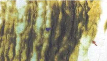
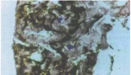
We demonstrated the presence of the 3 components of the elastic fiber system in the fascia transversalis of the Hesselbach ligaments. In young men, the elastic fibers showed a uniform dis-tribution among the connective tissue and ran parallel to the collagen fibers (Fig. 2). In the aged men, the elastic fi-bers were shortened, fragmented, and
thickened (Fig. 3).
The elastic fiber system was evalu-ated through the determination of linear density (LV) of these fibers in sections stained by selective methods for elastic fibers. LV values indicated the total length of mature, elaunin, and oxytalan fibers per unit area in WO-stained sec-tions, mature and elaunin fibers in W-stained sections, and mature fibers in V-stained sections. The mean values of LVV, LVW, and LVWO from the group studied are indicated in Table 1. There were significant positive linear correla-tions of LVV with age and LVW with agein the interfoveolar ligament. The equation curve for LVV was 0.00439 + 0.000112 * age (r = 0.83; F = 70.68; p < 0.001); the equation curve for LVW was 0.00611 + 0.000093 * age (r=0.83; F = 69.32; p < 0.001). In contrast, there was a significant negative linear corre-lation between age and LVWO; the equation curve for LVWO was 0.0162 – 0.000067 * age (r = 0.69; F = 28.78; p < 0.001. The regression curves for LVV, LVW, and LVWO are shown in figure 4.
DISCUSSION
In the elastic system, the fibers with elastin deposits are the elements re-sponsible for the reversible
distensibil-Figure 1- A test eyepiece reticule.
85
REV. HOSP. CLÍN. FAC. MED. S. PAULO 55(3):83-86, 2000 MAY-JUNE
ity of the connective tissue. Elastic fi-bers made up of microfibrils only (the oxytalanic fibers) have the mechanical resistance function17. It is well known that the network arrangement of elas-tic fibers contributes to tissue architec-ture, thus promoting passive with-drawal of the structure to its original size and shape when submitted to me-chanical stress18. Hence in the aorta, cardiovascular system, lung, and ten-dons, the relative concentration of elas-tic fibers is high19. The integration of elastic fibers with collagen fibers is necessary for the maintenance of nor-mal tissue resistance. If collagen and elastin composition, or its architectural arrangement is altered, tissue compla-cency will be jeopardized20.
Continuous elastin synthesis would promote an increase of complacency or of tissue elasticity. With aging, how-ever, elastic fibers undergo architec-tural distortion and strucarchitec-tural alter-ations, becoming tortuous, thickened, and fragmented, and they lose their original functionality21. These alter-ations seem to be promoted either by an increase in collagen, which com-presses the fibers through the action of tissue elastase and thus causes degen-eration of these fibers, or by the ar-rangement of collagen fibers, which produces architectural distortion.
In 1981, Cannon and Read2 showed evidence of increased elastolytic activity in plasma of patients around 60 years of age having direct inguinal hernias, as compared to the plasma elastolytic activ-ity of patients without hernias within the same age range. Elastic fibers, respon-sible for tissue elasticity, have been dem-onstrated to be structurally and quantita-tively altered with aging3,23. These struc-tural data corroborate the observations of Pinto24 that the low incidence of direct inguinal hernia in children, as well as its prevalence in adults — especially from fifty years of age onwards — suggest that aging humans are more susceptible to developing direct inguinal hernias.
Figure 3 - Histological section of the interfoveolar ligament from aged men showed shortened fragmented, and thickened elastic fibers. There were no thin elastic fibers. (Weigert-oxone, 400X).
Table 1 – Linear density of elastic fibers in the interfoveolar ligament from adult male cadavers, according to the staining method.
Staining method N Mean SD
Verhoeff 33 0.0088 0.00334
Weigert 33 0.0098 0.00281
Weigert-oxone 33 0.0134 0.00245
SD= Standard deviation.
Figure 4 - Elastic fibers as a function of age in the interfoveolar ligament from male cadavers. (a) LVV; (b) LVW; and (c) LVWO. The linear regression is shown by the line. Individual data are plotted at each age.
In order to better understand the etiology of indirect inguinal hernia, we looked for an explanation from the possible structural alterations with ag-ing—both qualitative and quantita-tive—suffered by the transversalis fas-cia within the deep inguinal ring region (Hesselbach’s interfoveolar ligament). We found evidence that aging is
86
REV. HOSP. CLÍN. FAC. MED. S. PAULO 55(3):83-86, 2000 MAY-JUNE
ligament (caused by aging) would pre-dispose individuals to the enlargement of the deep inguinal ring, where exte-riorization of the peritoneal sac and of the contents of the abdominal cavity and indirect inguinal hernia.
responsible for resistance. Decrease of oxytalanic fibers, and structural and ar-chitectural alterations in the elastic fi-bers that occur with age would expose the collagenous fibrous system to de-formation tensions higher than it
nor-mally endures. As a consequence, col-lagen fibers would be submitted to greater deformation, and since they represent a resistance element, they would end up by rupturing. This weak-ening of Hesselbach’s interfoveolar
RESUMO RHCFAP/3008
QUINTAS ML e col. - Alterações do sistema de fibras elásticas relacio-nadas ao envelhecimento do liga-mento interfoveolar. Rev. Hosp. Clín. Fac. Med. S. Paulo 55
(3):83-86, 2000.
Com o objetivo de avaliar as alte-rações do sistema de fibras elásticas relacionadas ao envelhecimento do li-gamento interfoveolar estudou-se o anel inguinal profundo em 33 cadáve-res do sexo masculino com idade vari-ando de recém-nascido a 76 anos.
Mé-todos de coloração seletiva para as fi-bras elásticas foram empregados para diferenciar fibras oxitalânicas, fibras elaunínicas e fibras elásticas maduras. Verificaram-se alterações quantitativas significantes no sistema de fibras elás-ticas do ligamento interfoveolar com o envelhecimento: uma redução progres-siva de fibras oxitalânicas, responsáveis pela resistência do tecido conjuntivo, e um aumento de fibras elásticas madu-ras e elaunínicas, responsáveis pela elasticidade tecidual. Demonstraram-se alterações estruturais, como
espes-samento, encurtamento e encurva-mento das fibras elásticas maduras e elaunínicas. Estas alterações induzem a perda de função das fibras elásticas, o que certamente levará a perda de complacência do ligamento inter-foveolar, predispondo à hérnia inguinal indireta, que freqüentemente aparece em homens adultos ou idosos, especi-almente acima de 50 anos de idade.
DESCRITORES: Hérnia inguinal.
Envelhecimento. Matriz extracelular. Elastina. Fascia transversal.
REFERENCES
1. CONDON RE - The anatomy of the inguinal region and its relationship to groin hernias. In: NYHUS LM & CONDON RE – Hernia, 2a.
ed. Philadelphia, Lippincott, 1978. p. 14-53.
2. WHITE HJ, SUN CN & READ RC - Inguinal hernia: A true collagen disease. Lab Invest 1977; 36:359.
3. RODRIGUES JUNIOR AJ, CASTRO DE TOLOSA EM & FERRAZ DE CARVALHO CA - Electron microscopic study on the elastic and elastic related fibers in the human fascia transversalis at different ages. Gegenbaurs Morphol Jahrb 1990; 136:645-652. 4. READ RC - A review: the role of protease-antiprotease imbalance in
the pathogenesis of herniation and abdominal aortic aneurism in certain smokers. Post Gen Surg 1992; 4: 161-5.
5. LICHTENSTEIN IL, SHULMAN AG & AMID PK - The tension-free hernioplasty. Am J Surg 1988;157:188-193.
6. GROSFELD JL - Current concepts in inguinal hernia in infants and children. World J Surg 1989;13:506-155.
7. RATHAUSER F - Historical overview of the bilateral approach to pediatric inguinal hernias. Am J Surg 1985;50:527-532. 8. SINAIDO ES - A method to facilitate hernial repair in children. Surg
Gynecol Obstet 1970;30:353-355.
9. LYTLE WJ - The internal inguinal ring. Br J Surg 1945;32:441. 10. DRYE JC - Intraperitonial pressure in the human. Surg Gynec Obst
1948;87:472-475.
11. DANNEMAN A - Etiopatogenese das hérnias inguinais diretas. Rev Col Bras Cir 1976;3(2):66-80.
12. NYHUS LM, CONDON RE & HARKINS HN - Clinical experiences with preperitoneal hernial repair for all types of hernia of the groin. Am J Surg 1960;100:234-244.
13. VERHOEFF FH - Some new staining methods of wide applicabality including a rapid differential stain for elastic tissue. J Am Med Assoc 1908;50: 876-877.
14. WEIGERT C - Uber eine Methode zur Farbung elastischer Fasern. Zbl Allg Path Anat 1898;9:289-292.
15. FULLMER HM & LILLIE RD - The oxytalan fiber: a previously undescribed connective tissue fiber. J Histochem Cytochem 1958;6:425-430.
16. NIEWOEHNER DE & KLEINERMAN J - Morphometric study of elastic fibers in normal and emphysematous human lungs. Am Rev Resp Dis 1977;115:15-21.
17. ROSS R - The elastic fiber. A review. J Histochem Cytochem 1973;21:199-208.
18. IMAYAMA S & BRAVERMAN IM - A hypothetical explanation for the aging of skin. Am J Pathol 1989;134:1019-1025. 19. UITTO J, RYHANEN L, ABRAHAM PA et al. - Elastin in diseases.
J Invest Dermatol 1982;79:160s-168s.
20. La BOURENE JI, COLES JG, JOHNSON DJ et al. - Alterations in elastin and collagen related to the mechanism of progressive pulmonary venous obstruction in a piglet model. A hemodynamic, ultrastructural and biochemical study. Circ Res 1990:66:438-456. 21. FUKUDA Y, MASUDA Y & ISHIZAKI M - Morphogenesis of abnormal elastic fibers in lungs of patients with panacinar and centriacinar emphysema. Hum Pathol 1989;20:652-659. 22. CANNON DR & READ RC - Metastatic emphysema. Mechanism
for acquiring inguinal herniation. Ann Surg 1981;94:270-277. 23. RODRIGUES CJ - Influência do envelhecimento sobre o músculo
diafragma. São Paulo, 1994. [Doutorado – Faculdade de Medicina, Universidade de São Paulo].
24. CARVALHO PINTO VA - Patologia do conduto peritônio-vaginal. Rev Ass Méd Bras 1961;7:251-255.