From the Division of Urology, Hospital das Clínicas, Faculty of Medicine, University of São Paulo.
PHEOCHROMOCYTOMA TREATED BY
LAPAROSCOPIC SURGERY
Lísias Nogueira Castilho, Paulo José de Medeiros, Anuar Ibrahim Mitre, Francisco Tibor Dénes, Antonio Marmo Lucon and Sami Arap
RHCFAP/3010 CASTILHO L N et al. - Pheochromocytoma treated by laparoscopic surgery. Rev. Hosp. Clín. Fac. Med. S. Paulo 55 (3):93-100,
2000.
SUMMARY: Objective: To evaluate the results of the laparoscopic technique in the treatment of adrenal pheochromocytoma.
Method: Ten patients, 7 men and 3 women, between 10 and 67 years of age (mean 48) with pheochromocytoma underwent
transperitoneal laparoscopic adrenalectomy and were evaluated retrospectively, based on clinical, laboratory, and pathological diagnosis. In all cases there was a solid unilateral adrenal tumor, 5 on the left side and 5 on the right side, whose greater diameter varied from 7 to 80 mm (mean 32). Nine of the 10 patients were chronically hypertensive or had already had hypertensive crises. One patient was normotensive, but presented metabolic alterations suggestive of adrenergic hyperfunction.
Results: No deaths occurred in this series. There were two (20%) conversions to open surgery, one due to venous bleeding and
one due to the difficulty of dissection behind the vena cava in a patient presenting a partially retro-caval tumor. Surgical time in the 8 non-converted cases ranged from 70 to 215 minutes (mean 136). One patient (10%) received blood transfusion, and another (10%) presented two complications – acute renal failure and a subcutaneous infection. Both had been converted to open surgery. None of the non-converted cases was transfused or presented complications. Hospital discharge occurred between the 2nd and 11th post-operative day (mean 3). The pathological exam of the surgical specimens confirmed the diagnoses of pheochromocytoma in all 10 cases, one of them associated with an aldosterone-producing cortical tumor.
Conclusions: Laparoscopic adrenalectomy for selected patients presenting pheochromocytoma is feasible and provides good results.
DESCRIPTORS: Pheochromocytoma. Adrenal tumor. Laparoscopic adrenalectomy.
Laparoscopic adrenalectomy was originally described by Canadian and Japanese authors, in 19921,2. Since then, according to the Index Medicus,
approximately 300 studies on this sub-ject have been published, totaling more than 1000 performed adrenalectomies, which reflects not only significant in-ternational experience, but also a grow-ing interest by urologists and surgeons who operate on endocrine glands. Laparoscopic adrenalectomy has been considered for some time to be the
gold standard treatment for benign
adrenal tumors of small volume, func-tioning or not3, due to the better results
obtained when compared to those of open surgery, in all aspects – morbid-ity, hospitalization, convalescence, bleeding, pain, etc., including costs4-15. Concerning operative time in laparoscopic surgeries, although it was initially longer than for open surgery, bringing doubts and criticisms, cur-rently, it is approximately the same since the learning curve has been climbed16.
The treatment of
pheochromocy-toma by laparoscopy has been the sub-ject of studies and controversies since 1992, when it was first successfully per-formed1. Laparoscopic technique pre-sents several demonstrated advantages over open surgery, both in unilateral and bilateral pheochromocytoma17-27.
We present below our experience with laparoscopic treatment of pheo-chromocytoma.
METHOD
adrenalec-tomy, with different diagnoses, be-tween May 1994 and December 1999. Sent from private clinics and from the Division of Urology of the Hospital das Clínicas da FMUSP, all patients were initially evaluated by endocrinologists and sent to us only after surgical indi-cation was firmly established. Exclu-sion criteria for laparoscopic surgery were: clinical-radiological suspicion of primary malignancy of the adrenal gland (of difficult characterization), moderate or severe respiratory insuffi-ciency, moderate or severe cardiac or coronary insufficiency, uncorrected coagulopathy, intraperitoneal or ab-dominal wall infections, and refusal of the patient or endocrinologist to accept laparoscopic surgery. Previous abdomi-nal surgery, obesity, and abdomiabdomi-nal wall hernias were not criteria for exclu-sion. Sixty-one patients fulfilled our pre-requisites and underwent laparoscopic procedures.
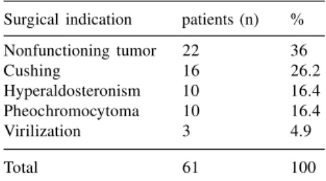
After rigorous clinical, laboratory, and radiologic investigations per-formed by endocrinologists, the 61 pa-tients were selected for laparoscopic surgery with the following pre-opera-tive diagnoses (Table 1): non-function-ing tumor (22), Cushnon-function-ing’s syndrome or disease (16), hyperaldosteronism (10), pheochromocytoma (10) and virilizing tumor (3). All patients underwent echography (US) and computerized to-mography (CT) of the abdomen. Some additionally underwent magnetic nuclear resonance (MRI) procedures for better characterization of small nodules. All patients with evidence from these tests of pheochromocytoma, also underwent radioisotopic exam with MIBG (meta-iodine-benzyl-guanidine). Patients presenting non-functioning tumors and previous his-tory of cancer were also investigated with bone scan and chest radiograms.
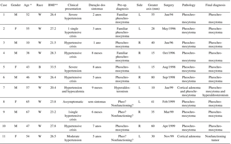
We retrospectively analyzed all cases with an initial diagnosis of pheo-chromocytoma, comparing them with the cases that had a diagnosis of
pheo-chromocytoma confirmed (Table 2). Of the 10 pheochromocytoma cases confirmed by pathologic examination, 7 were from male and 3 from female patients. The majority (90%) were of white color. Age varied from 10 to 67 years (mean 48). All patients presented the disease unilaterally, with a normal contralateral gland. Three cases pre-sented family history of pheochro-mocytoma.
Pre-operative clinical preparation of the patients consisted basically of rest, low sodium diet, and administra-tion of the alpha-blocking agent prasozine, in doses of 4 to 20 mg per day during the weeks prior to surgery. Surgical preparation conformed to the following general norm, adapted for each case: light diet two days before surgery, and a liquid diet and enema, with 500 ml, to empty the rectum, in the night preceding the procedure; ab-dominal trichotomy immediately be-fore surgery; and prophylactic admin-istration of wide spectrum antibiotic during the induction of anesthesia.
Under general anesthesia, the pa-tients were positioned on the surgical table in 45o lateral decubitus. Pads, ad-hesive tapes and, in some cases, elas-tic socks, were adequately placed to avoid ischemic injuries, burns, nervous lesions, and venous thrombosis.
Standard procedures were executed and the following technical steps were followed:
1st) CO
2 insufflation of the peritoneal cavity through the introduction of the Veress needle in the abdomen, either on the midline, on the lower border of the umbilicus, or on the midclavicular line on the same side the adrenalec-tomy would be performed. In all cases the transperitoneal approach was em-ployed.
2nd) Once the pneumoperitoneum was obtained, four 11 mm trocars were in-serted in the abdomen according to the pattern in figure 1. In the only child in the group, and in very thin patients, two 11 mm and two 5 mm trocars were used.
3rd) Once the cavity was inspected, the equipment and the patient’s position were adjusted and we proceeded to the medial mobilization of the colon, and exposure of the renal fascia and the great vessels–renal vein on the left and vena cava on the right.
4th) The central adrenal vein, tributary of the vena cava on the right side and of the renal vein on the left, was then identified and sectioned between me-tallic clips before manipulation of the gland to avoid hemodynamic distur-bances due to adrenergic discharges. 5th) Once the central vein, almost al-ways the sole one, was cut, the gland was freed of neighboring structures with care, in order to avoid visceral or vascular lesions. Small arterial, lym-phatic or venous vessels were simply cauterized.
6th) Once completely freed, the surgi-cal specimen was removed from the abdomen inside a special plastic bag, by means of an enlargement of one of the incisions in the abdominal wall, usually the most inferior one, close to the iliac spine.
7th) Once the specimen was removed, we proceeded to the revision of the cavity and suturing of the surgical wounds in two layers.
All surgical specimens were sent to pathologic examination. Those that raised doubts about their diagnoses Table 1 – Distribution of 61 patients treated in
accordance to the clinical-laboratorial indication of laparoscopic surgery.
Surgical indication patients (n) % Nonfunctioning tumor 22 36
Cushing 16 26.2
Hyperaldosteronism 10 16.4
Pheochromocytoma 10 16.4
Virilization 3 4.9
Table 2 – Tabulation of all the cases with clinical-laboratorial suspition of pheochromocytoma and of those with pathological confirmation.
Case Gender Age * Race BMI** Clinical Duração dos Pre-op. Side Greater Surgery Pathology Final diagnosis presentation sintomas diagnosis axis (mm)
1 M 52 W 26.4 Severe 2 anos Familiar L 55 Jun/94 Pheochro- Pheochro-hypertension
pheochro-mocytoma mocytoma mocytoma 2 F 55 W 27.2 1 single 3 anos Familiar L 28 May/1996 Pheochro- Pheochro-hypertensive pheochro- mocytoma mocytoma
crisis mocytoma
3 M 10 W 21.5 Hypertensive 1 ano Pheochro- R 40 Jun/96 Pheochro-
Pheochro-crisis mocytoma mocytoma mocytoma
4 M 38 W 28.3 Hypertensive 8 meses Familiar R 15 Oct/1996 Pheochro- Pheochro-crisis
pheochro-mocytoma mocytoma mocytoma 5 F 43 B 33.5 Severe 8 anos Pheochro- L 15 Aug/1998 Pheochro- Pheochro-hypertension mocytoma mocytoma mocytoma 6 M 46 W 26.4 Hypertensive 5 anos Pheochro- R 80 Sep/1998 Pheochro-
Pheochro-crisis mocytoma mocytoma mocytoma
7 M 57 W 20.4 Hypertension 9 meses Hyperaldos- L 10 Jan/99 Cortical adenoma Pheochro-and hypocalemia teronism and pheochr- mocytoma and
mocytoma hyperaldosteronism 8 F 65 W 23.8 Assymptomatic sem sintomas Pheo? L 41 Feb/1999 Pheochro-
Pheochro-Nonfunctioning? mocytoma mocytoma 9 M 67 W 23.2 1single 6 meses Pheo? R 35 Mar/99 Pheochro- Pheochro-hypertensive Nonfunctioning? mocytoma mocytoma
crisis
10 M 47 W 27.8 Hypertensive 7 anos Pheochro- R 60 Apr/1999 Pheochro-
Pheochro-crisis mocytoma mocytoma mocytoma
11 F 54 W 26.5 Moderate 5 anos Pheo? L 30 Nov/99 Cortical adenoma Nonfunctioning
hypertension Nonfunctioning? tumor
* Years; **Body Mass Index.
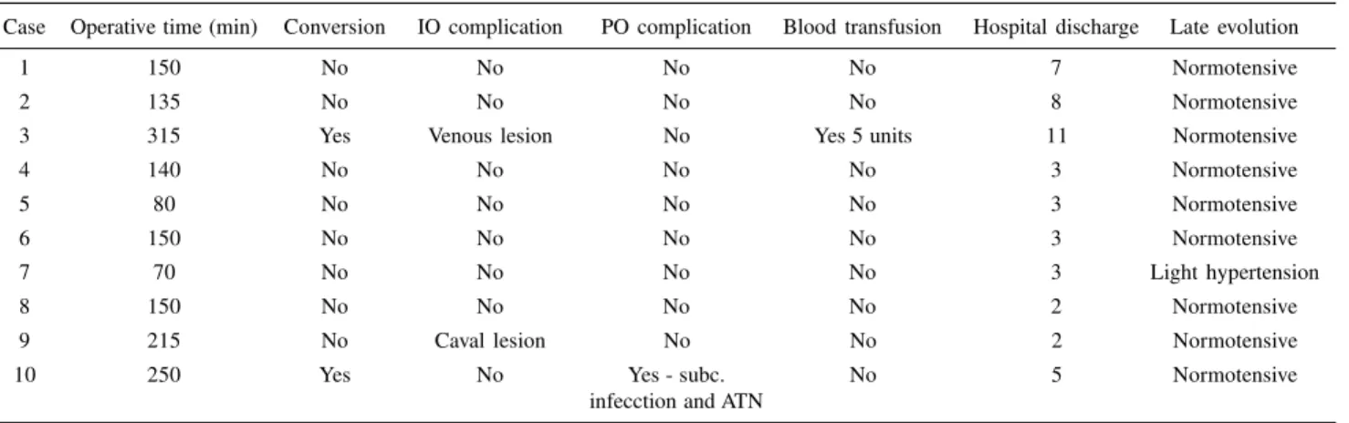
Table 3 - Results.
Case Operative time (min) Conversion IO complication PO complication Blood transfusion Hospital discharge Late evolution
1 150 No No No No 7 Normotensive
2 135 No No No No 8 Normotensive
3 315 Yes Venous lesion No Yes 5 units 11 Normotensive
4 140 No No No No 3 Normotensive
5 80 No No No No 3 Normotensive
6 150 No No No No 3 Normotensive
7 70 No No No No 3 Light hypertension
8 150 No No No No 2 Normotensive
9 215 No Caval lesion No No 2 Normotensive
10 250 Yes No Yes - subc. No 5 Normotensive
infecction and ATN
were submitted to immunohistochemi-cal exam.
RESULTS
No deaths occurred in this series, ei-ther during the hospitalization period or during the follow-up period, which ranged from 1 to 62 months (mean 22). Case 7 did not have a clinical or labo-ratory diagnosis of pheochromocytoma, but of hyperaldosteronism, despite pre-senting severe hypertension. The patho-logic examination with immune-his-tochemistry confirmed the co-existence of pheochromocytoma. Case 11 had a doubtful diagnosis because some tests, such as the MIBG and serum dosages of catecholamines were abnormal, but borderline; the pathologic examination showed a cortical adenoma, probably non-functioning and unrelated to the moderate arterial hypertension pre-sented by the patient, which remained unaltered in the post-operative period. Therefore, we considered the results of the 10 patients that had the final diag-nosis of pheochromocytoma, excluding case 11 (Table 3).
In two cases (3 and 10), there was need for conversion to open surgery (20%), the first one due to a lesion of the adrenal vein, and the other due to difficulty in finding a dissection plane
between the tumor, measuring 6 cm, and the vena cava (Table 3).
Surgical time varied from 70 to 315 minutes. Excluding the two cases that were converted, average surgical time was 136 min. (range, 70 min. to 215 min.).
There were intra-operative occur-rences in two cases. In case 3, a sec-ondary adrenal vein that drained to the liver was accidentally severed, and we were unable to attain hemostasia. This was the only case that required blood transfusion, and it was converted to open surgery. In case 9, a small lesion of the vena cava was sutured without difficulty, and the laparoscopic proce-dure was completed.
There were two complications on the same patient (case 10): acute renal failure, treated with diuretics and dopamine, with no need for dialysis, and subcutaneous infection after hos-pital discharge, drained spontaneously. In this patient laparoscopic surgery had been converted to open surgery.
Only case 7 remained hypertensive in the late post-operative period, al-though under easy control. This patient had undergone two kidney transplants, had a chronic nephropathy, and had been hypertensive for over 20 years. All others became normotensive with-out medication. All 10 patients under-went image exams with MIBG, US and
CT, in addition to metabolic exams. In none were alterations compatible with pheochromocytoma found during the follow-up period.
All patients were fed and walked in the first post-operative day. Hospital discharge occurred between the 2nd and 11th post-operative day (mean = 3 days).
Pathologic examination of the 10 specimens demonstrated the presence of a single solid tumor in all of them: the smallest specimen, a gland with a 7 mm diameter nodule, weighed 5 g; the largest, a tumor with a 80 mm di-ameter, weighed 250 g. Only the adre-nal gland of case 7 presented a differ-ent result: a cortical nodule (associated to the hyperaldosteronism) and med-ullar hyperplasia. The diagnosis of pheochromocytoma in the hyperplastic zone was established only through im-munohistochemical exams.
DISCUSSION
al-terations observed during the CO2 in-sufflation28. However, with increasing experience, it has been verified that the metabolic and hemodynamic alter-ations observed are of small impor-tance and do not augment the risk in laparoscopic surgery20,27,29,30,31. On the contrary, the laparoscopic approach re-vealed itself superior to open surgery for tumors of up to 6 cm at the largest diameter, being specially indicated in cases of greater risk, such as bilateral tumors20,25,26, tumors in pregnancy20,23, multiple tumors20 and patients with congestive heart disease32. Actually, neither the laparoscopic or the open approach are risk factors themselves, if adequately employed, meaning avoid-ing the main risk factors of the proce-dure by controlling the adrenal vein at the beginning, and doing minimal ma-nipulation of the tumor. The risks are inherent to the disease itself, especially when blood pressure levels are very high, and also to the clinical and anes-thetic precautions and procedures in-volved. Pre-operative preparation with alpha-blockers, in our preference prasozine, immediate pre-operative hyperhydration and rigorous hemody-namic control in the surgery room are the main factors determining the suc-cess of the procedure. The patient di-agnosed with precision, clinically pre-pared with alpha-blockers, well anes-thetized and operated on with the cor-rect technique, open or laparoscopic, rarely presents serious complications. The advantages observed by us and several other authors, concerning the laparoscopic approach over the open approach, are, essentially: shorter hos-pital stay, pain practically absent, lower morbidity, shorter convalescence time, and better aesthetic results8, 33-35.
The choice we made for a transperitoneal approach, instead of the extraperitoneal approach others use36,37, is due to two facts: first, our greater fa-miliarity with the transperitoneal ap-proach; second, the greater ease of
ob-taining control over the adrenal vein in the beginning of the procedure, a task that is more troublesome in the extraperitoneal approach33. However, the extraperitoneal approach has been used with increasing frequency and success, particularly for small tumors38. The choice of the approach depends on the personal experience of the surgeon and on the dimensions of the tumor. For larger tumors, the transperitoneal approach should be chosen7,8. For smaller tumors, apparently both the transperitoneal and the extraperitoneal approaches are equivalent.
The surgical time we recorded, 136 minutes on average, similar to that of other authors using the same tech-nique7,19,20,24, and in the experience of our team, is approximately the same as in open surgery. Although others have observed a significantly greater surgi-cal time in laparoscopic surgery17,33, we believe that with increasing experience, times tend to even out, as happened in our personal experience with nephrec-tomy, nephroureterectomy and radical nephrectomy (unpublished data). Even those authors who have found a signifi-cant difference in surgical times be-tween laparoscopic and open proce-dures, consider, as a whole, the results obtained through laparoscopic surgery to be much superior to those of open surgery17.
The conversion-to-open-surgery ra-tio that we had in pheochromocytoma, 20%, is far above our ratio in the whole group of 61 patients we treated, 8%, and this difference probably reflects the small number of pheochromocy-toma cases in our series. However, in retrospect, at least one of our two con-verted cases (Case 3) could have been treated laparoscopically, if we had larger experience, since we sutured a lesion of the vena cava during laparoscopy in a more recent case without great difficulty. Actually, with more experience, the number of con-versions to open surgery approaches
zero in surgery for pheochromocy-toma20.
One of our patients (10%) pre-sented two complications, one of small repercussion, a subcutaneous infection, but another more important, acute re-nal failure. Reviewing the case, which required conversion due to the retro-caval position of the tumor, greatly in-creasing the difficulty of the operation, we observed that immediate pre-opera-tive hyperhydration, which we rou-tinely used, was not correctly followed. This omission must have contributed to the hypotension that occurred immedi-ately after the ligation of the adrenal vein while still in the operating room. Patients with pheochromocytoma usu-ally present hypervolemia. Once the intense adrenergic activity is sup-pressed during the operation, the ten-dency of these patients is to present pre-renal renal insufficiency, hence the importance of expanding the intravas-cular bed with saline and colloids hours before the intervention. Authors very experienced with the laparoscopic treatment of pheochromocytoma have obtained complication ratios of 8% to 16%20,33.
It is relevant that the only patient who underwent blood transfusion and the only one who presented complica-tions were converted. Out of the 8 non-converted cases, none presented com-plications or needed blood transfu-sions.
Although we have operated on 3 cases of familiar pheochromocytoma, a condition considered ideal for the ex-ecution of partial adrenalectomy20,38,39, we preferred a total adrenalectomy and follow-up of the cases. In case of recurrence in the remaining gland, we intend to perform a partial resection of the gland, a technically simple proce-dure, especially for tumors of small volume.
confidence, either because of normal or borderline exams, in cases of function-ing pheochromocytomas, or in non-functioning tumors. In our series, out of 3 doubtful cases, the diagnosis was not confirmed in one. On the other hand, pheochromocytoma is eventually a pathological finding, especially in the non-functioning cases. We did not have any cases on which the pheochromocy-toma was completely nonfunctioning. In case 8 there were no symptoms, but urinary vanilmandelic acid was slightly above normal. Despite this being the only biochemical alteration found, the patient was prepared for the procedure as having a functioning pheochro-mocytoma, and pathologic examination confirmed the diagnosis. In case 7, di-agnosis of hyperaldosteronism was well established, and a pheochromocy-toma was found in the immunohis-tochemical exam; however, the pre-op-erative biochemical exams were only slightly altered, not leading to the pre-sumptive diagnosis of pheochromocy-toma associated with the cortical tumor causing hyperaldosteronism, a rare situation that we never before
ob-served. This patient was the only one who remained hypertensive in the late post-operative period (10%), what is in accordance with the information from the literature about pheochromocy-toma, in which about 90% of treated patients become normotensive immedi-ately or after a few weeks40. However, due to the relevant nephrologic history of the patient, it is possible that other factors have contributed to his hyper-tension.
The largest tumor diameter mea-sured by CT or MRI was 80 mm, but the average of the 10 cases was 32 mm. Pheochromocytomas of large volume are uncommon. Most are less than 6 cm in their greatest diameter, which emphasizes even more the pref-erence for laparoscopic surgery. How-ever, in tumors of greater volume, the determinant factor for the indication of laparoscopic surgery is the experi-ence of the surgeon, and not an abso-lute anatomical aspect. Therefore, some authors have successfully re-moved, by laparoscopy, tumors of 10, 12 and even 15 cm in diameter7,8. Our
experience suggests that tumors of up to 10 cm on the left side can be suc-cessfully approached by laparoscopy, but on the right side, due to the prox-imity of the vena cava, the limit must be established around 6 cm for most cases.
CONCLUSIONS
Considering our personal experi-ence and data accumulated in interna-tional literature since 1992, it can be said that laparoscopic surgery of pheo-chromocytoma can be safely and effi-ciently performed in adrenal tumors of small volume, with advantages over open surgery. Laparoscopy can also be indicated in selected cases of bilateral tumors, larger tumors (>6 cm in the longer diameter), paragangliomas, and tumors in pregnancy. Partial adrenalec-tomy should be considered in cases of single gland tumors and in patients who have a good chance to develop bi-lateral disease, as familiar pheochro-mocytoma and other genetic disorders.
RESUMO RHCFAP/3010
CASTILHO L N e col. – Feocro-mocitoma tratado por cirurgia laparoscópica. Rev. Hosp. Clín. Fac. Med. S. Paulo 55 (3): 93-100, 2000.
Objetivo: Avaliar os resultados da uti-lização da técnica laparoscópica no trata-mento do feocromocitoma de supra-renal. Método: Dez pacientes, sete ho-mens e três mulheres, entre 10 e 67 anos de idade (média 48), com feocro-mocitoma, foram operados por via
laparoscópica transperitoneal e avalia-dos retrospectivamente, com base nos diagnósticos clínico-laboratorial e anátomo-patológico. Em todos os ca-sos havia um tumor sólido unilateral de supra-renal, cinco à direita e cinco à esquerda, cujo maior eixo variou de 7 a 80 mm (média 32). Nove dos dez pa-cientes eram hipertensos crônicos ou tinham história de picos hipertensivos. Um paciente era normotenso, mas apresentava alterações metabólicas su-gestivas de hiperfunção adrenérgica.
REFERENCES
1. GAGNER M, LACROIX A & BOLTE E - Laparoscopic adrenalectomy in Cushing’s syndrome and pheochromocytoma. N Engl J Med 1992; 327: 1003-1006.
2. HIGASHIHARA E, TANAKA Y, HORIE S et al. - A case report of laparoscopic adrenalectomy. Jap J Urol 1992; 83:1130-1133. 3. YOSHIDA O, TERACHI T, MATSUDA T et al. - Complications in
369 laparoscopic adrenalectomies: a multi-institutional study in Japan. J Urol 1997; 157 (supp): 282, 1098A.
4. BABA S, ITO K, YANAIHARA H et al. - Retroperitoneoscopic adrenalectomy by a lumbodorsal approach: clinical experience with solo surgery. World J Urol 1999; 17: 54-58.
5. BENDINELLI C, MATERAZZI G, PUCCINI M et al. - Laparoscopic adrenalectomy: a retrospective comparison with traditional methods. Minerva Chir 1998; 53: 871-875.
6. DUDLEY NE & HARRISON BJ - Comparison of open posterior versus transperitoneal laparoscopic adrenalectomy. Br J Surg 1999; 86: 656-660.
7. FILIPPONI S, GUERRIERI M, ARNALDI G et al. - Laparoscopic adrenalectomy: a report on 50 operations. Eur J Endocrionol 1998; 138: 548-553.
8. GAGNER M, POMP A, HENIFORD BT et al. - Laparoscopic adrenalectomy: lessons learned from 100 consecutive procedures. Ann Surg 1997; 226: 238-246.
9. GASMAN D, DROUPY S, KOUTANI A et al. - Laparoscopic adrenalectomy: the retroperitoneal approach. J Urol 1998; 159: 1816-1820.
10. HERRERA MF, TORRES G, GAMINO R et al. - Laparoscopic adrenalectomy in a Mexican institution. Rev Invest Clin 1998; 50: 399-404.
11. MIYAKE O, YOSHIMURA T, YOSHIOKA T et al. - Laparoscopic adrenalectomy. Comparison of the transperitoneal and retroperitoneal approach. Eur Urol 1998; 33: 303-307. 12. SHEN WT, LIM RC, SIPERSTEIN AE et al. - Laparoscopic vs open
adrenalectomy for the treatment of primary hyperaldosteronism. Arch Surg 1999; 134: 628-631.
13. SHICHMAN SJ, HERNDON CD, SOSA RE et al. - Lateral transpe-ritoneal laparoscopic adrenalectomy. World J Urol 1999; 17: 48-53. 14. SMITH CD, WEBER CJ & AMERSON JR - Laparoscopic adrenalectomy: new gold standard. World J Urol 1999; 23: 389-396.
15. YOSHIMURA K, YOSHIOKA T, MIYAKE O et al. - Comparison of clinical outcomes of laparoscopic and conventional open adrenalectomy. J Endourol 1998; 12: 555-559.
16. TERACHI T, MATSUDA T, TERAI A et al. - Transperitoneal laparoscopic adrenalectomy: experience in 100 patients. J Endourol 1997; 11: 361-365.
17. MÖBIUS E, NIES C & ROTHMUND M - Surgical treatment of pheochromocytomas: laparoscopic or conventional? Surg Endosc 1999; 13: 35-39.
18. SCHELL SR, TALAMINI MA & UDELSMAN R. - Laparoscopic adrenalectomy for nonmalignant disease: improved safety, morbidity, and cost-effectiveness. Surg Endosc 1999; 13: 30-34. 19. PIATEK S, MANGER T, KUNZ D et al. - Laparoscopic transperitoneal adrenalectomy – technique and personal experiences. Zentralbl Chir 1997; 122: 1103-1107.
20. JANETSCHEK G, FINKENSTEDT G, GASSER R et al. -Laparoscopic surgery for pheochromocytoma: adrenalectomy, partial resection, excision of paragangliomas. J Urol 1998; 160: 330-334.
21. TAKAMI H, MIYOSHI H, KODAIRA S et al. - Laparoscopic adrenalectomy in asymptomatic pheochromocytoma. Am Surg 1997; 63: 820-822.
22. COL V, DE CANNIÈRE L, COLLARD E et al. - Laparoscopic adrenalectomy for pheochromocytoma: endocrinological and surgical aspects of a new therapeutic approach. Clin Endocrinol (Oxf) 1999; 50: 121-125
23. DEMEURE MJ, CARLSEN B, TRAUL D et al. - Laparoscopic removal of a right adrenal pheochromocytoma in a pregnant woman. J Laparoendosc Adv Surg TechA 1998; 8: 315-319.
24. CHIGOT JP, MOVSCHIN M, EL BARDISSI M et al. - Comparative study between laparoscopic and conventional adrenalectomy for pheochromocytomas. Ann Chir 1998; 52: 346-349.
25. TERAI A, TERACHI T, INOUE T et al. - Laparoscopic adrenalectomy for bilateral pheochromocytoma: a case report. Int J Urol 1997; 4: 300-303.
26. MANGER T, PIATEK S, KLOSE S et al. - Bilateral laparoscopic transperitoneal adrenalectomy in pheochromocytoma. Langenbecks Arch Chir 1997; 382: 37-42.
27. MICCOLI P, BENDINELLI C, MATERAZZI G et al. - Traditional versus laparoscopic surgery in the treatment of pheochromocytoma: a preliminary study. J Laparoendosc Adv Surg Tech A 1997; 7:167-171.
28. TAKEDA M, GO H, IMAI T et al. - Experience with 17 cases of laparoscopic adrenalectomy: use of ultrasonic aspirator and organ beam coagulator. J Urol 1994; 152:902-905.
29. JORIS JL, HAMOIR EE, HARSTEIN GM et al. - Hemodynamic changes and catecholamine release during laparoscopic adrenalectomy for pheochromocytoma. Anesth Analg 1999; 88: 16-21.
30. FERNÁNDEZ CRUZ L, SÁENZ A, TAURÁ P et al. - Helium and carbon dioxide pneumoperitoneum in patients with pheochromocytoma undergoing laparoscopic adrenalectomy. World J Surg 1998; 22: 1250-1255.
Ambos foram convertidos para cirurgia aberta. Nenhum dos casos não-conver-tidos recebeu transfusão ou apresentou complicação. A alta hospitalar foi con-cedida entre o 2o e o 11o PO (mediana 3). O exame anátomo-patológico das
peças cirúrgicas confirmou o feocro-mocitoma em todos esses dez casos, num deles associado a um tumor cortical produtor de aldosterona.
Conclusões: A supra-renalectomia laparoscópica para casos selecionados
de feocromocitoma é factível e apre-senta bons resultados.
31. PRETORIUS M, RASMUSSEN GE & HOLCOMB GW -Hemodynamic and catecholamine responses to a laparoscopic adrenalectomy for pheochromocytoma in a pediatric patient. Anesth Analg1998; 87: 1268-1270.
32. COL V, DE CANNIÈRE L, MESSAOUDI L et al. - Heart failure induced by pheochromocytoma: laparoscopic treatment and intraoperative changes of several new cardiovascular hormones. Horm Res 1999; 51: 50-52.
33. GILL IS & NOVICK AC - Laparoscopic versus open adrenal surgery. AUA Update Series 1999; 28:258-263.
34. THOMPSON GB, GRANT CS, VAN HEERDEN JA et al. -Laparoscopic versus open posterior adrenalectomy: a case-control study of 100 patients. Surgery 1997; 122: 1132-1136.
35. LINOS DA, STYLOPOULOS N, BOUKIS M et al. - Anterior, posterior, or laparoscopic approach for the management of adrenal diseases? Am J Surg 1997; 173: 120-125.
36. WALZ MK, PEITGEN K, HOERMANN R et al. - Posterior retroperitoneoscopy as a new minimally invasive approach for adrenalectomy: results of 30 adrenalectomies in 27 patients. World J Surg 1996; 20: 760-774.
37. GILL IS, ABDELMALAK B, BRAVO EL et al. - Laparoscopic versus open adrenalectomy for pheochromocytoma. J Endourol 1998; 12: S205 (abstract P18-4).
38. WALZ MK, PEITGEN K, SALLER B et al. - Subtotal adrenalectomy by posterior retroperitoneoscopic approach. World J Surg 1998; 22: 621-627.
39. IMAI T, TANAKA Y, KIKUMORI T et al. - Laparoscopic partial adrenalectomy. Surg Endosc 1999; 13: 343-345.
40. LUCON AM, PEREIRA MAA, MENDONÇA BB et al. - Pheochro-mocytoma: study of 50 cases. J Urol 1997; 157: 1208-1212.