From the Dentistry Division, Hospital das Clínicas, Faculty of Medicine, University of São Paulo and the Pediatric Rheumatology Unit, Children’s Institute, Hospital das Clínicas, Faculty of Medicine, University of São Paulo/SP, Brazil.
E-mail: [email protected]
Received for publication on September 17, 2003.
ORIGINAL RESEARCH
DENTAL AND FACIAL CHARACTERISTICS OF
PATIENTS WITH JUVENILE IDIOPATHIC ARTHRITIS
Cynthia Savioli, Clovis A.A. Silva, Lin H. Ching, Lucia M.M.A. Campos, Eliane F.B.G. Prado and José Tadeu T. Siqueira
SAVIOLI C et al. Dental and facial characteristics of patients with juvenile idiopathic arthritis. Rev. Hosp. Clín. Fac. Med. S. Paulo 59(3):93-98, 2004.
OBJECTIVE: It has been shown that the temporomandibular joint is frequently affected by juvenile idiopathic arthritis, and this degenerative disease, which may occur during facial growth, results in severe mandibular dysfunction. However, there are no studies that correlate oral health (tooth decay and gingival diseases) and temporomandibular joint dysfunction in patients with juvenile idiopathic arthritis. The aim of this study is to evaluate the oral and facial characteristics of the patients with juvenile idiopathic arthritis treated in a large teaching hospital.
METHOD: Thirty-six patients with juvenile idiopathic arthritis (26 female and 10 male) underwent a systematic clinical evaluation of their dental, oral, and facial structures (DMFT index, plaque and gingival bleeding index, dental relationship, facial profile, and Helkimo’s index). The control group was composed of 13 healthy children.
RESULTS: The mean age of the patients with juvenile idiopathic arthritis was 10.8 years; convex facial profile was present in 12 juvenile idiopathic arthritis patients, and class II molar relation was present in 12 (P = .032). The indexes of
plaque and gingival bleeding were significant in juvenile idiopathic arthritis patients with a higher number of superior limbs joints involved (P = .055). Anterior open bite (5) and temporomandibular joint noise (8) were present in the juvenile
idiopathic arthritis group. Of the group in this sample, 94% (P = .017) had temporomandibular joint dysfunction, 80% had
decreased mandibular opening (P = 0.0002), and mandibular mobility was severely impaired in 33% (P = .015).
CONCLUSION: This study confirms that patients with juvenile idiopathic arthritis a) have a high incidence of mandibular dysfunction that can be attributed to the direct effect of the disease in the temporomandibular joint and b) have a higher incidence of gingival disease that can be considered a secondary effect of juvenile idiopathic arthritis on oral health.
KEY WORDS: Juvenile idiopathic arthritis. Dental pain. Teeth. Temporomandibular joint juvenile. Rheumatoid arthritis.
Juvenile idiopathic arthritis (JIA) is a chronic, inflammatory, systemic dis-ease. Beginning before 16 years of age, it affects 1 or more joints of the body. It is characterized predominantly by idiopathic peripheral arthritis with an immunoinflammatory pathogenesis. According to the expression at onset and during the first 6 months of the disease, the most common types of JIA onset are: oligoarticular (4 or fewer in-volved joints), polyarticular (5 or more involved joints), and systemic
(pres-ence of arthritis and severe systemic involvement). The characteristic joint manifestations are chronic synovitis, arthralgia, and impaired joint mobility. Extra-articular manifestations of
dis-ease include fever, rheumatoid rash, cardiac disease, chronic uveitis, and others1,2.
as micrognathia, retrognathia, facial asymmetry, and anterior open bite, also occur due to condylar involvement3-7.
The teeth and gingiva can be in-directly affected by JIA due to physi-cal limitations in the superior limbs of these patients, which make performing adequate oral hygiene difficult, con-tributing to the higher incidence of dental and gingival pathology8,9.
Until recently, there have been no studies that evaluate the various com-ponents of masticatory system in chil-dren with JIA. The aim of this study is to evaluate the dental, gingival, and facial characteristics, including TMJ dysfunction, of patients with JIA seen at a tertiary teaching hospital.
PATIENTS AND METHOD
Patient selection
Thirty-six patients attending the Pediatric Rheumatology Unit at a large teaching hospital who fulfilled the diag-nostic criteria for JIA as proposed by the ILAR (International League of Associa-tions of Rheumatology)10 were evaluated
for their dental and facial characteristics, including temporomandibular joint (TMJ) dysfunction. In order to ensure consistency in the interview methods, the diagnoses were confirmed by a clini-cal examination performed by a trained and calibrated dentist. A control group consisted of 13 healthy children who were attended to by the Dentistry De-partment of the same hospital. The study was approved by the Ethics Commission of the hospital.
Clinical evaluation
The diagnostic protocol was applied to all patients equally. It consisted of a standardized interview and systematic evaluation of cervical, cranial, facial, oral, and dental structures. Considered were: a) age and gender; b) age of onset
of JIA; c) duration of JIA; d) type of on-set and evolution of JIA; e) Steinbrocker’s functional classification11
(classes I to IV, according to the capac-ity to perform daily activities); f) number of affected joints in superior limbs (swelling and limitation of mo-tion); g) the facial profile (determined by facial analysis and classified into 3 pat-terns—concave, convex, and straight profile); h) molar relationship deter-mined according to the Angle classifica-tion12 (classes l to lll, according to the
position of the superior first molar in re-lation to the inferior first molar); i) tooth decay index13 (used to record the
de-cayed, missing, and filled permanent and primary teeth—DMFT index); j) dental plaque index14 (used to evaluate
the level of oral hygiene, which was cal-culated according to the number of den-tal surfaces stained by plaque-detection tablets multiplied by 100 and divided by the total number of surfaces); k)
gingival bleeding index15 (used to
evaluate gingival inflammation as deter-mined by the number of bleeding sur-faces after survey with a periodontal probe, multiplied by 100 and divided by the total number of surfaces); and l) tem-poromandibular joint dysfunction index (Helkimo16), which consists of the
clini-cal dysfunction index (CDI) with evalu-ation of 5 clinical signs of dysfunction (jaw mobility, impaired TMJ function, jaw muscle pain, TMJ pain, and pain on mandibular movement), and the man-dibular movement index (MMI) consist-ing of an evaluation of mandibular movements (maximum interincisal opening, right and left movements, and protrusion), and the presence of TMJ noises on movement.
Statistical analysis
To evaluate the nature of the dis-tribution of the values of the variables or the variability of the measurements, parametric and nonparametric tests were performed. The following tests
were applied: Student t test for 2 in-dependent samples, chi-squared parti-tion test for 2XN tables, chi-squared test for 2X2 tables, and Fischer exact test. In all statistical tests, the level of significance was set at 5% (P≤ .05).
RESULTS
Twenty-six girls and 10 boys with JIA were evaluated. The median age of the JIA patients was 10.8 years (ranging from 4.7 to 20 years). The control group consisted of 9 girls and 4 boys with the median age of 9.4 years (ranging from 5.4 to 14 years). The median age of JIA onset was 4.6 years (ranging from 1 to 13 years). The median duration of JIA was 7 years (ranging from 1 to 16 years). Twenty-two patients showed systemic onset, 7 had polyarticular onset with rheumatoid factor negative, and 7 had oligoarticular onset, with an extended course in 6. Thirty-five patients showed polyarticular evolution, and only 1 had oligoarticular evolution. The following functional classes were observed accord-ing to Steinbrocker’s functional classifi-cation: Cl I in 20 patients, Cl ll in 9, Cl III in 4, and Cl IV in 3 patients. Eight-een patients had up to 2 affected joints, and the other 18 had between 3 and 8 joints affected by the JIA (Table 1).
Orofacial pain or jaw restriction
When interviewed, 10 JIA patients (28%) complained of some problem of the orofacial region, including tooth-ache (3), pain on chewing or during mouth opening (6), and difficulty in opening the mouth (1). Only 1 child in the control group reported pain on chewing.
Facial profile, dental relationship (occlusion)
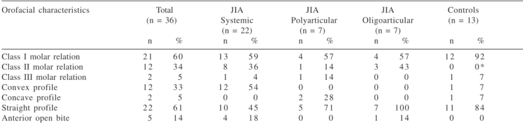
profile (5%), and 22 had a straight pro-file (61%). In the control group, 1 had a concave profile (7%), 11 had a straight profile (84%), and 1 had a convex profile (7%). A Class I molar relationship was more prevalent among controls (92%) compared with JIA patients (60%). A Class II molar re-lationship was observed only in pa-tients with JIA (34%); this difference was statistically significant (P = .032). A Class III molar relation was observed in 2 patients with JIA and 1 patient in the control group. In 1 patient with JIA, it was not possible to determine the molar relationship due to the ab-sence of molars and canines (Table 2).
Anterior open bite
Anterior open bite was observed in 5 patients (14%) with JIA, and 4 of
these patients presented with a sys-temic onset of the disease (Table 2). Three of these patients presented with a convex profile, and 2 presented with a straight profile. Three reported com-plaints in the orofacial region, with only 1 presenting with TMJ noise (bi-laterally crepitus).
Dental and gingival conditions
Patients in the JIA group had higher values of the DMFT index, dental plaque index, and gingival bleeding index than those of the trol group, with the worst dental con-ditions in patients with JIA. Patients with polyarticular JIA and with rheu-matoid factor negative onset had a gingival bleeding index of 29% and DMFT index of 6.6. These values were statistically significant (P = .047 and
P = .046, respectively) compared with
the control group. There was a progres-sive increase in the dental plaque in-dex and gingival bleeding inin-dex in the group with JIA in proportion to a higher number of involved superior limb joints in these patients, with a sta-tistically significant difference for the
gingival bleeding index (P = .055)
(Table 3).
Temporomandibular joint dysfunction
Evaluation of the Helkimo’s index (CDI) showed dysfunction among 34 patients (94%) with JIA that was more frequent in systemic onset, as
com-pared with the control group (P =
.017). Eleven patients with JIA (30%) presented with severe clinical dysfunc-tion, while this degree of dysfunction was not observed in patients in the control group. Regarding the man-dibular mobility index, severely im-paired mandibular mobility was ob-served in 12 patients (33%) with JIA compared with no patient in the
con-trol group (P = .015) (Table 4). The
mandibular range of movement of pa-tients with JIA was smaller compared with the controls. The maximum interincisal mouth opening of the pa-tients with JIA was 38.7 ± 7.3 mm com-pared with 47 ± 5.3 mm of the control
group (P = 0.0002). The mean right
Table 2 - Molar relationship and facial profile in patients with juvenile idiopathic arthritis according to the type of onset, compared with the control group.
Orofacial characteristics Total JIA JIA JIA Controls
(n = 36) Systemic Polyarticular Oligoarticular (n = 13)
(n = 22) (n = 7) (n = 7)
n % n % n % n % n %
Class I molar relation 2 1 6 0 1 3 5 9 4 5 7 4 5 7 1 2 9 2
Class II molar relation 1 2 3 4 8 3 6 1 1 4 3 4 3 0 0 *
Class III molar relation 2 5 1 4 1 1 4 0 0 1 7
Convex profile 1 2 3 3 1 2 5 4 0 0 0 0 1 7
Concave profile 2 5 0 0 2 2 8 0 0 1 7
Straight profile 2 2 6 1 1 0 4 5 5 7 1 7 100 1 1 8 4
Anterior open bite 5 1 4 4 1 8 0 0 1 1 4 0 0
*P < 0.032. The statistical significance is related to the comparison of control group and total of patients with juvenile idiopathic arthritis.
Table 1 - Clinical characteristics of 36 patients with juvenile idiopathic arthritis.
Sex Male: 10; female: 26
Age range 4 y 7 mo to 20 y (median= 10 y 8 m)
Age at onset of the disease 4 y 6 mo (median) Disease duration at time of study entry 1 y to 16 y (median = 7 y)
Type of onset Systemic: 22
Polyarticular with rheumatoid factor negative: 7 Oligoarticular: 7 (extended course in 6)
Type of evolution Polyarticular course: 35
Oligoarticular course: 1 Steinbrocker’s functional class I: 20
and left jaw mobility was also reduced in patients with JIA (8.2 ± 2.9 mm and 7.3 ± 2.9 mm, respectively); in rela-tionship to the control group (9.8 ± 1.7 and 10 ± 1.2 mm, respectively), this difference was statistically signifi-cant (P = .024 and P =.001, respec-tively). In the patients with JIA, the mean values for protrusion were also smaller (4.5 ± 2.5 mm); these differ-ences were statistically significant compared with the control group (6.2
± 2.2 mm) (P = .044). Only patients
with JIA (8) presented with TMJ noise upon movement (22%).
DISCUSSION
Although 94% of the children with JIA presented with clinical signs of temporomandibular dysfunction, they
did not present with spontaneous com-plaints of pain in the orofacial region. However, upon questioning, 28% in-dicated having pain in the face or teeth or restricted chewing. Since TMJ pain is not normally included in the primary complaints of patients with JIA, its in-volvement is usually fairly asympto-matic compared with other joints3,6,16
and rarely disables complete mandibu-lar function, thus leading to under-di-agnosis, mainly in the initial stages of the disease.17,18 JIA is a physically
lim-iting disease that impairs the patient’s quality of life.3,17 Nevertheless, there
are few studies that have evaluated re-strictions of mandibular function of these children. Our study shows a higher index of clinical dysfunction and mandibular mobility in patients with JIA and confirms TMJ impairment in these patients. While dental and
TMJ abnormalities due JIA have been studied in the past, this is the first study that evaluates both the signs and symptoms of dental and TMJ problems of JIA patients in the same study. It demonstrates a wider prevalence of the damage to the stomatognathic system provoked directly or indirectly by the JIA.
Patients with JIA can present with a decrease in the mandibular growth, re-sulting in facial alterations such as con-vex profile, micrognathia, retrognathia, and anterior open bite.4,19,20 Convex
profile is a relevant physical aspect in children with JIA,4,5,6,21 and 33% of the
JIA patients presented this condition. This type of profile is usually associ-ated with advanced TMJ disease, with partial resorption of the condyle and micrognathia, which are common find-ings of this degenerative joint disease.22,
Table 4 - Clinical dysfunction index, mandibular mobility index, and temporomandibular joint noise in patients with juvenile idiopathic arthritis according to the type of onset, compared with the control group.
Index Total JIA JIA JIA Controls
(n = 36) Systemic Polyarticular Oligoarticular (n = 13)
(n = 22) (n = 7) (n = 7)
n % n % n % n % n %
Dysfunction 3 4 9 4 2 1 9 5 6 8 5 7 100 4 30*
Severe 1 1 3 0 7 3 9 2 2 8 2 2 8 0 0
Moderate 5 1 3 3 1 3 1 1 4 1 1 4 1 7
Mild 1 8 5 0 1 1 5 0 3 4 3 4 5 7 3 2 3
Normal 2 5 1 4 1 1 4 0 0 9 6 9
Decreased mobility 2 9 8 0 1 7 7 7 5 7 1 7 100 9 6 9
Severe 1 2 3 3 8 3 6 3 4 3 1 1 4 0 0 *
Mild 1 7 4 7 9 4 1 2 2 8 6 8 5 9 6 9
Normal 6 1 6 5 2 2 1 1 4 0 0 4 3 0
TMJ Noise 8 2 2 5 2 2 2 2 8 1 1 4 0 0
*P < 0.015. The statistical significance is related to the comparison of the control group and total of patients with juvenile idiopathic arthritis.
Table 3 - Dental and gingival findings in patients with juvenile idiopathic arthritis according to the number of affected joints in superior limbs, compared with the control group.
Mean Index Juvenile idiopathic arthritis Juvenile idiopathic arthritis Controls
0 to 2 affected joints in 3 to 8 affected joints in (n = 13)
superior limbs (n = 18) superior limbs (n = 18)
DMFT index 3.82 6.72 3.6
Plaque index (%) 47.5 62.7 42.4
Gingival bleeding index (%) 15.0 27.7* 4.07
23 An anterior open bite was found in 5
patients with JIA (14%). However, 2 of these patients presented with a straight profile. This finding indicates that care must be exercised regarding the asso-ciation between a convex facial profile with anterior open bite and microg-nathia, because the soft tissues can mimic this condition. In addition, TMJ noises were clinically detected in 27% of the patients with JIA, although in 4 of the patients with micrognathia an anterior open bite, TMJ noises were not detected even though they presented with severe TMJ dysfunction. The level of the compromised and limited TMJ movement accounts for the difficulty in detecting articular sounds.
Complaints of dental pain may not result from JIA but can be a conse-quence of the physical limitations of the patients, mainly when functional impairment of the superior limbs joints is present.2,8,9 Children with JIA cannot
perform appropriate oral hygiene, and this can result in decay and gingivi-tis. The plaque index was similar be-tween the 2 groups; however, on analyzing the gingival bleeding index (GBI) of our sample, it was observed
that the GBI increased as the child pre-sented with a higher number of in-volved joints. When JIA is detected by clinicians, they should counsel and encourage their patients to maintain good oral hygiene. The parents also should be trained to help with oral hy-giene. Dietary recommendations, par-ticularly regarding ingestion of sugar, and periodic dental consultations will aid in the management of associated dental conditions. This strategy will help in avoiding the presence of op-portunistic infectious diseases, such as caries or gingival diseases, which are potentially due to the continued activ-ity of JIA.20
Our data reinforces the need for standardized investigation of not only TMJ but also the dental, gingival, and facial aspects in patients with JIA. Fur-ther standardized studies would help to define the TMJ involvement and es-tablish preventive dental and gingival procedures. More visits to dentist may be necessary for these children. Aware-ness of the dental and TMJ alterations arising from JIA, as well early evalua-tion of the stomatognathic system of children with this disease, is necessary
to identify these patients. These stud-ies would lead to the establishment of treatment plans that minimize the po-tential orofacial morbidity associated with this disease, thereby achieving an improvement in the quality of these children’s lives.
CONCLUSION
This study confirms that JIA signifi-cantly affects jaw function and indi-rectly causes dental abnormalities. The increased levels of the tooth decay in-dex, plaque inin-dex, and gingival bleed-ing index among patients with JIA, as-sociated with their TMJ dysfunction, indicates the need for frequent dental and TMJ evaluations for these chil-dren.
ACKNOWLEDGMENTS
The authors wish to acknowledge the assistance of Dr. Cibele Nasri and Dr. Gary M. Heir for the revision of this text.
RESUMO
SAVIOLI C e col. Características dentárias e faciais de pacientes com artrite idiopática juvenil. Rev. Hosp. Clín. Fac. Med. S. Paulo 59(3):93-98, 2004.
OBJETIVO: A articulação tempo-romandibular é freqüentemente afeta-da pela artrite idiopática juvenil, e esta doença degenerativa, durante o cresci-mento facial, resulta em disfunção mandibular grave. No entanto, não há estudos que avaliam conjuntamente alterações na saúde oral (cáries e do-enças gengivais) e na articulação temporomandibular decorrentes da
ar-trite idiopática juvenil. O objetivo deste estudo é avaliar a condição dentária e a função mandibular de pa-cientes com artrite idiopática juvenil tratados em um hospital escola.
MÉTODO: Trinta e seis pacientes com artrite idiopática juvenil (26 me-ninas e 10 meninos) foram submetidos a uma avaliação clínica sistemática de suas estruturas dentárias, orais e faciais (índice CPO-D, índice de placa e sangramento gengival, relação dentária, perfil facial e índice de Helkimo para articulação temporo-mandibular). O grupo controle foi composto por 13 crianças saudáveis.
apresen-tou 94% dos pacientes com disfunção da articulação temporomandibular (p=0,017), além de amplitude mandi-bular diminuída (p=0,0002) e mobili-dade mandibular gravemente compro-metida em 33% (p=0,015).
CONCLUSÃO: Este estudo
confir-ma que pacientes com artrite idio-pática juvenil: a) têm alto índice de disfunção mandibular, que pode ser atribuído ao efeito direto da doença sobre a articulação temporomandibular e b) maior índice de doença gengival, que pode ser considerado como efeito
indireto da artrite idiopática juvenil na saúde oral.
UNITERMOS: Artrite Idiopática
Juvenil. Dor dentária. Dentes. Articu-lação temporomandibular. Artrite reumatóide juvenil.
REFERENCES
1. Cassidy JT, Petty RE. Juvenile rheumatoid arthritis. In: Cassidy JT, Petty RE. Textbook of Pediatric Rheumatology. 4rd ed.
Philadelphia, B. Saunders Co, 2001. p. 218-322.
2. Brewer EJ, Giannini EH, Person DA. Artrite Reumatóide Juvenil. 2.ed. São Paulo, Ed. Manoele 1984. p. 1-59.
3. Grosfeld O. The orthodontist in the team-treatment for children with rheumatoid arthritis. European J Ortho 1989; 11(2):120-124.
4. Kjellberg H. Craniofacial growth in juvenile chronic arthritis. Acta Odontol Scand 1998; 56(6): 360-365.
5. Kjellberg H, Kiliaridis S, Thilander B. Dentofacial growth in orthodontically treated and untreated children with juvenile chronic arthritis (JCA). A comparison with Angle Cl II division 1 subjects. European J Ortho 1995;17(5):357-373.
6. Ronchezel MV, Hilário MOE, Goldenberg J, Lederman HM, Faltin KJr, Azevedo MF et al. Temporomandibular joint and mandibular growth alterations in patients with juvenile rheumatoid arthritis. J Rheumatol 1995;22(10):1956-1961. 7. Stabrun AE. Impaired mandibular growth and micrognathic
development in children with juvenile rheumatoid arthritis. A longitudinal study of lateral cephalographs. European J Ortho 1991;13 (5):423-34.
8. Tanchyk AP. Dental considerations for the patient with juvenile rheumatoid arthritis. General Dentistry 1991;39(5):300-332. 9. Walton AG, Welbury RR, Foster HE, Thomason JM. Juvenile chronic arthritis: a dental review. Oral Diseases 1999;5(1): 68-75.
10. Petty RE, Southwood TR, Baum J, Bhettay E, Glass DN, Manners P et al. Revision of the proposed classification criteria for juvenile idiopathic arthritis: Durban 1997. J Rheumatol 1998;25(11):1991-1994.
11. Steinbrocker O, Traeger CH, Batterman RC. Therapeutic criteria in rheumatoid arthritis. JAMA 1949;140(6):650-662. 12. Vellini FF. Ortodontia – Diagnóstico e Planejamento Clínico 1a
ed. São Paulo, Ed. Artes Médicas,1997. p. 75-96.
13. World Health Organization – Oral health surveys: basic methods. 4th ed. Geneva, WHO, 1997.
14. Ainamo J, Bay I. Problems and proposals for recording gingivitis and plaque. International Dental Journal 1975; 25(2):229-235.
15. O’Leary TJ. The periodontal screening examination. J Periodontol 1967; 38(5):617-624.
16. Helkimo H. Studies on function and dysfunction of the masticatory system. II. Index for anamnestic dysfunction and occlusal state. Swed Dent J 1974; 67(2):101-119.
17. Harper RP, Brown CM, Triplett MM, Villasenor A, Gatchel RJ. Masticatory function in patients with juvenile rheumatoid arthritis. Pediatr Dent 2000; 22(3):200-206.
18. Pedersen TK, Jensen JJ, Melsen B, Herlin T. Resorption of the temporomandibular condylar bone according to subtypes of juvenile chronic arthritis. J Rheumatol 2001; 28(9):2109-2115.
19. Marini I, Vecchiet F, Spiazzi L, Capurso U. Stomatognathic function in juvenile rheumatoid arthritis and in developmental open-bite subjects. J Dent Child 1999;66(1): 30-5.
20. Savioli C, Silva CAA, Siqueira JTT. Functional Morphologic Characteristics of the Stomatognathic System in Patients Carriers of Arthritis Juvenile Rheumatoid. JBO 2000; 4(25):70-78. 21. Simões WA. An orthodontic challenge. Juvenile rheumatoid
arthritis: examination protocol. World J Orthod 2001;2(1):56-68.
22. Siqueira JTT. Dor articular-Anormalidades na ATM. In: Siqueira JTT, Teixeira MJ. Dor Orofacial, Diagnóstico, Terapêutica e Qualidade de Vida. 1a ed. Curitiba, Ed. Maio, 2001. p.
447-487.
23. Kopp S. Degenerative and inflammatory temporomandibular joint disorders: Clinical perspectives. In: Sessle BJ. Temporomandibular Disorders and related Pain Conditions, Progress in Pain Research and Management .4th ed. Seattle,