From the Biomedical Research Institute and the Department of Pediatrics, São Lucas Hospital, Pontifícia Universidade Católica of Rio Grande do Sul (PUCRS) - Porto Alegre, Rio Grande do Sul, Brazil.
E-mail: [email protected] Received for publication on
September 17, 2003.
ORIGINAL RESEARCH
EFFECT OF CLARITHROMYCIN ON THE CELL
PROFILE OF BRONCHOALVEOLAR LAVAGE FLUID
IN MICE WITH NEUTROPHIL-PREDOMINANT LUNG
DISEASE
Leonardo Araújo Pinto, Camila Camozzato, Monique Avozani, Denise Cantarelli Machado, Marcus Herbert Jones, Renato Tetelbom Stein and Paulo Márcio Condessa Pitrez
PINTO LA et al. Effect of clarithromycin on the cell profile of bronchoalveolar lavage fluid in mice with neutrophil-predominant lung disease. Rev. Hosp. Clín. Fac. Med. S. Paulo 59(3):99-103, 2004.
OBJECTIVE: Macrolide antibiotics have anti-inflammatory properties in lung diseases. The aim of this study was to investigate the effect of clarithromycin in pulmonary cellular inflammatory response in mice.
METHOD: Eight adult Swiss mice were studied. All animals received an intranasal challenge (80 µL) with dead Pseudomonas aeruginosa (1.0 x 10¹² CFU/mL). Bronchoalveolar lavage was performed 2 days later, with total cell count
and differential cell analysis. The study group (n = 4) received clarithromycin treatment (50 mg/kg/day, intraperitoneal) for 5 days. Treatment was initiated 2 days before intranasal challenge.
RESULTS: There was no significant difference in total cell count between the groups (mean: 2.0 x 10⁶ and 1.3 x 10⁶,
respectively). In both groups, there was a predominance of neutrophils. However, the study group had a higher percentage of lymphocytes in the bronchoalveolar lavage than the control group (median of 19% vs 2.5%, P = .029).
CONCLUSION: Clarithromycin alters the cytological pattern of bronchoalveolar lavage of Swiss mice with neutrophil pulmonary inflammation, significantly increasing the percentage of lymphocytes.
KEY WORDS: Clarithromycin. Lung inflammation. Lung. Mice. Neutrophil.
Macrolides have been used for the treatment of different bacterial infec-tions. They are frequently prescribed in clinical practice, particularly for res-piratory infections. Erythromycin, clarithromycin, and azithromycin are the most common macrolides used, and they are recognized as the first line treatment for infections caused by Mycoplasma sp., Chlamydia sp., Ureaplasma sp., and other bacteria.1
Recently, studies have been emerg-ing demonstratemerg-ing an anti-inflamma-tory effect of these drugs in lung dis-eases.2 Clinical trials have shown a
benefit of macrolide therapy in some
pulmonary disorders, such as diffuse panbronchiolitis (DPB), asthma, and cystic fibrosis.6-8
Macrolides may inhibit neutrophil recruitment and interleukin (IL)-8 pro-duction.3-5 Other studies have shown
that macrolides may inhibit corticos-teroid metabolism and may increase the treatment effect in asthmatic
pa-tients.7,9 However, the
anti-inflamma-tory mechanisms of macrolides in pul-monary diseases are not fully under-stood.
Little data has been published about the effect of macrolides on cel-lular inflammation in neutrophil-pre-dominant lung disease in animal mod-els. One previous study using mice with chronic infection by Pseu-domonas aeruginosa (P. aeruginosa) and treated with clarithromycin has demonstrated a reduction on lymphocyte cell counts in bronchoalveolar lavage (BAL).10 This
aeruginosa for inducing lung disease. On the other hand, Sugiyama et al. found no significant changes in differ-ential cell counts of in vitro lipopoly-saccharide (LPS)-stimulated BAL in rats treated with erythromycin.11
The aim of this study is to analyze the effect of clarithromycin on the pat-tern of pulmonary cellular response in BAL of mice with neutrophil-pre-dominant lung disease that is induced using dead P. aeruginosa.
METHOD
Animals
Eight adult (6 to 8 weeks old) male Swiss mice from our University were used. Animals were provided by the University and were kept at Institute during the study period.
Protocol for induction of neutrophil pulmonary disease
The Microbiology Department provided the P. aeruginosa sample in a culture plate. After being scraped from the plate, the sample was diluted in phosphate-buffered saline (PBS), to a concentration of 1 x 10¹² CFU/mL.
All P. aeruginosa were frozen to – 20°C.
While under sedation, the animals from both the study group (n = 4) and control group (n = 4) received 1 intra-nasal challenge (80 mL) with P. aeruginosa solution.14,15 Sedation, per-formed to allow pulmonary aspiration of the instilled solution by suppress-ing upper airway reflexes, consisted of intraperitoneal administration of 0.1 mL of a solution of ketamine (0.4 mL), xylazine (0.1 mL), and normal saline (0.5 mL).
There was no control group with-out P. aeruginosa in this study. The authors of this study have previously shown a significant induction of
neu-trophil-predominant lung disease in mice using this protocol.13
Clarithromycin treatment
A dose of 15 mg/kg/day is used for clinical treatment of infections in hu-mans. Based on previous study protocols14,15 in experimental models,
we used a dosage of 50 mg/kg/day. The treatment was initiated 2 days be-fore the intranasal challenge with P. aeruginosa.
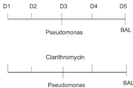
The study group received clarithromycin treatment over a 5-day period, with a single daily intraperito-neal (i.p.) dose. The protocol used in the study is illustrated in figure 1.
Bronchoalveolar lavage
Bronchoalveolar lavage was per-formed 2 days after intranasal chal-lenge. Mice were anesthetized with an i.p. injection of 0.2 mL of the same ketamine and xylazine solution that was used at the intranasal challenge. After anesthesia, a tracheotomy was performed, with trachea cannulation and tube fixation. One mL of normal saline was instilled. After 5 seconds, the material was aspirated. This proce-dure was repeated 3 times with the same solution.
Total cell count and differential cytological test
The BAL was weighed and centri-fuged (2,000 rpm for 2 minutes). The precipitate was diluted in 1 mL of PBS. Determination of TCC and cell viability was performed in a Neubauer chamber (Boeco, Germany), with trypan blue staining.
For differential cytology, 40mL of the sample was cytocentrifuged (FANEM, São Paulo, Mod. 218), at 500 rpm, for 5 minutes. Slides were fixed with methyl alcohol and stained with May-Grunwald-Giemsa stain. Cells were analyzed according to their morphology. The cell types at light microscopy were expressed as a per-centage after counting 200 cells.
Statistical analysis
Values are described as mean and median, and the statistical difference was calculated using the Mann-Whitney or t test, depending on the sample distribution. Differences were considered significant when P <.05.
Ethics
The study was approved by the Ethics Committee for Animal Research
of the Institution and was based on animal models research guidelines.
RESULTS
The BAL was performed success-fully in all studied mice. The mean of returned volume of BAL was 66%. Mice did not present adverse reactions associated with clarithromycin treat-ment.
The mean TCC in all mice studied was 1.65 x 10⁶ cells/mL. The mean
TCC of the study and the control group were 2.01 x 10⁶ cells/mL and 1.3
x 106 cells/mL, respectively. There was
no significant difference between the groups.
All mice had increased neutrophil counts in the BAL. The differential cell count results are presented in ta-ble 1. The study group had a signifi-cantly higher percentage of lymphocytes in the BAL compared with the control group, with medians of 19% and 2.5% (P = .029), respec-tively (Fig. 2). Neutrophil counts were not different between the groups stud-ied (P = .097) (Fig. 3).
DISCUSSION
In the present study, the BAL of mice having a neutrophilic pulmonary disorder that was induced by dead P. aeruginosa and treated with clarithromycin had a significant in-crease in the percentage of lymphocytes compared with control group.
We used a small number of mice. Since the groups are homogeneous and the bias factors are smaller com-pared with human studies, the practice of using small numbers of animals is usually sufficient.
The pulmonary inflammatory ab-normalities in the BAL of mice follow-ing aspiration of P. aeruginosa have been previously described. Pinto et
Table 1 - Comparison of differential cell counts in the bronchoalveolar lavage (BAL) between the groups studied.
All Control group Macrolide group x 106 cells/mL % x 106 cells/mL % x 106 cells/mL %
Total cell count 1.30 100 1.21 100 1.29 100
Neutrophils 1.00 74.6 1.00 81.9 0.87 67.2
Lymphocytes 0.10 8.30 0.03 2.90 0.29 22.5
Macrophages 0.20 12.8 0.18 15.2 0.13 10.2
Figure 2 - Comparison of lymphocyte percentage in the bronchoalveolar lavage (BAL)between the groups studied.
al.13 showed that mice with pulmonary
inflammation induced by dead P. aeruginosa had an increase in TCC and in the percentage of neutrophils in the BAL. Thus, this experimental model may be used for studies analyzing the macrolide anti-inflam-matory effect in neutrophilic pulmo-nary diseases.
There are a few mechanisms that may explain the anti-inflammatory ef-fect of macrolides. Some studies have shown a decreased neutrophil recruit-ment to the lungs following treatrecruit-ment with erythromycin. Kadota et al. dem-onstrated this effect in mice that re-ceived an intratracheal injection of li-popolysaccharide.3 Furthermore, a
de-crease in neutrophil count, 8, and IL-1 was observed in the BAL of patients with DPB, who received treatment with erythromycin or roxythromycin.5-12
Therefore, these drugs may inhibit neu-trophil recruitment.4
Yanagihara et al. showed that mice chronically infected by P. aeruginosa, which have similar pathologic changes to DPB, had a progressive reduction of the lymphocyte count in the BAL when treated with clarithromycin.10
However, in 1 previous study analyzing cellular response in BAL following lipopolysaccaride challenge in rats treated with erythromycin, no
difference in differential cell percent-age was detected.11 The present study
has shown an increased lymphocyte percentage in the BAL of mice with neutrophilic pulmonary disease and treated with clarithromycin. Therefore, our results demonstrate that macrolides alter the differential cell count in the BAL in this illness. How-ever, our results should not be consid-ered particularly an anti-inflammatory effect of macrolides and should be in-terpreted with caution. In spite of this limitation, we speculate that macrolide treatment could both stimulate an ear-lier onset of an adaptive cellular re-sponse or inhibit an acute neutrophilic response (with a consequent increase in lymphocyte percentage), resulting in a quicker resolution of the airway inflammation. On the other hand, the increased number of lymphocytes in the airways of the treated group could be interpreted as a marker of increased pulmonary inflammatory response.
Studies with a larger number of animals may reveal more significant alterations both in lymphocytic and neutrophilic response. Moreover, the bacterial concentration used for intra-nasal challenge was very high (1x10¹²
CFU/mL). Studies with lower concen-trations of P. aeruginosa in the intra-nasal challenge may show greater
ef-fects of macrolides on the BAL. Therefore, additional studies using larger numbers of mice, lower concen-trations of P. Aeruginosa, and includ-ing the measurement of some interleukin levels (IL-8, IL-10, or al-pha-tumor necrosis factor) in the BAL are essential for a better understanding of the anti-inflammatory mechanisms of these medications. Furthermore, the present experimental model using dead intranasal P. aeruginosa for in-ducing neutrophil lung inflammation have been shown to be low-cost and effective and consequently should be used for the development of further studies with macrolides.
In conclusion, our study demon-strates that macrolide treatment affects lymphocyte response in the BAL of mice with neutrophilic lung disease. Further studies are required to better in-vestigate the mechanisms of the anti-inflammatory action of macrolides in neutrophilic pulmonary diseases, which will be useful in treatment of many prevalent diseases with high morbidity.
ACKNOWLEDGEMENTS
The study was supported by CNPq and Pontifícia Universidade Católica of Rio Grande do Sul (PUCRS).
RESUMO
PINTO AL e col. Efeito da claritro-micina na celularidade do lavado broncoalveolar em camundongos com doença pulmonar neutrofílica induzida. Rev. Hosp. Clin. Fac. Med. S. Paulo 59(3):99-103, 2004.
OBJETIVO: Os antibióticos macrolídeos podem apresentar um efeito antiinflamatório em doenças pulmona-res. O objetivo deste estudo é investi-gar o efeito da claritromicina na
respos-ta inflamatória celular pulmonar em ca-mundongos Swiss. MÉTODO: Foram utilizados 8 camundongos Swiss adul-tos (6-8 semanas). Todos os animais re-ceberam um desafio intranasal (80 µL) com Pseudomonas aeruginosa mortas (1 x 10¹² UFC/mL). Dois dias após o
de-safio, foi realizado lavado bronco-alveolar (LBA) com contagem total de células (CTC) e exame citológico dife-rencial. O grupo em estudo (n=4) rece-beu tratamento com claritromicina
(50mg/kg/dia, intraperitoneal) por 5 dias, sendo iniciado o tratamento 2 dias antes do desafio intranasal. O grupo controle (n=4) não recebeu tratamento com claritromicina. RESULTADOS:
Não houve diferença significativa na CTC entre os grupos (média de 2x10⁶
e 1,3x10⁶, respectivamente). Em ambos
maior de linfócitos no LBA (mediana de 2,5% vs 19%, p=0,029). CONCLU-SÃO: O uso de claritromicina altera o exame citológico diferencial do lavado
bronco-alveolar de camundongos Swiss com inflamação pulmonar neutrofílica, aumentando significativamente o nú-mero percentual de linfócitos.
UNITERMOS: Claritromicina, Inflamação. Doenças pulmonares. Modelos animais. Neutrófilo.
REFERENCES
1. Klein JO. History of macrolide use in pediatrics. Pediatr Infect Dis J 1997;16:427-31.
2. Jaffé A, Bush A. Anti-inflammatory effects of macrolides in lung disease. Pediatric Pulmonology 2001;31:464-73.
3. Kadota J, Sakito O, Kohno S, Sawa H, Mukae H, Oda H, et al. A mechanism of erythromycin treatment in patients with diffuse panbronchiolitis. Am Rev Respr Dis 1993;147:153-59. 4. Oda H, Kadota J, Kohno S, Hara K. Erythromycin inhibits
neutrophil chemotaxis in bronchoalveoli of diffuse panbronchiolitis. Chest 1994;106:1116-23.
5. Takizawa H, Desaki M, Ohtoshi T, Kawasaki S, Kohyama T, Sato M, et al. Erythromycin modulates IL-8 expression in normal and inflamed human bronchial epithelial cells. Am J Respir Crit Care Med 1997;156:266-71.
6. Krishnan P, Thachil R, Gillego V. Diffuse panbronchiolitis: a treatable sinobronchial disease in need of recognition in the United States. Chest 2002;121:659-61.
7. Spahn JD, Fost DA, Covar R, Martin RJ, Brown EE, Szefler SJ, et al. Clarithromycin potentiates glucocorticoid responsiveness in patients with asthma: results of a pilot study. Ann Allergy Asthma Immunol 2001;87:501-05.
8. Wolter J, Seeney S, Bell S, Bowler S, Masel P, McCormack J. Effect of long term treatment with azithromycin on disease parameters in cystic fibrosis: a randomised trial. Thorax 2002;57:212-16.
9. Fost DA, Leung DY, Martin RJ, Brown EE, Szefler SJ, Spahn JD. Inhibition of methylprednisolone elimination in the presence of clarithromycin therapy. J Allergy Clin Immunol 1999;103:1031-15.
10. Yanagihara K, Tomono K, Sawai T, Hirakata Y, Kadota J, Koga H, et al. Effect of clarithromycin on lymphocytes in chronic respiratory Pseudomonas aeruginosa infection. Am J Respir Crit Care Med 1997;155:337-42.
11. Sugiyama Y, Yanagisawa K, Tominaga SI, Kitamura S. Effects of long-term administration of erythromycin on cytokine production in rat alveolar macrophages. Eur Respir J 1999;14:1113-16.
12. Sakito O, Kadota J, Kohno S, Abe K, Shirai R, Hara K. Interleukin 1 beta, tumor necrosis factor alpha, and interleukin 8 in bronchoalveolar lavage fluid of patients with diffuse panbronchiolitis: a potential mechanism of macrolide therapy. Respiration 1996;63:42-48.
13. Pinto LA, Camozzato C, Avozani M, Machado DC, Jones MH, Stein RT, et al. Desenvolvimento de um modelo experimental de indução de resposta pulmonar neutrofílica em camundongos. J Pneumol 2003;29(4):213-6.
14. Konishi T, Nakao M. Antibiotic treatment in mice infected with Japanese Borrelia garinii: efficacy of ceftriaxone for eradicating the infection induced by Ixodes persulcatus tick bites. Microbiol Immunol 1997;41(2):165-8