From the Departments of Neuropsychiatry and Medical Psychology, Faculty of Medicine of Ribeirão Preto, University of São Paulo - São Paulo/SP, Brazil. E-mail: rfsanches@uol.com.br
Received for publication on November 14, 2003.
REVIEW
PROTON MAGNETIC RESONANCE SPECTROSCOPY
OF THE FRONTAL LOBE IN SCHIZOPHRENICS: A
CRITICAL REVIEW OF THE METHODOLOGY
Rafael Faria Sanches, José Alexandre de Souza Crippa; Jaime Eduardo Cecílio Hallak; David Araújo; Antonio Waldo Zuardi
SANCHES RF et al. Proton magnetic resonance spectroscopy of the frontal lobe in schizophrenics: a critical review of the methodology. Rev. Hosp.Clín. Fac. Med. S. Paulo 59(3):145-152, 2004.
Schizophrenic patients undergoing proton magnetic resonance spectroscopy show alterations in N-acetyl aspartate levels in several brain regions, indicating neuronal dysfunction. The present review focuses on the main proton magnetic resonance spectroscopy studies in the frontal lobe of schizophrenics. A MEDLINE search, from 1991 to March 2004, was carried out using the key-words spectroscopy and schizophrenia and proton and frontal. In addition, articles cited in the reference list of the studies obtained through MEDLINE were included.As a result, 27 articles were selected. The results were inconsistent, 19 papers reporting changes in the N-acetyl aspartate levels, while 8 reported no change. Methodological analysis led to the conclusion that the discrepancy may be due the following factors: (i) number of participants; (ii) variation in the clinical and demographic characteristics of the groups; (iii) little standardization of the acquisition parameters of spectroscopy. Overall, studies that fulfill strict methodological criteria show N-acetyl aspartate decrease in the frontal lobe of male schizophrenics.
KEY WORDS: Spectroscopy. Proton. Frontal. Schizophrenia. Review.
Neuroimaging techniques were in-troduced in schizophrenia research by the pioneering work of Jakobi and Winkler1, showing an enlargement of
the lateral ventricles in chronic schizo-phrenics. During the last eighty years there was strong advance in this field, from the improvement of existing tech-niques to the introduction of new re-search capabilities. Among the latter stands magnetic resonance spectros-copy (MRS).
MRS is a non-invasive, non-radio-active procedure that allows quantifi-cation of several metabolites in spe-cific regions of the human brain2-5.
Bloch and Purcell originally described its basic principles in 1946, but it was only in 1980 that Ackerman and
coworkers developed the in vivo
tech-nique6-7. Since 1991 MRS has been
used to identify chemical changes in the brain of schizophrenic patients8.
Phosphorus and hydrogen are the most used atoms in MRS. While phos-phorus spectroscopy (P31 MRS) makes
possible the research of cell energy metabolism and neurodevelopment, hydrogen spectroscopy (better known as proton spectroscopy, H1 MRS),
pro-vides information about
neurotrans-mitter levels and neuronal integrity, in addition to measures of energy me-tabolism9-11.
Among the substances identifiable by H1 MRS, N-acetyl aspartate (NAA)
- an amino-acid found in neurons - has been the most investigated compound, because its concentrations were found altered in various neuro-psychiatric pathologies12-14. A decrease in NAA
levels has been associated to neuronal death, energetic deficit in the cell body, and axonal injury or lesion15-16.
con-sistent with theoretical assumptions about an abnormal development of neuronal pathways in these brain re-gions.
Stanley et al.21 have suggested that
many inconsistencies found in re-ported results with MRS are due to fast and complex methodological changes. Thus, critical methodological analysis may lead to a better understanding of these results.
OBJECTIVE
This review aims at discussing methodological aspects of reported findings on frontal lobe H1 MRS of
schizophrenic patients.
METHOD
Empirical articles in English were searched through MEDLINE, between 1991 and March 2004 and only hu-man studies were included. The key words were spectroscopy and schizo-phrenia and proton and frontal. In
ad-dition, articles cited in the reference list of the studies obtained through MEDLINE were included. As a result, 27 articles were reviewed.
The studies were divided into two groups for the analysis of clinical, de-mographic and procedural variables: the group of studies showing some kind of NAA alteration was named G+ and the group not evidencing any al-teration of NAA levels, even if show-ing alteration of other metabolites was called G-. The group means of vari-ables such as age, sex, use of anti-psychotics, and echo time were com-pared.
RESULTS
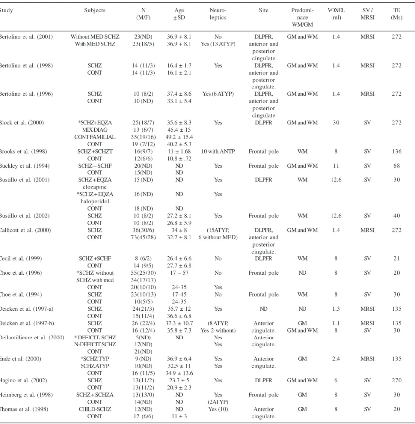
The data analyzed are summarized on Table 1 (G+) and Table 2 (G-).
Twenty-six studies included healthy volunteers as controls, while the re-maining study used intra-subject com-parisons. The results of the latter study showed an increase of the NAA/Cr ra-tio during anti-psychotic medicara-tion, as compared to a period without medi-cation22. Among the 26 studies that
have included healthy volunteers as controls, eight did not evidence a sig-nificant difference between the groups, concerning NAA, 13 found NAA de-crease in patients, as compared to healthy controls; the remaining five evidenced NAA changes in subgroups of patients, only.
The alterations most often found in the overall comparison of patients with healthy volunteers were a decrease of the NAA/Cr ratio17-18, 23-29 and of the
NAA absolute value19, 30,31. Regarding
subgroup comparisons, Heimberg et al.32, Bustillo et al.33 and Ende et
al.34found NAA differences between
schizophrenics treated with atypical anti-psychotics as compared to schizo-phrenics under typical neuroleptics, and between each subgroup and its control. Buckley et al.4 evidenced NAA
decrease in male schizophrenics as compared to normal controls and to fe-male schizophrenics. Dellamillieure et al.24 demonstrated NAA decrease in
schizophrenics with deficit syndrome, as compared to patients without such deficit and to controls.
Clinical and Demographic Characteristics
Age
It is important to control age be-cause there are suggestions showing that NAA decreases with aging35-36.
Therefore, NAA alterations could be due to age differences in non-paired groups. In the studies presently re-viewed there were no significant dif-ferences concerning age between pa-tients and controls, indicating meth-odological rigor in most of them.
Thomas el al.28, Brooks et al.27 and
Bertolino et al.18 did not find any
cor-relation between age and NAA levels. Such findings disagree with the results reported by Omori et al.37, Ende et al.34
and Block et al.38 who observed
nega-tive correlation between age and NAA in schizophrenic patients.
Ende et al.34, suggest that the
nega-tive correlation between NAA concen-tration and age may be due to in-creased partial volume of cerebrospi-nal fluid (CSF) in the voxel and or to decreased neuronal density. According to the same author, a possible expla-nation for such correlation is the pro-gressive character of schizophrenia, leading to cortical atrophy and result-ing NAA decrease.
When the studies showing NAA al-teration (G+) were compared with those not evidencing such alteration (G-), no significant difference in age was found (29.87 and 33.54 years of age, respectively).
Sex
One of the evidences supporting a possible effect of gender on schizo-phrenia is given by the study by Buckley et al.4. In their results, male
schizophrenic patients presented sig-nificant NAA decrease, as compared to male controls and to female patients. Such results are consistent with neuroimaging findings, in which al-terations in brain morphology are more frequent in male than in female schizophrenics. It has been suggested that these findings are due to a greater vulnerability among men to neurodevelopmental types of schizo-phrenia39-40.
Fukuzako et al.41 state that a factor
that may have contributed to the lack of NAA decrease in schizophrenics shown by their results is the predomi-nance of females in their sample (11 women and 4 men).
Table 1 - Studies of proton spectroscopy in the frontal lobe of schizophrenic patients showing NAA decrease.
Study Subjects N Age Neuro- Site Predomi- VOXEL SV / TE (M/F) + SD leptics nace (ml) MRSI (Ms)
WM/GM
Bertolino et al. (2001) Without MED SCHZ 23(ND) 36.9 + 8.1 No DLPFR, GM and WM 1.4 MRSI 272 With MED SCHZ 23(18/5) 36.9 + 8.1 Yes (13 ATYP) anterior and
posterior cingulate
Bertolino et al. (1998) SCHZ 14 (11/3) 16.4 ± 1.7 Yes DLPFR, GM and WM 1.4 MRSI 272 CONT 14 (11/3) 16.1 ± 2.1 anterior and
posterior cingulate.
Bertolino et al. (1996) SCHZ 10 (8/2) 37.4 ± 8.6 Yes (6 ATYP) DLPFR, GM and WM 1.4 MRSI 272 CONT 10 (ND) 33.1 ± 5.4 anterior and
posterior cingulate
Block et al. (2000) *SCHZ+EQZA 25(18/7) 35.6 ± 8.3 Yes DLPFR GM and WM 30 SV 272 MIX DIAG 13 (6/7) 45.4 ± 15
CONT FAMILIAL 35(19/16) 49.2 ± 15.4 CONT 19 (7/12) 40.2 ± 5.3
Brooks et al. (1998) SCHZ +SCHZT 16(9/7) 11 ± 1.68 10 with ANTP Frontal pole WM 8 SV 136 CONT 12(6/6) 10.8 ± .72
Buckley et al. (1994) SCHZ + SCHF 20(ND) ND Yes Frontal pole GM and WM 11 SV 68 CONT 15(ND) ND
Bustillo et al. (2001) SCHZ + EQZA 15 (ND) ND Yes DLPFR WM 12.6 SV 30 clozapine
*SCHZ + EQZA 16 (ND) ND Yes haloperidol
CONT 18 (ND) ND
Bustillo et al. (2002) SCHZ 10 (8/2) 27.2 ± 8.1 Yes Frontal pole WM 12.6 SV 40 CONT 10 (8/2) 26.8 ± 5.9
Callicott et al. (2000) SCHZ 36(30/6) 34 ± 8 (15ATYP, DLPFR, GM and WM 1.4 MRSI 272 CONT 73(45/28) 32.2 ± 8.1 6 without MED) anterior and
posterior cingulate.
Cecil et al. (1999) SCHZ +SCHF 8 (6/2) 26.4 ± 6.6 No DLPFR WM 8 SV 21 CONT 14 (9/5) 27.7 ± 6.8
Choe et al. (1996) *SCHZ without 55(25/30) 17 – 57 No Frontal pole ND 8 SV 20 SCHZ with med 34(17/17)
CONT 20(10/10) 24-35 Yes
Choe et al. (1994) SCHZ 23(10/13) 17-45 No Frontal pole WM 8 SV 30 CONT 10(5/5) 24-35
Deicken et al. (1997-a) SCHZ 24(21/3) 35.7 ± 12 Yes ND ND 1.3 MRSI 135 CONT 15(11/4) 36.6 ± 6.8
Deicken et al. (1997-b) SCHZ 26 (22/4) 37.3 ± 10.7 (8 ATYP, Anterior GM 1.1 MRSI 135 CONT 16 (12/4) 35.8 ± 7.3 Yes 2 without) cingulate. GM and WM 8 SV 30 Dellamillieure et al. (2000) * DEFICIT- SCHZ 5(ND) ND Yes Anterior
N-DEFICIT SCHZ 17(ND) Yes cingulate. CONT 21(ND)
Ende et al. (2000) *SCHZ TYP 9 (ND) 36.9 ± 6.4 Yes Anterior GM 2.4 MRSI 135 SCHZ ATYP 10(ND) 32.5 ± 11 Yes cingulate.
CONT 16 (11/5) 34.9 ± 13.6
Hagino et al. (2002) SCHZ 13(11/2) 23.7 ± 5 Yes DLPFR GM and WM 6 SV 270 CONT 13(11/2) 20.9 ± 2.3
Heimberg et al. (1998) SCHZ + SCHZA 13(13/0) ND Yes Frontal pole GM 8 SV 30 CONT 14(ND) ND (2ATYP)
Thomas et al. (1998) CHILD-SCHZ 12(ND) ND Yes (10) Anterior GM 8 SV 20 CONT 12 (6/6) 11 ± 3 cingulate.
SCHZ=schizophrenics; CONT=healthy controls; SCHF=schizophreniforms; SCHZT=schizotypicals; SCHZA=schizoaffectives; CHILD-SCHZ=childhood-onset schizophrenia; MIX DIAG=mixed psychiatric diagnoses; ±SD=standard deviation; SV= single voxel. MRSI=functional spectroscopy. TE=echo time; DLPFR=dorsolateral prefrontal region; WM=white matter; GM=gray matter. ND=not described. *=NAA statistical difference as compared to control group. ANTP=anti-psychotics. ATYP=atypical anti-psychotics. TYP=typical anti-psychotics. MED=medication
determinant factor in the results pre-sented, it seems that male schizophren-ics are more likely to present decrease in the NAA levels.
Medication
The strongest evidence of NAA
de-crease is found in chronic schizo-phrenic patients. One reason, already discussed, is the possible negative cor-relation between NAA and age. An-other possibility is that chronic pa-tients generally have a history of pro-longed use of anti-psychotics,
al-though the consequences of such treat-ment on neuronal functioning are un-known. Some studies have concen-trated on that variable, such the men-tioned intra-subject study by Bertolino et al.22, which found a NAA/
patients. Nevertheless, several limita-tions were pointed out in this study by Bustillo et al.33: it is a naturalistic,
non-controlled study; used NAA/Cr ratio, and the NAA change observed was only 9%, which can be seen as a normal variation, although reaching statistical significance in the dorsola-teral pre-frontal cortex (DLPFC). In their own results, Bustillo et al.33
re-ported NAA decrease in schizophren-ics treated with haloperidol, as com-pared to controls and patients taking clozapine, raising the hypothesis of an association of typical anti-psychotics with neuronal toxicity.
Two other studies have analyzed if the type of anti-psychotic drug may interfere with the NAA levels. Omori et al.37 did not find any differences in
the frontal lobe, when they compared typical anti-psychotic drugs (n=13) to atypical ones (n=5). In contrast, Heimberg et al.32 reported NAA/Cr
in-crease in patients taking atypical anti-psychotic drugs (n=2), as compared to schizophrenics taking typical neuroleptics (n=4). Nevertheless, the last result should be seen with caution,
due to the small number of subjects participating in the study.
Still about the effects of anti-psy-chotic drugs on NAA, this review found five well-designed studies. Three of them used schizophrenic patients under no medication and, two of the studies were longitudinal. Cecil et al.25
and Choe et al.17 studied
non-medi-cated patients and found NAA/Cr de-crease. However, Bartha et al.42 did not
find any NAA alteration in non-medi-cated schizophrenics. In a longitudinal follow-up study, Choe et al.29 observed
that schizophrenic patients (n=55) had decreased levels of NAA/Cr, even though such levels did not change with anti-psychotic drugs during a treatment period of up to six-months (n=34). The comparison of this study with the other longitudinal investiga-tion reviewed (Bertolino et al.22) is
questionable, because of critical meth-odological differences. In another lon-gitudinal study, Bustillo et al.31 found
NAA decrease during the second scan (after the use of anti-psychotic drugs). It is noteworthy that five stud-ies4,24,27,34,43 have reported no
signifi-cant differences in NAA levels be-tween subgroups of non-medicated and medicated patients. Accordingly, the results obtained by Callicott et al.23, Bertolino et al.18,26, Deicken et al. 19,30 and Fukuzako et al.41 did not show
any correlation between NAA concen-tration and dosage of anti-psychotic drug.
Because of the difficulty in per-forming experiments with non-medi-cated schizophrenic patients and the diversity of results found, the query about the real interference of medica-tion on the NAA levels is still to be answered.
Experimental Design
Sample Size
Studies in schizophrenic patients using spectroscopy are generally car-ried out with a small number of pa-tients. There are several reasons for that, from the difficulty of recruiting patients and the high costs of the pro-cedure to the time spent in each exami-nation. Because of that, a question raised about such studies is whether
Table 2 - Studies of frontal lobe proton spectroscopy in schizophrenic patients who did not show NAA decrease.
Study Subjects N Age Neuro- Site Predomi- VOXEL SV / TE (M/F) + SD leptics nace (ml) MRSI (Ms)
WM/GM
Bartha et al. (1997) SCHZ 10 (8/2) 26.3 ± 6.4 No Cingulate GM 4.5 SV 20 CONT 10 (8/2) 24.4 ± 5.1
Bertolino et al. (2000 SCHZ 8(ND) 40.1 ± 8.7 No DLPFR GM 1.4 MRSI 272 CONT 7 (5/2) 36.4 ± 7.3
Callicott et al. (1998) SCHZ + SCHZA 47 (43/4) 34.2 ± 8.8 8 without DLPFR, GM and
CONT 66 (42/24) 32.9 ± 8.2 anterior WM 1.4 MRSI 272 and posterior
cingulate
Fukuzako et al. (1995) SCHZ 15(4/11) 39.3 ± 7.6 Yes Frontal pole. ND 27 SV 135 CONT 15(4/11) 38.8 ± 7.8
Omori et al. (2000) SCHZ 20 (12/8) 23–43 Yes Frontal pole. WM 8 SV 136 CONT 16 (10/6) (5 ATYP,
2 without)
Sigmundsson et al. (2003) SCHZ 25 (24/1) 34.9 ± 8 Yes DLPFR. WM 2 SV 136 CONT 26 (22/4) 31.8 ± 6.7
Stanley et al. (1996) SCHZ 13(11/2) 26 ± 7 No DLPFR. WM (70%) 8 SV 20 SCHZ 12(10/2) 26 ± 7 Yes acute GM (30%)
SCHZ 12(11/1) 41 ± 5 Yes chronic CONT 24(24/0) 32 ± 11
Steel et al. (2001) SCHZ 10 (5/5) 34 ± 14 Yes Frontal pole. WM 15 SV 145 CONT 10 (4/6) 35 ± 7
the number of subjects used is enough to minimize type II errors44. Steel et
al.45 and Bertolino et al.46 admit that
the lack of significant NAA decrease observed in their studies may be due to the small number of subjects used, 10 and 8 respectively.
Considering the difficulties in per-forming such kind of study in large number of subjects, the ones, which achieve bigger samples, should be ap-preciated.
Voxel Size and Location
Even though spectroscopy has many advantages, there are some short-comings to be surmounted, such as for example, its low anatomical defini-tion. By using the 1.5 Tesla magnetic field, H1 MRS gets a good resolution,
with voxels measuring from one to eight ml. When compared to P31 MRS,
H1 MRS allows the selection of smaller
voxels, because proton sensitivity is fifteen times larger than that of phos-phorus. As far as the intensity of mag-netic fields can be increased, voxels with still smaller volumes would be selected. The advantage of small voxels is the decrease of the partial volume effect, such as the ratio of white/gray matter or CSF. On the other hand, smaller voxels decrease the sig-nal-to-noise ratio and thus, the spec-trum quality is lowered43.
A pertinent query is whether NAA changes observed in some studies are due to decrease of the volume of the structure, with resulting presence of CSF, white matter (WM) or gray (GM) from neighboring structures. Several authors 4,22,32,41-42,45 raise the possibility
of the influence of results by adjacent areas. Stanley et al.43 for instance,
ad-mit that the lack of change in metabolites shown by their results may be explained by the fact that 70% of the voxel was composed by WM, bear-ing in mind that the differences in metabolites only can be found in the GM.
Among the articles reviewed, there was a considerable variation in size among the voxels selected, ranging from 1.1 ml to 30 ml.
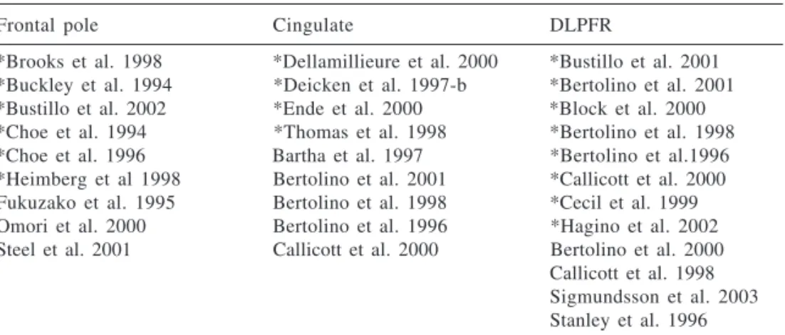
In addition, there was large varia-tion in the localizavaria-tion of voxels, even though they were all inside the frontal lobe. Table 3 shows the distribution of studies by voxel localization in the frontal lobe (dorsolateral prefrontal gion, frontal pole, cingulate). In the re-viewed studies, NAA decrease was found in the three sub-regions, as pointed out before18.Taking into
con-sideration that NAA changes are not limited to a specific region, it is even more necessary to be able to decrease voxel size without impairing the signal-to-noise ratio. Moreover, with the devel-opment of techniques for separating WM from GM and to minimize CSF in-terference in the voxel, more reliable results will certainly be obtained.
Laterality
Some spectroscopy studies re-viewed in this article (N=11) did not evaluate the frontal region bilaterally. Among 1618-19,22-26,28-30,34,44-48 articles
that have evaluated bilateral frontal lobe, in ten18-19,22-26,28-29,34 the
abnor-malities were the same bilaterally and in two30,47 studies NAA differences
were found only on the left side. In the other four44-46,48 studies no differences
were found. When only one side was selected, the left frontal lobe was clearly preferred for investigation (N= 104,27,31-33,37-38,41-43) as compared to the
right frontal lobe (N=117). There were
NAA abnormalities in 184,18-19,22-34,38,47
(69%) out of 26 articles that evaluated the left side, whereas NAA differences occurred in 1117-19,22-26,28-29,34 (64%) out
of 17 articles that studied the right side. Therefore, these results suggest that the NAA abnormalities in the fron-tal lobe are not influenced by brain laterality.
MRS Parameters
The variation of acquisition pa-rameters in spectroscopy, as well as the physicochemical proprieties of the measured substances may distort the results obtained. Several authors30,41,43
admit the possibility of interference of the relaxation times T1 and T2 in their results. T1 is the time the atom nucleus takes to return to its low energy basal state, which is more stable, while T2, transversal relaxation time, is the time the nucleus takes to become out of phase (such as clocks from several countries, winding in the same fre-quency, but showing different times). Times T1 and T2 are determined by the molecular environment around the atom nucleus.
Table 3 - Distribution of proton spectroscopy studies in schizophrenic patients paired with healthy controls among the frontal lobe sub-regions.
Frontal pole Cingulate DLPFR
*Brooks et al. 1998 *Dellamillieure et al. 2000 *Bustillo et al. 2001 *Buckley et al. 1994 *Deicken et al. 1997-b *Bertolino et al. 2001 *Bustillo et al. 2002 *Ende et al. 2000 *Block et al. 2000 *Choe et al. 1994 *Thomas et al. 1998 *Bertolino et al. 1998 *Choe et al. 1996 Bartha et al. 1997 *Bertolino et al.1996 *Heimberg et al 1998 Bertolino et al. 2001 *Callicott et al. 2000 Fukuzako et al. 1995 Bertolino et al. 1998 *Cecil et al. 1999 Omori et al. 2000 Bertolino et al. 1996 *Hagino et al. 2002 Steel et al. 2001 Callicott et al. 2000 Bertolino et al. 2000
Other parameters previously de-fined by the authors, such as: Echo Time (TE – the time between the 90 degree pulse and the maximum in the echo in a spin-echo sequence), use or not of metabolite ratio, and predomi-nance of white or gray matter made it difficult the comparison of the results obtained.
The definition of TE depends on the metabolite of interest and so, a short TE is preferable when the focus is in substances such as glutamate. However, if NAA is the center of atten-tion, the NAA peak definition im-proves with a longer TE.
Block et al.38 found NAA/Cho
de-crease in schizophrenics, only when they used 272 ms TE. With 30 ms there was no difference, probably due to higher standard deviations. Fukuzako et al.41 reported that the NAA/Cr ratio
decreases when TE drops from 135 ms to 50 ms.
It can be seen that nine 4,17,24-25,28-29,31-33 of the 19 studies that found
NAA decrease used a short TE, indi-cating that TE is not the only deter-minant of the results obtained.
The best way to determine the brain concentration of a substance is its absolute value, but as such meas-urement is highly complex, the results are not always reliable. The determi-nation of NAA ratio with other sub-stances (NAA/Cr, NAA/Cr+Cho, NAA/ Cho) is, on the other hand, easily ob-tainable, does not vary with the re-laxation times T1 and T2, and is not affected by CSF influence. The disad-vantage is that the result is a function
of both the denominator and the nu-merator.
Even when the quantification in absolute value is used, the technique employed in this situation, also uses a kind of “ratio”. For instance, in Heimberg et al.’s32 study, the water
con-centration was used as a reference for the quantification of metabolites of in-terest, assuming that the magnetic characteristics of the water do not change in pathological situations.
As many as 17 17-18,22-29,32,37-38,41,44,46-47 from the 27 articles reviewed, used
“ratio” instead of absolute values. Among them, 1317-18,22-29,32,38,47 studies
found decrease in the NAA ratio. From the eight studies that did not find NAA changes, two42-43 have been criticized
by Bertolino and Weinberger20. They
argued that the use of absolute values based on previous knowledge of metabolite concentration is unreliable, because measurements obtained in dif-ferent sessions are hard to replicate.
CONCLUSION
The main difficulty in analyzing these 27 articles resulted from the great variation in the methodological vari-ables discussed above. None of those aspects, by themselves, was able to predict the results obtained in the stud-ies.
Though the studies were rigorous in many ways, few reached satisfactory criteria, both in respect to the clinical and demographic characteristics, and in the parameters of image acquisition.
Small subject group sizes, samples with a high proportion of female schizophrenics, large voxel volume and short TE are factors likely to im-pair the detection of NAA change. Tak-ing into account the number of pa-tients studied (> 20), the predominance of male patients (>80%), the TE (> 135 ms), the voxel size (< 2 ml), six well designed studies can be selected. Four out of these six best-designed stud-ies19,22-23,30 showed a decrease in the
NAA levels in the frontal lobe of schizophrenics; and two44,48 reported
negative findings.
Thus, the results of the present re-view show that it is not clear if there is an association between NAA abnor-malities in the frontal lobe and schizo-phrenia. Since many aspects of this disorder are heterogeneous, standardi-zation of spectroscopic methodology and a more judicious selection of sub-jects are likely to generate more reli-able evidence concerning the role of NAA in schizophrenia.
ACKNOWLEDGEMENT
This work was supported by a grant from Fundação de Amparo à pesquisa do Estado de São Paulo (FAPESP). AWZ is the recipient of a Conselho Nacional de Desenvolvimento Cientí-fico e Tecnológico fellowship. JASC was recipient of Conselho Nacional de Desenvolvimento Científico e Tecno-lógico fellowship (grant 200984/01-2, 2002/2003) and is recipient of a CAPES fellowship (Prodoc, 2003/2005).
RESUMO
SANCHES RF e col. Espectroscopia de Próton por ressonância magnética de lobo frontal em esquizofrênicos – Revisão crítica da metodologia.
Rev. Hosp. Fac. Med.S. Paulo 59(3):145-152, 2004
Pacientes esquizofrênicos submeti-dos à espectroscopia de próton por res-sonância magnética demonstram alte-rações nos níveis de N-acetilaspartato em diversas regiões cerebrais, supor-tando a hipótese de disfunção
período entre 1991 e março de 2004, com o cruzamento dos termos spec-troscopy, schizophrenia, proton e fron-tal. Foram selecionados 27 artigos ori-ginais, cujos resultados mostram-se discordantes quanto à alteração nos valores de N-acetilaspartato (19 artigos apresentaram alterações nos níveis de N-acetilaspartato e oito estudos não
apresentam alterações). A presente re-visão sugere que esta diversidade de resultados pode ser atribuída aos se-guintes fatores: 1-número de partici-pantes; 2- variação nas características clínicas e demográficas dos grupos; 3-pouca padronização dos parâmetros de aquisição dos espectros. Os artigos que
satisfazem os critérios metodológicos mais rígidos sugerem diminuição de NAA no lobo frontal de esquizo-frênicos do sexo masculino.
UNITERMOS: Espectroscopia. Próton. Frontal. Esquizofrenia. Revi-são.
REFERENCES
1. Jakobi W, Winkler H. Encephalographische studien an chronisch schizophrenen. Arch Psychiat Nervenkrankh 1927; 81:299-332.
2. Waddington JL, O’Callaghan E, Larkin C, Redmond O, Stack J, Ennis JT. Magnetic resonance imaging and spectroscopy in schizophrenia. British Journal of Psychiatry 1990; 157(9): 56-65.
3. Urenjak J, Williams SR, Gadian DG, Noble M. Proton nuclear magnetic resonance spectroscopy unambiguously identifies different neural cell types. The JournalofNeuroscience 1993; 13(3): 981-989.
4. Buckley PF, Moore C, Long H, Larkin C, Thompson P, Mulvany F, et al. 1H Magnetic resonance spectroscopy of the left temporal
and frontal lobes in schizophrenia: clinical, neurodevelopmental, and cognitive correlates. Biol Psychiatry 1994; 36(12):792-800.
5. Lafer B, Amaral Jams. Espectroscopia de próton por ressonância magnética: aplicações em psiquiatria. Revista de Psiquiatria Clínica 2000; 27(3):154-63.
6. Maier M. In vivo magnetic resonance spectroscopy. applications in psychiatry. British Journal of Psychiatry 1995; 167(3): 299-306.
7. McClure RJ, Keshavan MS, Pettegrew JW. Chemical and physiologic brain imaging in schizophrenia. Psychiatr Clin North Am 1998; 21(1): 93-122.
8. Pettegrew JW, Keshavan MS, Panchalingam K, Strychor S, Kaplan DB, Tretta MG, Allen M. et al . Alterations in brain high-energy phosphate and phospholipid metabolism in first episode, drug naive schizophrenia. A pilot study of the dorsal prefrontal cortex by in vivo 31P NMR spectroscopy. Archives
of General Psychiatry 1991; 48(6):563-568.
9. Duncan JS. Imaging and epilepsy. Brain1997; 120(Pt2): 339-377.
10. Gonçales L, Yacubian J, Marques AP. A espectroscopia como método de investigação neurobiológica. Rev Psiquiatria Clin 1998; 25(1):43-45.
11. Matson GB, Weiner M - Spectroscopy. In: Stark DD, Bradley WG. Magnetic Resonance Imaging. 3rd ed. St Louis, Mosby, 1999. p. 181-214.
12. Taylor DL, Davies SE, Obrenovitch TP, Urenjak J, Richards DA, Clark JB,et al. Extracellular N-acetylaspartate in the rat brain: in vivo determination of basal levels and changes evoked by high K+. J. Neurochem1994; 62(6): 2349-2355.
13. Buckley PF, Friedman L. Magnetic resonance spectroscopy – briding the neurochemistry and neuroanatomy of schizophrenia (editorial). British Journal ofPsychiatry 2000; 176:203-205.
14. Deicken RF, Johnson C, Pegues M. Proton magnetic resonance spectroscopy of the human brain in schizophrenia. Rev Neurosci 2000; 11(2-3):147-158.
15. Taylor DL, Davies SE, Obrenovitch TP, Doheny MH, Patsalos PN, Clark JB, et al. Investigation into role of N-acetylaspartate in cerebral osmoregulation. J. Neurochem 1995; 65(1): 275-281.
16. Soares JC, Innis RB - Neurochemical brain imaging investigations of schizophrenia. Biol Psychiatry 1999; 46(5):600-615. 17. Choe BY, Kim KT, Suh TS, Lee C, Paik IH, Bahk YW, et al. 1H
Magnetic resonance spectroscopy characterization of neuronal dysfunction in drug-naive, chronic schizophrenia. Acad Radiol 1994; 1(3):211-216.
18. Bertolino A, Nawroz S, Mattay VS, Barnett AS, Duyn JH, Moonen CT, et al. Regionally specific pattern of neurochemical pathology in schizophrenia as assessed by multislice proton magnetic resonance spectroscopy imaging. Am JPsychiatry 1996; 153(12):1554-1563.
19. Deicken RF, Zhou L, Schuff N, Weiner MW. Proton magnetic resonance spectroscopy of the anterior cingulate region in schizophrenia. Schizophr Res 1997-b; 27(1):65-71. 20. Bertolino A, Weinberger DR. Proton magnetic resonance
spectroscopy in schizophrenia. European Journal of Radiology 1999; 30(2):132-141.
21. Stanley JA, Pettegrew JW, Keshavan MS. Magnetic resonance spectroscopy in schizophrenia: methodological issues and findings – Part I. Biol Psychiatry 2000; 48(5):357-368. 22. Bertolino A, Callicott JH, Mattay VS, Weidenhammer KM, Rakow
23. Callicott JH, Bertolino A, Egan MF, Mattay VS, Langheim FJ, Weinberger DR. Selective relationship between prefrontal N-acetylaspartate measures and negative symptoms in schizophrenia. Am J Psychiatry 2000; 157(10):1646-1651. 24. Delamillieure P, Fernandez J, Constans JM, Brazo P, Benali K,
Abadie P, et al. Proton magnetic resonance spectroscopy of the medial prefrontal cortex in patients with deficit schizophrenia: preliminary report. Am J Psychiatry 2000; 157(4):641-643.
25. Cecil KM, Lenkinski RE, Gur RE, Gur RC. Proton magnetic resonance spectroscopy in the frontal and temporal lobes of neuroleptic naive patients with schizophrenia. Neuropsychopharmacology 1999; 20(2):131-140.
26. Bertolino A, Kumra S, Callicott JH, Mattay VS, Lestz RM, Jacobsen L, et al. Common pattern of cortical pathology in childhood-onset and adult childhood-onset schizophrenia as identified by proton magnetic resonance spectroscopy imaging. Am JPsychiatry 1998; 155(10): 1376-1383.
27. Brooks WM, Hodde-Vargas J, Vargas LA, Yeo RA, Ford CC, Hendren RL. Frontal lobe of children with schizophrenia spectrum disorders: a proton magnetic resonance spectroscopy study. Biol Psychiatry 1998; 43(4):263-269.
28. Thomas MA, Ke Y, Levitt J, Caplan R, Curran J, Asarnow R, et al. Preliminary study of frontal lobe 1H MR spectroscopy in
childhood-onset schizophrenia. J Magn Reson Imaging 1998; 8(4):841-846.
29. Choe BY, Suh TS, Shinn KS, Lee CW, Lee C, Paik IH. et al. Observation of metabolic changes in chronic schizophrenia after neuroleptic treatment by in vivo hydrogen magnetic resonance. Invest Radiol 1996; 31(6):345-352.
30. Deicken RF, Zhou L, Corwin F, Vinogradov S, Weiner MW. Decreased left frontal lobe N- acetylaspartate in schizophrenia. Am J Psychiatry 1997-a; 154(5):688-690.
31. Bustillo JR, Lauriello J, Rowland LM, Thomson LM, Petropoulos H, Hammond R, et al. Longitudinal follow-up of neurochemical changes during the first year of antipsychotic treatment in schizophrenia patients with minimal previous medication exposure. Schizophr Res 2002; 58(2-3):313-321. 32. Heimberg C, Komoroski RA, Lawson WB, Cardwell D, Karson CN. Regional proton magnetic resonance spectroscopy in schizophrenia and exploration of drug effect. Psychiatry Res 1998; 83(2): 105-115.
33. Bustillo JR, Lauriello J, Rowland LM, Jung RE, Petropoulos H, Hart BL, et al. Effects of chronic haloperidol and clozapine treatments on frontal and caudate neurochemistry in schizophrenia. Psychiatry Research: Neuroimaging 2001; 107(3):135-149.
34. Ende G, Braus DF, Walter S, Weber-Fahr W, Soher B, Maudsley AA, et al. Effects of age, medication and illness duration on the N-acetylaspartate signal of the anterior cingulate region in schizophrenia. Schizophrenia Research 2000; 41(3):389-395. 35. Angelie E, Bonmartin A, Boudraa A, Gonnaud PM, Mallet JJ, Sappey-Marinier D. Regional differences and metabolic changes in normal aging of the human brain: proton MR spectroscopic imaging study. Am J Neuroradiol 2001; 22(1):119-127.
36. Sijens PE, den Heijer T, Origgi D, Vermeer SE, Breteler MM, Hofman A, et al. Brain changes with aging: MR spectroscopy at supraventricular plane shows differences between women and men. Radiology 2003; 226(3):889-896.
37. Omori M, Murata T, Kimura H, Koshimoto Y, Kado H, Ishimori Y, et al. Thalamic abnormalities in patients with schizophrenia revealed by proton magnetic resonance spectroscopy. Psychiatry Res 2000; 98(3): 155-162.
38. Block W, Bayer TA, Tepest R, Traber F, Rietschel M, Muller DJ, et al. Decreased frontal lobe of N-acetyl aspartate to choline in familial schizophrenia: a proton magnetic resonance spectroscopy study. Neuroscience Letters 2000; 289(2):147-151.
39. Castle DJ, Murray RM. The neurodevelopmental basis of sex differences in schizophrenia. Psychol Med 1991; 21(3):565-576.
40. Waddington JL. Schizophrenia: developmental neuroscience and pathobiology. Lancet 1993; 341(8844):531-536.
41. Fukuzako H, Takeuchi K, Hokazono Y, Fukuzako T, Yamada K, Hashiguchi T, et al. Proton magnetic resonance spectroscopy of the left medial temporal and frontal lobes in chronic schizophrenia: preliminary report. Psychiatry Res Neuroimag 1995; 61(4):193-200.
42. Bartha R, Williamson PC, Drost DJ, Malla A, Carr TJ, Cortese L, et al. Measurement of glutamate and glutamine in the medial prefrontal cortex of never-treated schizophrenic patients and healthy controls by proton magnetic resonance spectroscopy. Arch Gen Psychiatry 1997; 54(10): 959-965.
43. Stanley JA, Williamson PC, Drost DJ, Rylett RJ, Carr TJ, Malla A, et al. An in vivo proton magnetic resonance spectroscopy study of schizophrenia patients. Schizophrenia Bulletin 1996; 22(4):597-609.
44. Callicott JH, Egan MF, Bertolino A, Mattay VS, Langheim FJ, Frank JA, et al. Hipocampal N-acetylaspartate in unaffected siblings of patients with schizophrenia: a possible intermediate phenotype. Biological Psychiatry 1998; 44(10):941-950. 45. Steel RM, Bastin ME, McConnell S, Marshall I,
Cunningham-Owens DG, et al. Diffusion tensor imaging (DTI) and proton magnetic resonance spectroscopy 1H-MRS in schizophrenic
subjects and normal controls. Psychiatry Research: Neuroimaging 2001; 106(3):161-170.
46. Bertolino A, Breier A, Callicott JH, Adler C, Mattay VS, Shapiro M, et al. The relationship between dorsolateral prefrontal neuronal N-acetylaspartate and evoked release of striatal dopamine in schizophrenia. Neuropsychopharmacology 2000; 22(2):125-133.
47. Hagino H, Suzuki M, Mori K, Nohara S, Yamashita I, Takahashi T, et al. Proton magnetic resonance spectroscopy of the inferior frontal gyrus and thalamus and its relationship to verbal learning task performance in patients with schizophrenia: A preliminary report. Psychiatry ClinNeurosci 2002; 56(5):499-507. 48. Sigmundsson T, Maier M, Toone BK, Williams SC, Simmons A,

