*e-mail: [email protected] Recebido: 12/06/2012 / Aceito: 05/03/2013
Relationship between peak and mean amplitudes of the stimulating
output voltage for functional control of the knee by spinal cord
patients and healthy volunteers
Eddy Krueger*, Eduardo Mendonça Scheeren, Guilherme Nunes Nogueira-Neto, Eduardo Borba Neves,
Vera Lúcia da Silveira Nantes Button, Percy Nohama
Abstract Introduction: Functional electrical stimulation (FES) may evoke movements in people with movement impairments due to neurological lesion. The mean value of electrical current or voltage during FES depends on the stimulatory proile parameters. To investigate the relationship between peak and mean amplitudes of the stimulator output voltage while causing a knee extension angle change from 90° to 40° to choose the best and safest proile to be applied in people who have suffered a spinal cord injury. Methods: Healthy (N = 10) volunteers and those with spinal cord injuries (N = 10) participated in this study. Each FES proile (P1, P2, P3 and P4) had 1-kHz pulses (100 µs or 200 µs on and 900 µs or 800 µs off) with burst frequencies of 50 Hz (3 ms on and 17 ms off) or 70 Hz (3 ms on and 11 ms off) and peak amplitudes set between 53-125V for healthy volunteers and 68-198 V for volunteers with spinal cord injury. Results: The highest mean amplitude were obtained using a FES proile with active/total pulse period of 200 us/1000 us and burst frequency of 3ms/14ms. The best results of mean amplitude were observed using a FES proile duty cycle of 10% for pulses (100 µs/1000 µs) and 15% for bursts (3 ms/20 ms). Conclusion: The FES proile (100 µs – 50 Hz) seems to be the most suitable for both groups, inasmuch as it presents smaller mean amplitudes and peak amplitudes similar to other FES proiles.
Introduction
Functional electrical stimulation (FES) is the
application of electrical pulses to neural pathways
(Kesar
et al
., 2010). This technique can be used to
create functional movements artiicially for people
who have suffered spinal cord injuries (Kern
et al
.,
2010a). However, FES eficiency may be impaired due
to physiological alterations, such as muscular fatigue
(Enoka and Duchateau, 2008; Yu and Chang, 2010)
and/or motoneuron adaptation (Nordstrom
et al
., 2007).
In clinical application, the physical therapist
should have theoretical knowledge regarding the
best electrical stimulation pattern, which is directly
related to the success of FES application
(Krueger-Beck
et al.
, 2010a). Sometimes a poor choice of
electrical stimulation protocol can cause tissue
and neuromuscular damage or delay the patient’s
rehabilitation.
Several FES proiles have been used
(Krueger-Beck
et al
., 2010b). The most commonly used
FES active pulse periods vary from 100 µs up to
500 µs, whereas burst frequencies are adjusted from
20 to 100 Hz (Bailey
et al
., 2010; Baptista
et al
.,
2009; Fisekovic and Popovic, 2001; Fujita
et al
.,
1995; Gollee
et al
., 2004; Jezernik
et al
., 2004;
Langzam
et al
., 2007; Marsolais and Kobetic, 1988;
Marsolais and Kobetic, 1987; Matsunaga
et al
., 1999;
McAndrew
et al
., 2006; Thrasher
et al
., 2005, 2006).
Burst frequencies lower than 20 Hz may produce
fasciculation during muscular contractions (Petrofsky,
2004), although frequencies over 70 Hz may cause
discomfort during stimulation (Mesin and Merletti,
2008; Packman-Braun, 1988; Rabischong, 1996;
Rooney
et al.
, 1992).
The FES magnitude required to evoke artiicial
functional movements are higher in individuals who
have suffered spinal cord injuries than in healthy
people (Gollee
et al
., 2004). Due to the reduction
in voluntary muscle contraction, paraplegics have
decreased muscle mass, mainly in their fast ibres,
and this alteration in the proportions of slow and
fast ibres leads to a decrease in force production
(Andersen
et al.
, 1999).
Tissue impedance varies depending on the coupling
of the electrodes. Dry, intact skin has an impedance
of approximately 93.0 kΩ/cm² at 60 Hz (Bronzino,
1992). When surface electrodes (silicon-carbon) are
coupled to the skin with electrolyte gel, the impedance
reduces to 10.8 kΩ/cm² (Bronzino, 1992). FES is
delivered through bursts of voltage pulses (Ward
and Shkuratova, 2002), and the mean amplitude
is related to the energy inside these pulses. Tissue
impedance is inluenced by many variables, such as
the frequency of the electric current, electrochemical
processes, temperature, pH, hydration and the viscosity
of the biological tissue under analysis (Neves
et al
.,
2009). An inadequate stimulatory proile can result
in a high charge density and may create injuries in
peripheral nerves (Jezernik and Morari, 2005), as well
as in the central nervous system (McCreery
et al
.,
1990). Despite the enormous versatility of electrical
parameters in the available stimulators, only the
optimal settings will be safe and both physiologically
and biomechanically effective.
Using FES to control paralysed limbs, it is essential
to design safe stimulatory electrical proiles that will
evoke the best muscle response. To this end, we are
looking for a safe protocol that will achieve the most
eficient contraction while delivering less energy to the
patient. Thus, the goal of this study was to investigate
the relationship between peak and mean amplitudes
of the stimulator output voltage, while causing a
knee extension angle change from 90
°
to 40
°
and to
choose the safest and most effective proile among
the proiles evaluated in this experimental study.
Methods
Volunteers
Electrical stimulation parameters
A custom electrical stimulator (Ariana - 16 channels)
(Zagheni, 1998) was calibrated with a two-channel
oscilloscope Tektronix
®TDS 1002B and a 1-kΩ resistor
to simulate skin impedance (Bronzino, 1992). The
stimulatory waveform was a monophasic square wave
with four FES proiles shown in Table 1 conigured
with different duty cycles, frequencies, pulse periods
and burst periods.
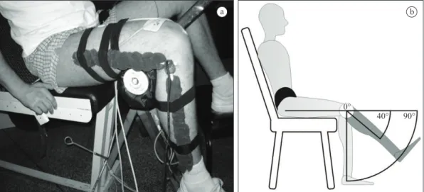
After trichotomy and skin cleaning procedures,
two self-adhesive electrodes (4.5 × 9.0 cm) were
positioned on the knee region (anode) and on the
femoral triangle (cathode) to stimulate the quadriceps
muscle (Figure 1).
Angular data acquisition
A custom monoaxial electrogoniometer built with a
10-kΩ linear potentiometer was placed laterally to the
knee to acquire the knee joint angle (Figure 1). All
signals and volunteer data were saved into European
Data Format (EDF) iles. The acquisition system
contained a DT300 series Data Translation™ board
working at a 1 kHz sampling rate.
Electrical stimulation protocol and data
acquisition
All FES proiles (Table 1) were applied to each
volunteer randomly, one proile per day, over four
testing days with a minimal interval of two rest days
(Kesar
et al
., 2008; Marion
et al
., 2010; Smith
et al
.,
1997; Stock
et al
., 2010) between the tests to avoid
physiological interference between consecutive
protocols. The volunteer was positioned on an
adapted chair with the hip and knee angles set to
70
°
(Matsunaga
et al.
, 1999) and 90
°
, respectively, as
illustrated in Figure 1. The electrogoniometer signal
was zeroed at the maximum knee extension (i.e.,
angle deined as 0
°
). After the zeroing step, the leg
was placed at the 90
°
initial rest position (as shown
in Figure 1). Then, the magnitude of the electrical
stimuli was adjusted according to the knee movement
range from 90
°
to 40
°
. When the knee joint reached
an angle of 40
°
, the corresponding stimulator output
amplitude was determined, and electrical stimulation
was ceased.
Table 1. FES proiles chosen for the experimental protocol.
Proile Pulse Burst
On (µs) Off (µs) Frequency (kHz) On (ms) Off (ms) Frequency (Hz)
P1 100 900 1 3 17 50
P2 100 900 1 3 11 70
P3 200 800 1 3 17 50
P4 200 800 1 3 11 70
On: active pulse duration; Off: inactive pulse duration; pulse on time: 100 µs, 200 µs (Jezernik et al., 2004); pulse frequency: 1 kHz (Ward and Robertson, 1998); burst frequency: 50 Hz and 70 Hz (Chou et al., 2005).
a b
Analysis
The mean amplitude was calculated by means of
Equation 1:
on Bon
Mean Peak
T BT
T T
V V
T T
= ×
(1)
where
•
V
Meanis the mean amplitude expressed in
volts (V);
•
V
Peakis the peak amplitude;
•
T
onis the active pulse period;
•
T
Tis the total pulse period;
•
T
Bonis the active burst period;
•
T
BTis the total burst period.
The application of the Kolmogorov-Smirnov test
showed that the data followed a Gaussian distribution.
The software PASW Statistics 18 was used to perform
the statistical analysis: (I) One-sample Student’s
t-tests were applied to compare peak and mean
amplitudes for HV and SCIV participants, split by
FES proiles; (II) Independent t-tests were applied
to compare HV and SCIV groups in terms of peak
and mean amplitude split by FES proiles; (III) A
one-way analysis of variance (ANOVA) with LSD
post-hoc test was applied to data split into HV and
SCIV groups to ind the proile which resulted in the
lowest peak and mean output amplitudes.
Ethical considerations
This study was performed according to the Declaration
of Helsinki and was approved by Pontifícia
Universidade Católica do Paraná’s (PUCPR) Human
Research Ethics Committee under register n. 2416/08.
Results
Six SCIVs were excluded from the initial group of
volunteers because they either did not tolerate the
sensation evoked by the electrical current or because
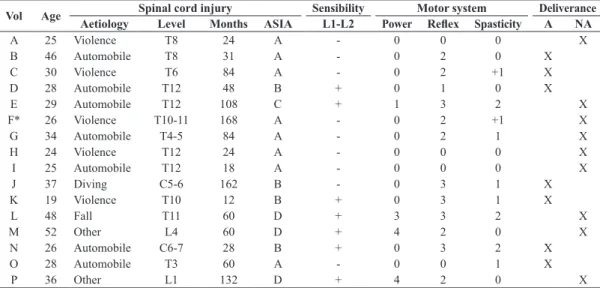
of denervation of motor units. Table 2 presents
demographic information for the participants and
indicates the motor response parameters relecting
their neuromuscular conditions. Sudden onset of
spasticity was not observed during the protocol due
to FES-induced inhibition.
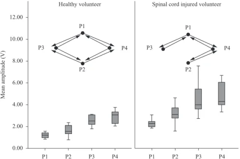
One-sample Student’s t-tests indicated that peak
and mean amplitudes are different (p < 0.01) for both
HV and SCIV groups. Independent t-tests showed
that peak and mean amplitudes necessary to stimulate
HVs were smaller than those for SCIVs across all FES
proiles. Table 3 shows the peak and mean voltage
amplitudes applied during FES for healthy and
spinal cord injured volunteers in different electrical
stimulation proiles. Figures 2 and 3 show post-hoc
comparisons of peak and mean voltages, respectively,
for different FES proiles in HVs and SCIVs.
Discussion
The one-sample Student’s t-tests demonstrated that
peak and mean amplitudes were different (P < 0.01)
for both HVs and SCIVs. According to the independent
t-test, the peak and mean amplitudes required for
raising the leg to 40
°
of knee lexion were higher
Table 2. Clinical data of volunteers with spinal cord injuries.
Vol Age Spinal cord injury Sensibility Motor system Deliverance
Aetiology Level Months ASIA L1-L2 Power Relex Spasticity A NA
A 25 Violence T8 24 A - 0 0 0 X
B 46 Automobile T8 31 A - 0 2 0 X
C 30 Violence T6 84 A - 0 2 +1 X
D 28 Automobile T12 48 B + 0 1 0 X
E 29 Automobile T12 108 C + 1 3 2 X
F* 26 Violence T10-11 168 A - 0 2 +1 X
G 34 Automobile T4-5 84 A - 0 2 1 X
H 24 Violence T12 24 A - 0 0 0 X
I 25 Automobile T12 18 A - 0 0 0 X
J 37 Diving C5-6 162 B - 0 3 1 X
K 19 Violence T10 12 B + 0 3 1 X
L 48 Fall T11 60 D + 3 3 2 X
M 52 Other L4 60 D + 4 2 0 X
N 26 Automobile C6-7 28 B + 0 3 2 X
O 28 Automobile T3 60 A - 0 0 1 X
P 36 Other L1 132 D + 4 2 0 X
Vol: volunteer; ASIA: American Spinal Injury Association impairment scale (A-E) (Maynard et al., 1997); nociceptive sensibility “-” absent, “+”
present; Power: Higuet scale (0-5) (Cipriano, 2003); Relex: Wexler scale (0-5) (Cipriano, 2003); Spasticity: Ashworth modiied scale (0-4) (Bohannon
in SCIVs than in HVs (Figures 2 and 3), which is
consistent with the indings of Gollee
et al
. (2004).
This result is most likely related to muscle atrophy
in SCIVs (Andersen
et al
., 1999; Kern
et al
., 2010a)
and the consequent difference in Ca
++activation of
cross bridges in sarcomere cells (Gobbo
et al
., 2006).
FES may have triggered a muscle cell recovery
process (Thrasher
et al
., 2006) and may also lead to
Table 3. Peak and mean voltages for healthy volunteers and volunteers with spinal cord injuries.P1 (V) P2 (V) P3 (V) P4 (V)
HV Vpeak 82.20±16.73 76.60±22.16 91.90±33.23 67.60±13.97
Vmean 1.23±0.25 1.64±0.47 2.75±0.99 2.90±0.59
SCIV Vpeak 161.40±36.39 154.60±41.96 150.80±49.51 121.80±53.55
Vmean 2.42±0.54 3.31±0.89 4.52±1.48 5.22±2.29
Vpeak: Peak amplitude; Vmean: Mean amplitude.
Figure 2. Peak amplitudes required to achieve 40° of knee lexion in four FES proiles and any statistically signiicant differences. Open circles: 35, 69 and 72 are outliers; Closed circles: control; Arrows: statistically signiicant differences (p ≤ 0.05).
hypertrophy (Kern
et al
., 2010b); thus, the stimulation
amplitude required to achieve a knee lexion angle
of 40
°
may decrease during the recovery process of
neuromuscular tissue.
Regarding peak amplitude, Figure 2 shows that
P4 required the weakest amplitudes to achieve 40° of
knee lexion for both HVs and SCIVs. The low peak
amplitude in P4 is due to the greater pulse duty cycle
(on time and its period ratio: 200 µs/1000 µs) and
also to the 22% burst duty cycle (3 ms/14 ms, i.e.,
the active and total burst period ratio), which allow
more energy to be released to the tissue compared
to the other FES proiles investigated (Table 1). This
inding may explain the fact that for HVs, there was
only a statistically signiicant difference (p < 0.05)
between P3 and P4 FES proiles. Laufer and Elboim
(2008) found that a 20% burst duty cycle (4 ms/20 ms)
was better than 50% burst duty cycle (10 ms/20 ms)
in minimising muscle fatigue while evoking strong
muscle contractions. The results of Laufer and Elboim
(2008) were different from those obtained in the
present study, in which the proiles with lower burst
duty cycles (P1 and P3, 15%) required different
mean amplitudes to achieve 40° of knee lexion. P1
required a weaker mean amplitude, even with the
same burst duty cycle as that of P3. In fact, the FES
proiles with low pulse duty cycles, as well as low
burst duty cycles, were better than the FES proiles
with only low burst duty cycles.
According to the results shown in Figure 3, P1
is the proile that required the weakest mean values
of electrical stimulation to achieve 40
°
of knee
lexion for both HVs and SCIVs; there was also no
signiicant difference at the p<0.05 level between the
active pulse ratios for P1 and P2 (100 µs/1000 µs).
Considering the different trends between peak and
mean amplitudes, P4 is the proile that required the
highest mean amplitude and the lowest peak amplitude
to achieve 40
°
of knee lexion for both HVs and
SCIVs. McLoda and Carmak (2000) studied different
burst duty cycles (10, 30, 50, 70 and 90%) and found
that 10% was the optimum for eliciting the strongest
muscle contraction. In the present study, the mean
amplitude necessary to elicit 40° of knee lexion was
minimised by the FES proile with a lower pulse duty
cycle and lower burst duty cycle.
According to Shannon (1992), safety limits for
electrical stimulation rely on several parameters,
including waveform shape, electrode sizes and charge
densities. These limits must be found for patient
safety during FES. The P4 proile applied higher
mean currents (Vanderthommen and Duchateau,
2007) to volunteers and, theoretically, may have
produced greater increases in skin temperature; greater
increases in skin temperature may cause lesions,
such as burns (Popovic
et al
., 2001), especially in
individuals with spinal cord injuries, who usually are
less sensitive to nociceptive stimuli (Maynard
et al
.,
1997). Because the P1 and P2 FES proiles did not
show signiicant differences, choosing one of them
would result in smaller applied mean currents, but P2,
with a burst frequency of 70 Hz, may cause sensorial
discomfort in patients with augmented nociceptive
sensitivity (Packman-Braun, 1988; Rabischong, 1996;
Rooney
et al
., 1992).
Due to the current research design, our study was
limited by the inability to control certain variables, such
as the skin-electrode interface, involuntary contraction
(healthy subjects) due to FES and possible diffusion
to other muscles (spill-over effect). However, the
results showed that the stimulating proiles studied
effectively generated functional muscular contractions
(in our protocol, knee lexion) and can be generalised
unless more detailed studies obtain different results.
In FES applications, peak and mean amplitudes
delivered by electrical stimulation exhibited different
values and responses. The peak and mean amplitudes
required for raising the leg and changing the knee
lexion angle from 90° to 40° were higher for SCIVs
than for HVs. For both SCIVs and HVs, the FES proile
with duty cycles of 200 µs/1000 µs and 3 ms/14 ms,
for pulse and burst, respectively, required smaller
peak amplitudes to achieve 40° of knee lexion. The
smallest mean amplitudes were obtained for proile P1
with duty cycles of 100 µs/1000 µs and 3 ms/20 ms for
pulse and burst, respectively. Therefore, P1 seems to
be the most suitable FES proile for HVs and SCIVs
because it presented the smallest mean amplitudes and,
consequently, presents smaller hypothetical increases
in skin temperature. Moreover, it can be postulated
that the P1 burst frequency (50 Hz) might have
caused less nociceptive sensation (low frequencies)
compared to other FES proiles used in this study.
Healthcare professionals involved with spinal cord
injury rehabilitation may use this information to plan
treatments using stimulation parameters that promote
functional movements effectively and safely with
regard to the energy transferred by the stimulating
current.
Acknowledgements
We would like to thank CNPq, CAPES and
SETI-PR for providing scholarships and inancial support.
References
ibres in human skeletal muscle. Journal of Applied Physiology. 1999; 86(2):455-60. PMid:9931176.
Bailey SN, Hardin EC, Kobetic R, Boggs LM, Pinault G, Triolo RJ. Neurotherapeutic and neuroprosthetic effects of implanted functional electrical stimulation for ambulation after incomplete spinal cord injury. Journal of Rehabilitation Research & Development. 2010; 47(1):7-16. http://dx.doi. org/10.1682/JRRD.2009.03.0034
Baptista RR, Scheeren EM, Macintosh BR, Vaz MA. Low-frequency fatigue at maximal and submaximal muscle contractions. Brazilian Journal of Medical and Biological Research. 2009; 42:380-5. PMid:19330267. http://dx.doi. org/10.1590/S0100-879X2009000400011
Bohannon RW, Smith M. Interrater reliability of a Modiied Ashworth Scale of muscle spasticity. Physical Therapy. 1987 Feb; 67(2):206-7. PMid:3809245.
Bronzino JD. Management of medical technology: a primer for clinical engineers. Boston: Butterworth-Heinemann; 1992. PMid:1596745.
Chou LW, Ding J, Wexler AS, Binder-Macleod SA. Predicting optimal electrical stimulation for repetitive human muscle activation. Journal of Electromyography and Kinesiology. 2005; 15(3):300-9. PMid:15763677. http:// dx.doi.org/10.1016/j.jelekin.2004.10.002
Cipriano JJ. Photographic manual of regional orthopaedic and neurological tests. 4th ed. Atlanta: Lippincott Williams & Wilkins; 2003.
Davoodi R, Andrews BJ. Fuzzy logic control of FES rowing exercise in paraplegia. IEEE Transactions on Biomedical Engineering. 2004 Mar; 51(3):541-3. PMid:15000386. http://dx.doi.org/10.1109/TBME.2003.821043
Enoka RM, Duchateau J. Muscle fatigue: what, why and how it inluences muscle function. The Journal of Physiology. 2008; 586(1):11-23. PMid:17702815 P M C i d : 2 3 7 5 5 6 5 . h t t p : / / d x . d o i . o rg / 1 0 . 111 3 / jphysiol.2007.139477
Fisekovic N, Popovic DB. New controller for functional electrical stimulation systems. Medical Engineering and Physics. 2001; 23(6):391-9. http://dx.doi.org/10.1016/ S1350-4533(01)00069-8
Fujita K, Handa Y, Hoshimiya N, Ichie M. Stimulus adjustment protocol for FES-induced standing in paraplegiausing percutaneous intramuscular electrodes. IEEE Transactions on Rehabilitation Engineering. 1995; 3(4):360-6. http://dx.doi.org/10.1109/86.481976
Gobbo M, Cè E, Diemont B, Esposito F, Orizio C. Torque and surface mechanomyogram parallel reduction during fatiguing stimulation in human muscles. European Journal of Applied Physiology. 2006; 97(1):9-15. PMid:16477444. http://dx.doi.org/10.1007/s00421-006-0134-8
Gollee H, Hunt KJ, Wood DE. New results in feedback control of unsupported standing in paraplegia. IEEE Transactions on Neural Systems and Rehabilitation
Engineering. 2004; 12(1):73-80. PMid:15068190. http:// dx.doi.org/10.1109/TNSRE.2003.822765
Jezernik S, Morari M. Energy-optimal electrical excitation of nerve fibres. IEEE Transactions on Biomedical Engineering. 2005; 52(4):740-3. PMid:15825876. http:// dx.doi.org/10.1109/TBME.2005.844050
Jezernik S, Wassink RGV, Keller T. Sliding mode closed-loop control of FES: controlling the shank movement. IEEE Transactions on Biomedical Engineering. 2004; 51(2):263-72. P M i d : 1 4 7 6 5 6 9 9 . h t t p : / / d x . d o i . o rg / 1 0 . 11 0 9 / TBME.2003.820393
Kern H, Carraro U, Adami N, Biral D, Hofer C, Forstner C, Modlin M, Vogelauer M, Pond A, Boncompagni S. Home-based functional electrical stimulation rescues permanently denervated muscles in paraplegic patients with complete lower motor neuron lesion. Neurorehabil Neural Repair. 2010a May; 24(8):709-21. PMid:20460493. http:// dx.doi.org/10.1177/1545968310366129
Kern H, Stramare R, Martino L, Gargiulo P, Carraro U. Permanent LMN denervation of human skeletal muscle and recovery by hb FES: management and monitoring. European Journal Translational Myology. 2010b; 20(3):91-104.
Kesar T, Chou LW, Binder-Macleod SA. Effects of stimulation frequency versus pulse duration modulation on muscle fatigue. Journal of Electromyography and Kinesiology. 2008 Aug; 18(4):662-71. PMid:17317219 PMCid:2562565. http:// dx.doi.org/10.1016/j.jelekin.2007.01.001
Kesar TM, Perumal R, Jancosko A, Reisman DS, Rudolph KS, Higginson JS, Binder-Macleod SA. Novel patterns of functional electrical stimulation have an immediate effect on dorsilexor muscle function during gait for people poststroke. Physical Therapy. 2010; 90(1):55-66. PMid:19926681 PMCid:2802826. http://dx.doi.org/10.2522/ptj.20090140
Krueger-Beck E, Scheeren E, Nogueira-Neto GN, Button VLdSN, Nohama P. Optimal FES parameters based on mechanomyographic eficiency index. In: Annual International Conference of the IEEE EMBC: Proceedings of the Annual International Conference of the IEEE Engineering in Medicine and Biology Society; 2010 Aug 31-Sept 4; Buenos Aires, Argentina. Buenos Aires: IEEE; 2010a. p. 1378-81. PMid:21096336.
Krueger-Beck E, Scheeren EM, Nogueira-Neto GN, Button VLdSN, Nohama P. Efeitos da estimulação elétrica funcional no controle neuromuscular artiicial. Revista Neurociências. 2010b; 1-11. In Press.
Langzam E, Nemirovsky Y, Isakov E, Mizrahi J. Muscle enhancement using closed-loop electrical stimulation: Volitional versus induced torque. Journal of Electromyography and Kinesiology. 2007; 17(3):275-84. PMid:16690326. http:// dx.doi.org/10.1016/j.jelekin.2006.03.001
Marion MS, Wexler AS, Hull ML. Predicting fatigue during electrically stimulated non-isometric contractions. Muscle & Nerve. 2010; 41(6):857-67. PMid:20229581. http://dx.doi. org/10.1002/mus.21603
Marsolais EB, Kobetic R. Development of a practical electrical stimulation system for restoring gait in the paralyzed patient. Clinical Orthopaedics and Related Research. 1988; 233:64-74. PMid:3261221.
Marsolais EB, Kobetic R. Functional electrical stimulation for walking in paraplegia. Journal of Bone and Joint Surgery. 1987; 69(5):728-33. PMid:3496340.
Matsunaga T, Shimada Y, Sato K. Muscle fatigue from intermittent stimulation with low and high frequency electrical pulses. Archives of Physical Medicine and Rehabilitation. 1999; 80(1):48-53. http://dx.doi.org/10.1016/ S0003-9993(99)90306-4
Maynard FM, Bracken MB, Creasey G, Ditunno JF, Donovan WH, Ducker TB, Garber SL, Marino RJ, Stover SL, Tator CH. International standards for neurological and functional classiication of spinal cord injury. Spinal Cord. 1997; 35(5):266-74. PMid:9160449. http://dx.doi. org/10.1038/sj.sc.3100432
McAndrew DJ, Rosser NAD, Brown JMM. Mechanomyographic measures of muscle contractile properties are inluenced by the duration of the stimulatory pulse. Journal of Applied Research. 2006; 6(1):142-52.
McCreery DB, Agnew WF, Yuen TGH, Bullara L. Charge density and charge per phase as cofactors in neural injury induced by electrical stimulation. IEEE Transactions on Biomedical Engineering. 1990; 37(10):996-1001. PMid:2249872. http://dx.doi.org/10.1109/10.102812
McLoda TA, Carmack JA. Optimal burst duration during a facilitated quadriceps femoris contraction. Journal of Athletic Training. 2000; 35(2):145-50. PMid:16558623 PMCid:1323410.
Mesin L, Merletti R. Distribution of electrical stimulation current in a planar multilayer anisotropic tissue. IEEE Transactions on Biomedical Engineering. 2008; 55(2):660-70. PMid:18270002. http://dx.doi.org/10.1109/ TBME.2007.902248
Neves EB, Pino AV, Souza MN. Comparison of two bioimpedance spectroscopy techniques in the assessment of body luid volumes. In: Annual International Conference of the IEEE EMBC: Proceedings of the 31th Annual International Conference of the IEEE Engineering in Medicine and Biology Society; 2009; Minneapolis, Minnesota. Minneapolis: IEEE; 2009. p. 853-6. PMid:19963476.
Nordstrom MA, Gorman RB, Laouris Y, Spielmann JM, Stuart DG. Does motoneuron adaptation contribute to muscle fatigue? Muscle & Nerve. 2007; 35(2):135-58. PMid:17195169. http://dx.doi.org/10.1002/mus.20712
Packman-Braun R. Relationship between functional electrical stimulation duty cycle and fatigue in wrist
extensor muscles of patients with hemiparesis. Physical Therapy. 1988; 68(1):51-6. PMid:3257300.
Petrofsky JS. Electrical stimulation: neurophysiological basis and application. Basic and Applied Myology. 2004; 14(4):205-13.
Popovic MR, Curt A, Keller T, Dietz V. Functional electrical stimulation for grasping and walking: indications and limitations. Spinal Cord. 2001; 39(8):403-12. PMid:11512070. http://dx.doi.org/10.1038/sj.sc.3101191
Rabischong E. Surface action potentials related to torque output in paraplegics’ electrically stimulated quadriceps muscle. Medical Engineering & Physics. 1996; 18(7):538-47. http://dx.doi.org/10.1016/1350-4533(96)00001-X
Rooney JG, Currier DP, Nitz AJ. Effect of variation in the burst and carrier frequency modes of neuromuscular electrical stimulation on pain perception of healthy subjects. Physical Therapy. 1992; 72(11):800-6. PMid:1409877.
Shannon RV. A model of safe levels for electrical stimulation. IEEE Transactions on Biomedical Engineering. 1992; 39(4):424-6. PMid:1592409. http:// dx.doi.org/10.1109/10.126616
Smith DB, Housh, Terry J., Stout JR, Johnson GO, Evetovich TK, Ebersole KT. Mechanomyographic responses to maximal eccentric isokinetic muscle actions. Journal of Applied Physiology. 1997; 82(3):1003-7. PMid:9074994.
Stock MS, Beck TW, DeFreitas JM, Dillon MA. Linearity and reliability of the mechanomyographic amplitude versus dynamic constant external resistance relationships for the biceps brachii. Physiological Measurement. 2010; 31:1487-98. PMid:20871133. http://dx.doi.org/10.1088/0967-3334/31/11/006
Tepavac D, Schwirtlich L. Detection and prediction of FES-induced fatigue. Journal of Electromyography and Kinesiology. 1997; 7(1):39-50. http://dx.doi.org/10.1016/ S1050-6411(96)00008-9
Thrasher A, Graham GM, Popovic MR. Reducing muscle fatigue due to functional electrical stimulation using random modulation of stimulation parameters. Artiicial Organs. 2005; 29(6):453-8. PMid:15926981. http://dx.doi. org/10.1111/j.1525-1594.2005.29076.x
Thrasher TA, Flett HM, Popovic MR. Gait training regimen for incomplete spinal cord injury using functional electrical stimulation. Spinal Cord. 2006; 44(6):357-61. PMid:16249784. http://dx.doi.org/10.1038/sj.sc.3101864
Uhlir JP, Triolo RJ, Kobetic R. The use of selective electrical stimulation of the quadriceps to improve standing function in paraplegia. IEEE Transactions on Rehabilitation Engineering. 2000; 8(4):514-22. PMid:11204043. http:// dx.doi.org/10.1109/86.895955
Ward AR, Robertson VJ. Variation in torque production with frequency using medium frequency alternating current* 1,* 2. Archives of Physical Medicine and Rehabilitation. 1998; 79(11):1399-404. http://dx.doi. org/10.1016/S0003-9993(98)90234-9
Ward AR, Shkuratova N. Russian electrical stimulation: the early experiments. Physical Therapy. 2002; 82(10):1019. PMid:12350217.
Williamson R, Andrews BJ. Sensor systems for lower limb functional electircal stimulation (FES) control. Medical
Engineering & Physics. 2000; 22(5):313-25. http://dx.doi. org/10.1016/S1350-4533(00)00038-2
Yu NY, Chang SH. The Characterization of contractile and myoelectric activities in paralyzed tibialis anterior post electrically elicited muscle fatigue. Artificial Organs. 2010; 34(4):E117-E21. PMid:20420602. http:// dx.doi.org/10.1111/j.1525-1594.2009.00956.x
Zagheni AL. Sistema de NMES multicanal controlado por computador para aplicações em locomoção artiicial [dissertação]. Curitiba: Universidade Tecnológica Federal do Paraná; 1998.
Authors
Eddy Krueger*, Percy Nohama
Programa de Pós-graduação em Engenharia Elétrica e Informática Industrial – CPGEI, Laboratório de Engenharia de Reabilitação, Universidade Tecnológica Federal do Paraná – UTFPR, Av. Sete de Setembro, 3165, CEP 80230-901, Curitiba, PR, Brasil.
Eduardo Mendonça Scheeren
Programa de Pós-graduação em Tecnologia em Saúde – PPGTS, Escola de Saúde e Biociências e Escola Politécnica, Pontifícia Universidade Católica do Paraná – PUCPR, CEP 80250-901, Curitiba, PR, Brasil.
Guilherme Nunes Nogueira-Neto, Vera Lúcia da Silveira Nantes Button
Departamento de Engenharia Biomédica – DEB, Faculdade de Engenharia Elétrica e Computação – FEEC, Centro de Engenharia Biomédica – CEB, Universidade Estadual de Campinas – UNICAMP, Cidade Universitária “Zeferino Vaz”, CEP 13084-971, Campinas, SP, Brasil.
Eduardo Borba Neves