The Zwitterionic Cell Wall Teichoic Acid of
Staphylococcus aureus
Provokes Skin Abscesses in Mice
by a Novel CD4
+
T-Cell-Dependent Mechanism
Christopher Weidenmaier, Rachel M. McLoughlin, Jean C. Lee*
Channing Laboratory, Department of Medicine, Brigham and Women’s Hospital and Harvard Medical School, Boston, Massachusetts, United States of America
Abstract
Zwitterionic polysaccharide (ZPS) components of the bacterial cell envelope have been shown to exert a major histocompatibility complex (MHC) II-dependent activation of CD4+T cells, which in turn can modulate the outcome and
progression of infections in animal models. We investigated the impact of zwitterionic cell wall teichoic acid (WTA) produced byStaphylococcus aureuson the development of skin abscesses in a mouse model. We also compared the relative biological activities of WTA and capsular polysaccharide (CP), important S. aureus pathogenicity factors, in abscess formation. Expression of both WTA and CP markedly affected the ability ofS. aureusto induce skin abscess formation in mice. Purified wild-type zwitterionic WTA was more active in inducing abscess formation than negatively charged mutant WTA or purified CP8. To assess the ability of purified native WTA to stimulate T cell proliferation in vitro, we co-cultivated WTA with human T-cells and antigen presenting cells in the presence and absence of various inhibitors of MHC-II presentation. Wild-type WTA induced T cell proliferation to a significantly greater extent than negatively charged WTA. T cell activation was dependent on the presentation of WTA on MHC II, since inhibitors of MHC II-dependent presentation and antibodies to MHC II significantly reduced T cell proliferation. T cells activated in vitro with wild-type WTA, but not negatively charged WTA, induced abscess formation when injected subcutaneously into wild-type mice. CD42/2 mice similarly injected with WTA failed to develop abscesses. Our results demonstrate that the zwitterionic WTA ofS. aureus
induces CD4+T-cell proliferation in an MHCII-dependent manner, which in turn modulates abscess formation in a mouse
skin infection model. An understanding of this novel T cell-dependent host response to staphylococcal abscess formation may lead to the development of new strategies to combatS. aureusskin and soft tissue infections.
Citation:Weidenmaier C, McLoughlin RM, Lee JC (2010) The Zwitterionic Cell Wall Teichoic Acid ofStaphylococcus aureusProvokes Skin Abscesses in Mice by a Novel CD4+T-Cell-Dependent Mechanism. PLoS ONE 5(10): e13227. doi:10.1371/journal.pone.0013227
Editor:Michael Otto, National Institutes of Health, United States of America
ReceivedJuly 7, 2010;AcceptedSeptember 1, 2010;PublishedOctober 7, 2010
Copyright:ß2010 Weidenmaier et al. This is an open-access article distributed under the terms of the Creative Commons Attribution License, which permits unrestricted use, distribution, and reproduction in any medium, provided the original author and source are credited.
Funding:CW was supported by a fellowship from the Deutsche Forschungsgesellschaft (http://www.dfg.de/en/index.jsp). The funders had no role in study design, data collection and analysis, decision to publish, or preparation of the manuscript.
Competing Interests:The authors have declared that no competing interests exist.
* E-mail: [email protected]
Introduction
Wall teichoic acid (WTA) ofStaphylococcus aureusis a zwitterionic cell wall glycopolymer composed of ,40 ribitol phosphate repeating units modified withN-acetylglucosamine and D-alanine [1,2]. The negatively charged phosphate groups in the ribitol repeating unit and the ester-linked D-alanine residues contribute to the zwitterionic charge of WTA (Fig. 1). The presence of WTA-like molecules in most Gram-positive bacteria prompted scientists to focus on the role of WTA in cell wall function [3,4] and turn-over [5]. More recently, defined mutants of S. aureus that lack WTA or have altered WTA structures have facilitated experiments to elucidate in greater detail the role of WTA in staphylococcal pathogenesis [6–10]. WTA has been implicated in the adhesion of S. aureus to human epithelial and endothelial cells [9–11], and expression of WTA has been shown to be essential forS. aureus nasal colonization of cotton rats [9,11]. AS. aureusmutant lacking WTA showed attenuated virulence in a rabbit model of endocarditis [10]. Moreover, purified WTA was able to induce intraabdominal abscesses when rats were inoculated by the intraperitoneal route [12]. S. aureus mutants in the dlt operon,
which mediates D-alanylation of WTA, exhibit a negatively charged cell surface and are more sensitive to cationic antimicro-bial peptides than the parental strain [13,14].
WTA is expressed constitutively [1], whereasS. aureuscapsular polysaccharides (CPs) are expressed in a tightly regulated manner [15,16]. Most clinical isolates ofS. aureus express either capsule type 5 (CP5) or 8 (CP8). However, 20–25% of clinical isolates produce no capsule due to a variety of mutations within the conserved capsule biosynthesis operon [17,18].
model, McLoughlin et al. demonstrated that the presence of CD4 T cells modulated CXC chemokine production at the S. aureus infection site in an IFN-c dependent manner, which led to a massive recruitment of neutrophils, and this effect was mediated by CP8 [26]. These findings suggest that staphylococcal ZPS are important factors for T cell dependent immune stimulation, which strongly influences the outcome of infections.
Skin and soft tissue infections are the most common types ofS. aureus infections and occur in the hospital as well as in the community, affecting hosts without predisposing risk factors [27,28]. Since bacterial ZPS are associated with abscess formation [12,29], we explored the contribution of zwitterionic WTA and CPs in a mouse skin infection model. We demonstrate here that WTA activates T cells in a MHC II dependent manner, and that T cells activated in vitro by WTA induce skin abscesses in mice. These novel findings link staphylococcal skin infections to recent discoveries of glyco-immunology and may lead to novel approach-es to combat skin infections byS. aureus.
Results
WTA and Capsule Impact Abscess Formation When Live Bacteria Are Injected Subcutaneously into the Flanks of Mice
We used a mouse model to study the ability of variousS. aureus strains to induce skin infections that closely resemble staphylococ-cal skin infections in humans. S. aureuscells mixed with cytodex beads were injected subcutaneously into the flanks of mice. The cytodex beads allow for formation of a localized abscess even at low inocula or with low concentrations of purified WTA.
To assess the relative roles played byS. aureusWTA and CPs in skin infections, we transduced thetagO mutation into the CP5+
strain Newman and its isogenic acapsular cap5O mutant. The resultingtagO deletion mutants lacked WTA in their cell wall as evidenced by the low phosphorus content of cell wall fractions, but their doubling times were similar to that of the parental strains under in vitro conditions (Fig. S1). The NewmantagOmutant was complemented by introducing a wild-type copy oftagOon plasmid prBtagO. In addition, we used previously described mutants of the acapsular SA113 strain that either lack WTA (tagO) [9] or the D-alanine esters (dltA) [13] in the repeating WTA subunits. ThedltA mutation was transduced into Newman, and the phenotype of the resulting mutant was consistent with the absence of positive charges in the teichoic acids (Fig. S1).
S. aureuswild-type strains SA113 and Newman at inocula ranging from 106to 104cfu were potent inducers of subcutaneous abscesses in mice (Fig. 2). The tagO mutants of both bacterial strains were
significantly attenuated in their ability to induce abscesses, whereas the dltAmutants showed an intermediate phenotype at the lower inocula (Figs. 2A and 2B and Supplemental Fig. 2). Consistent with previous data with a CP8+strain [22], the acapsular Newmancap5Omutant was also attenuated for subcutaneous abscess formation (Fig. 2C). When the bacteria were grown on Columbia salt agar to enhance capsule expression (Fig. 2C), strain Newman provoked a higher bacterial burden per abscess than when it was grown on tryptic soy agar (Fig. 2B). The Newman tagO and cap5O mutants were both significantly attenuated for abscess induction when compared to the parental strain. A double mutant of Newman that expresses neither capsule nor WTA was more attenuated for abscess formation than the single mutants (Fig. 2C), but the differences did not reach statistical significance. These data suggest that both WTA and CP promoteS. aureusskin abscess formation when live bacteria are used as the inocula.
Purified WTA Induces Skin Abscesses More Efficiently than Purified CP8
To further assess its biological activity, we purified WTA from the acapsular strains SA113 wild-type (zwitterionic WTA) and SA113 dltA (anionic WTA). WTA purity was assessed by 1H NMR, phosphate content, the absence of nucleic acids (,0.1%), protein (,0.1%), and endotoxin (,1 EU/mg). Sterile abscesses were induced by injection of sterile cytodex beads mixed with purified wt WTA at doses ranging from 200 to 0.02mg. After 48 h, abscesses were excised and quantified by their weight. Purified wt WTA was a potent inducer of abscess formation at doses as low as 0.02mg (Fig. 3A). WTA purified from the dltA mutant or beads alone showed minimal activity under these conditions (Fig. 3A). Administration ofdltAWTA resulted in the recruitment of fewer neutrophils in the abscessed tissue than wt WTA (Fig. S2). We quantified the host response by measuring neutrophil accumulation at the infection site with the myeloper-oxidase (MPO) assay (Fig. 3B) [30]. MPO is present in the azurophilic granules of neutrophils and is routinely used to assess the tissue inflammatory responses [31] and abscess formation. MPO activity in abscesses provoked by 20mg wt WTA after 48 h was significantly (P,0.0001) higher than that induced by 20mg dltA WTA. Despite its potent activity in the induction of intraabdominal abscesses [12], purified CP8 exhibited only minimal potency in the s.c. abscess model, even at a 50mg dose.
Purified WTA Fails to Induce Neutrophil Chemotaxis or Proinflammatory Cytokines
To investigate whether purified WTA exhibited direct chemotactic activity, we assayed chemotaxis of human neutrophils in response to Figure 1.Staphylococcus aureusWTA is a zwitterionic cell wall polymer composed of,40 ribitol phosphate repeating units modified withN-acetylglucosamine and D-alanine.The negatively charged phosphodiester bonds and the positively charged D-alanine residues are responsible for the zwitterionic nature of the polymer.
WTA in a transwell assay. WTA purified fromS. aureusSA113 or the SA113dltAmutant failed to induce significant chemotaxis compared to the authentic chemoattractant fMLP (Fig. 4A).
Activation of pro-inflammatory responses via pattern recogni-tion receptors like Toll-like receptors (TLRs) could also lead to pronounced inflammation and modulate abscess formation. Therefore, we tested whether purified WTA stimulates pro-inflammatory cytokines in macrophages or in HEK293 cells transfected with TLR-2. These experiments not only interrogated WTA proinflammatory activity but also enabled us to exclude functional contaminants in our purified WTA preparation. We measured production of TNF-a by human monocytes (THP-1 cells) or mouse macrophages (RAW 264.7 cells) after stimulation with WTA or with the TLR agonists LPS, MALP-2, or Pam3Cys as positive controls. We stimulated HEK293 cells transfected with TLR-2 with WTA, MALP-2, or Pam3Cys, and assayed for IL-8 production in the culture supernatants. Purified WTA at concentrations as high as 100mg/ml showed no proinflammatory activity in THP-1 or RAW 264.7 cells (Fig. 4B) or in TLR-2 expressing HEK293 cells (Fig. 4C). This is consistent with previous reports that demonstrated the inability of WTA to directly activate host cells in a manner typical of classical pathogen associated molecular pattern molecules [32].
WTA activates proliferation of human T cells in a MHC II dependent manner
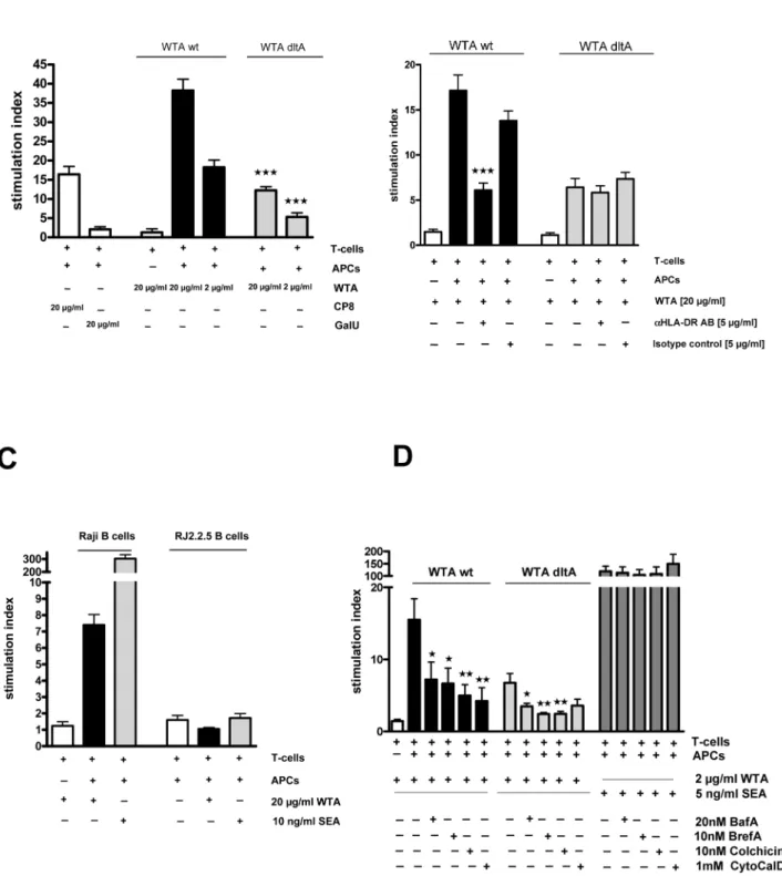
The induction of intraperitoneal abscesses by other zwitterionic polysaccharides is based on their ability to directly activate T-cells, which in turn modulate cellular responses at the infection site [14,28]. Therefore, we cultured nylon wool-purified human T-cells or column-purified CD4+ T cells with irradiated antigen-presenting cells (APCs) and wt ordltAWTA.S. aureusenterotoxin A (SEA) and polygalacturonic acid (GalU) were used as positive and negative controls, respectively. Wt WTA elicited a potent dose-dependent proliferative response in human T cells on day 6 (Fig. 5A). The T-cell response to wt WTA was significantly higher than that exhibited by CP8,dltAWTA, or beads alone (Fig. 5A). The negatively charged polygalacturonic acid failed to induce T-cell proliferation, and theS. aureus superantigen SEA induced a significantly higher T-cell response than proliferation induced by either of the WTA antigens (Fig. 5D).
To determine whether T-cell proliferation induced by purified WTA was mediated by MHC class I or II molecules, we performed assays in the presence of blocking antibodies (Abs) and isotype control Abs. Wt WTA induced T-cell proliferation was dependent on the MHC class II molecule HLA-DR since T cell proliferation was reduced significantly in the presence of blocking Abs to DR (Fig. 5B), but not with Abs to DP, HLA-DQ, or HLA-A, -B, or –C (data not shown). The residual activity of thedltAWTA was not inhibited by any of the Abs. In assays utilizing the human Burkitt lymphoma cell line (Raji) and its MHC class II transcriptional mutant cell line (RJ2.2.5), WTA stimulated T-cell proliferation only when the MHC-II–expressing Raji cells, but not the RJ2.2.5 cells, were incubated with T-cells and WTA (Fig. 5C). Inhibitors of the intracellular antigen-processing pathways that are required for MHC II-dependent antigen Figure 2. Strains with no WTA or structurally altered WTA are deficient in induction of s.c. abscesses.106to 104CFUS. aureuswas injected s.c into the flanks of mice. After 48 h, the mice were euthanized, and each abscess was excised, homogenized, and cultured quantitatively. Horizontal bars represent group median values, n = 8. A) The Sa113dltA mutant (negatively charged teichoic acids) and thetagO mutant (no WTA) were attenuated compared to the wt (allp-values are 0.0002). B) NewmantagO(p= 0.0002 at 106;p= 0.0003 at 105,p= 0.0002 at 104) and thedltA mutant (p= 0.0047 at 106;p= 0.0006 at 105,p= 0.007 at 104) were significantly less virulent in the abscess model compared to the wt, n = 8. Allp -values were determined by Mann-Whitney analysis. C) Newman wt,tagO, isogenic acapsular mutantcap5Oand thecap5O/tagOdouble mutant were grown on CSA plates to enhance CP production. The NewmantagO(p,0.05 at 106;p,0.01 at 105,p,0.01 at 104) andcap5O(p,0.01 at 106;p,0.05 at 105,p.0.05 at 104) mutants and the double mutant (p,0.001 at 106;p,0.001 at 105,p,0.001 at 104) were significantly inhibited in abscess formation compared to the wt. The mutants showed no significant difference when compared to each other. P-values for Figure 2C were determined by Kruskal-Wallis one-way ANOVA with Dunn’s multiple comparison test.wp,0.05;wwp,0.01,wwwp,0.001.
doi:10.1371/journal.pone.0013227.g002
Figure 3. Purified WTA is a potent abscess inducer.WTA was purified from wt SA113 or thedltAmutant, mixed with cytodex beads, and injected s.c. into mouse flanks. A) The weights of the abscesses were determined after 48 h and expressed as mean6SEM, n = 8. The zwitterionic WTA purified from SA113 induced well-defined abscesses, whereas the negatively chargeddltAWTA was only a weak abscess inducer (wt WTA compared to beadsp= 0.0062 at 200mg;p= 0.0381 at 20mg.dltAWTA compared to beads yielded p values.0.05 at all doses. Allp-values were determined by two-tailed Student t-tests). B) 20mg WTA purified from wild-type SA113 or thedltAmutant was mixed with cytodex beads and injected into the flanks of mice. After 48 h, the abscesses were excised, homogenized, and MPO activity was determined to assess the intensity of the host response. Wt WTA induced strong MPO activity, indicative of neutrophil recruitment, whereas dltAWTA and CP8 were considerably less active (p-values,0.0001 by Mann-Whitney analyses).wp,0.05;wwp,0.01,wwwp,0.001.
presentation were used at concentrations that had no effect on the stimulatory activity of the S. aureussuperantigen SEA (Fig. 5D). The inhibitors Bafilomycin A (BafA), Brefeldin A (BrefA), Colchicine, and Cytochalasin D (CytoCalD) reduced the T-cell proliferation induced by wt WTA and dltA WTA, providing evidence that, unlike superantigens, WTA requires intracellular processing for efficient MHC II presentation to T cells.
WTA Stimulated Mouse T Cells Modulate Abscess Formation in the S.C Abscess Model
The role of T-cell activation in the modulation of WTA-induced abscess formation was assessed by T-cell transfer experiments.S. aureuswt WTA stimulated a potent proliferative response in mouse T cells after 6 days, whereasdltAWTA was less active (Fig. 6A). In T cell transfer assays, purified mouse CD4+T cells were cultured in vitro for 6 days with WTA and irradiated APCs. The stimulated T cells were then purified, assayed for purity by FACS analysis, mixed with cytodex beads, and injected s.c. into mouse flanks. Abscess formation was quantified by MPO activity in the abscessed tissue after 48 h. In dose-dependency experiments, we found that 36105 activated T cells were sufficient to induce an abscess after 48 h (data not shown). T cells stimulated with wt WTA induced abscess formation in mice, whereas T cells stimulated withdltAWTA, naive T-cells, or APCs incubated with WTA in the absence of T cells were significantly less potent (Fig. 6B).
To lend further evidence to the premise that T cells are crucial forS. aureusabscess formation, we used CD4 T cell deficient mice. As predicted, WTA provoked abscesses in C57Bl/6 mice, but not in CD4-deficient C57Bl/6 tm1mac mice (Fig. 6C). These in vivo data clearly demonstrate the link between WTA dependent T cell activation and the modulation of abscess formation by activated CD4 T cells.
Discussion
Ribitol-phosphate WTA is a conserved cell wall-associated ZPS produced by S. aureus. CP produced by the majority of staphylococcal strains may mask WTA on the bacterial surface. However, neither CP5 nor CP8 is expressed by 20–25% of clinical isolates [17,18]. Even in encapsulated strains, WTA is most likely surface exposed during the logarithmic growth phase when little CP is produced [16,17,33]. WTA is present both in the logarithmic and stationary phases of bacterial growth, and it appears to be expressed constitutively by the bacterial cell. Structural genes encoding enzymes of WTA biosynthesis machin-ery can be upregulated under certain in vivo conditions, such as the early phases of nasal colonization, as demonstrated in the cotton rat model [34].
We were interested in the role of WTA in the development of skin infections caused byS. aureus,since these are among the most frequent staphylococcal infections. Moreover, WTA was shown to induce abscesses in a rat model of intraabdominal abscess formation [12]. Our data demonstrate that S. aureusWTA plays a critical role in the development of staphylococcal skin lesions.
Mutants that lack WTA (tagO) or have an altered WTA structure (dltA) are significantly impaired in the mouse s.c. abscess model. The attenuated phenotype of thedltA mutant may be partially attributed to its susceptibility to killing by neutrophils and cationic microbial peptides [9,13], which are highly abundant in skin infections [35]. The absence ofS. aureusWTA or the presence of an altered WTA structure influences the molecular events that modulate abscess formation since purified wt WTA induced abscesses more efficiently thandltAWTA. T cells have previously been shown to play a role in animal models of S. aureus intraperitoneal and subcutaneous abscess formation [12,22] and in a staphylococcal surgical wound infection model [22]. In this study we identify a novel role for zwitterionic WTA, together with CP, in the induction of subcutaneous abscess formation through a mechanism dependant upon T-cell activation. We demonstrated that purified WTA stimulated CD4+T-cell proliferation in vitro, and that this was dependent upon the zwitterionic charge of WTA. WTA was unable to induce T cell proliferation when APCs lacking MHC II were used in co–culture, and the stimulatory activity of wt WTA, but not the residual activity of thedltAWTA, was diminished by HLA-DR blocking Abs. In contrast, inhibitors of intracellular processing pathways blocked the activity of wt WTA and the residual activity ofdltA WTA. This argues for a charge-dependency of WTA presentation but not intracellular processing, which is consistent with reports characterizing other bacterial ZPS polymers [36]. Thus, WTA qualifies as a bacterial ZPS that is processed and presented by APCs via the MHCII pathway [19]. Having defined a mechanism by which WTA can activate T-cells, we then established that the T cell stimulatory activity of WTA could modulate skin abscess formation. CD4 T cells activated by purified wt WTA were sufficient to provoke abscesses when injected s.c. into the flanks of healthy mice. Moreover, CD42/2mice injected with WTA failed to develop abscesses. Taken together, we present a novel function of WTA as a ZPS that can modulate CD4 T cell-dependent development of skin abscesses in mice.
S. aureusmutants defective in CP production were attenuated in the s.c. abscess model of infection, but purified CP8 was less active than WTA in T-cell proliferation assays and in the skin abscess model. Both CP8 and WTA showed similar potency in the rat intraperitoneal abscess model [12], suggesting that the underlying molecular mechanisms leading to abscess formation differ between the skin and peritoneal cavity. USA300 strains that are responsible for most SSTIs in the U.S. are negative for CP production [37], and thus WTA may be a critical factor that promotes USA300 abscess formation in humans.
skin abscesses through MHC II–dependent activation of CD4+T cells. A recent study by Cho et al. demonstrated the importance of
cd T cells in controlling neutrophil recruitment and influencing the outcome of intradermalS. aureusinfections in mice [38]. The discrepancies between the results of Cho et al. and our own results may reflect subtle differences between the two infection models, as well as characteristics of theS. aureusstrains utilized by the different labs. The immune mechanisms modulating the onset of infection likely differ depending on the site of infection, the specific virulence factors produced by the microbe, and the repertoire of effector cells responding to the bacterial insult.
Multiple virulence determinants likely affectS. aureusinduction of SSTIs [39], and the host response to infection is currently under investigation by several laboratories. WTA is present in allS. aureus strains, and synergistic interactions among leukocidins, CPs, a -toxin [40], phenol-soluble modulins [41] and WTA may increase the frequency or severity of staphylococcal SSTIs. The ribitol phosphate WTA produced by S. aureus differs from the glycerol phosphate WTA polymer synthesized byStaphylococcus epidermidis, the most common staphylococcal species found on human skin [42,43]. Whether the biological properties of the two polymers differ is a question that has not yet been addressed.
Our results demonstrate a novel function for the zwitterionic WTA polymer ofS. aureus. We link the novel finding that WTA can stimulate CD4+T cells to the development of staphylococcal SSTIs in anin vivoinfection model. We offer new insights into the pathology of SSTIs caused byS. aureusand propose a novel host mechanism involved in staphylococcal skin infections.
Methods
Ethics Statement
Human blood was collected from healthy volunteers giving written informed consent for venipuncture, as approved by the Institutional Review Board of The Brigham and Women’s Hospital (Human Subject Assurance Number 00000484). Animal experiments were performed in accordance with the guidelines of the Harvard Medical School Standing Committee on Animals (Animal Welfare Assurance Number A3431-01) under approved protocol 86-02131. The Harvard Medical School animal man-agement program is accredited by the American Association for Accreditation of Laboratory Animal Care and meets National Institutes of Health standards as set forth in ‘‘Guide for the Care and Use of Laboratory Animals’’ (DHSS Publication No. (NIH) 85-23 Revised 1985). The institution also accepts as mandatory the Public Heath Service ‘‘Policy on Humane Care and Use of Laboratory Animals by Awardee Institutions’’ and NIH ‘‘Princi-ples for the Utilization and Care of Vertebrate Animals Used in Testing, Research and Training.’’
Bacterial Strains and Growth Conditions
S. aureus SA113 (ATCC 35556) is a previously described laboratory strain [44]. The WTA-deficient Sa113tagOmutant was generated by replacing the tagO gene with an erythromycin resistance cassette [9]. TheDdltAmutant ofS. aureusSA113 was generated by replacing the dltA gene with a spectinomycin resistance cassette [13], and this mutation was transduced into strain Newman with phage 80a. The phenotype of thedltAmutant was assessed by its ability to bind more positively charged cytochrome C (Sigma) than the wildtype strain, as described
previously [45].S. aureusNewman and the isogenic Newmancap5O mutant were described previously [46]. The tagO::tet mutation from RN4220tagO::tet geh::tagO(kindly provided by Drs. Timothy Meredith and Suzanne Walker, Harvard Medical School) was introduced into strain Newman by transduction with phage 80a. The strain NewmantagOmutation was verified by PCR, and the absence of WTA was confirmed by measuring the phosphorus content of cell wall fractions prepared from the isogenic wild-type and mutant strains [9]. For genetic complementation studies, the mutants were complemented with a plasmid containing a wild-type copy of thetagO gene (pRBtagO) [9]. Bacterial strains were grown in tryptic soy broth (BBL) or B-Medium BM [9] unless otherwise noted.
WTA Isolation and Purification
Staphylococcal WTA was isolated and analyzed as described [9]. The phosphorus content in WTA samples was determined by colorimetric assays [9,13]. WTA was then dialyzed against 20 mM Bis/Tris buffer and run on a Q-Sepharose column with a NaCl gradient (0–1 M) followed by a Sephacryl S-300 size exclusion column. All buffers and water were pyrogen-free. The instruments and devices used in the WTA purification process were deproteinated by treatment with sulfuric acid and depyrogenated by heat inactivation for 4 h at 240uC or by treatment with a 1– 2 M sodium hydroxide buffer. WTA was analyzed for protein by the Biorad method and by UV absorbance at 280 nm; for nucleic acid by UV absorbance at 260 nm and by agarose gel electrophoresis; and for endotoxin by the Limulus amebocyte lysate test (Charles River Laboratories, Charleston, SC). 1 H-nuclear magnetic resonance spectroscopy (NMR) was performed for structural evaluation.
Mouse Model of Subcutaneous Abscess Formation
Male Swiss Webster ND4 mice (4–6 weeks old) were purchased from Charles River or Harlan, and C57Bl/6 and C57Bl/6 tm1mac CD42/2 mice were purchased from The Jackson Laboratory. 104to 106CFUS. aureusor purified WTA was mixed with sterile dextran beads (Cytodex 1, Sigma), and the mixture (0.2 ml) was injected s.c. into the shaved flanks of mice as described previously [47]. The abscesses were either weighed or homogenized in TSB for quantitative culture. Alternatively, the myeloperoxidase (MPO) activity in abscess homogenates was measured with a colorimetric assay [48]. For histological analysis, excised tissue was fixed in formalin, embedded in paraffin, and stained with hematoxylin and eosin for microscopic examination.
T-Cell Activation, Neutrophil Chemotaxis, and Cytokine Production Assays
Mononuclear cells were purified by density gradient centrifuga-tion in Polymorphoprep (Axis-Shield) from blood drawn from various healthy human donors. Total T cells were purified from the mononuclear cell fraction with nylon wool columns (Polysciences, Inc.), and CD4 T cells were purified with CD4 enrichment columns (R&D Systems). Purity was assessed by FACS with CD3 and CD4 antibody staining. T cells (16105) were cultured with 16105 irradiated antigen-presenting cells and S. aureus WTA. Dose-dependency experiments were performed using WTA at concen-trations of 20 and 2mg/ml using SEA (5 ng/ml) and polygalac-turonic acid (20mg/ml) as positive and negative controls, BrefA:p= 0.0026; dltWTA with Colchicine:p= 0.0032;dltWTA with CytoCalD:p= 0.0527). Shown are means6SEM, n = 4. All p-values were determined by the two-tailed Student t-test.wp,0.05;wwp,0.01,wwwp,0.001.
respectively. Cellular proliferation was measured after 6 days by a
3
H-thymidine incorporation method [21]. In some experiments splenic mouse T cells were purified over nylon wool columns and stimulated with WTA as described above. In addition, T-cell proliferation assays were performed in the presence of blocking Abs to the MHC class I molecules HLA-A, HLA-B, and HLA-C, the MHC class II molecules HLA-DR, HLA-DP, and HLA-DQ, and their respective isotype controls. The human Burkitt lymphoma cell line (Raji) expressing MHC class II and MHC class I molecules and its MHC class II transcriptional mutant cell line (RJ2.2.5) were used as APCs in certain experiments [21]. WTA was incubated with T cells purified from human blood and either MHCII+/+Raji B-cells or MHCII2/2RJ 2.5.5 cells for 6 days, and cellular proliferation was measured as described above. WTA was tested for chemotactic activity on human neutrophils as previously described [49].
For cytokine detection assays HEK293 cells were cultured in Iscove’s Modified Dulbecco’s Medium (Invitrogen) with 10% fetal calf serum. THP-1 human monocytes were cultured in RPMI 1640 medium supplemented with 2 mM L-glutamine and 10% fetal calf serum. RAW 264.7 mouse macrophages were cultured in Dulbecco’s modified Eagle’s medium (GIBCO) with 10% fetal calf serum.
After stimulation with purified WTA, LPS, MALP-2 or Pam3Cys for 8 h (TNF alpha) or 10 h (IL-8), supernatants were harvested and stored at 220uC until assayed by ELISA (R&D Biosystems) according to the manufacturer’s instructions. All assays were performed with triplicate or quadruplicate replicas.
Adoptive Transfer Assays
The role of T-cell activation by WTA in the modulation of abscess formation was determined by transferring T cells stimulated with WTA into mice. Mouse T cells purified with nylon wool columns were cultured in vitro for 6 d with irradiated APCs and either 20mg wt or dltA WTA. CD4+ T cells were isolated from the co-culture with mouse CD4+ enrichment columns (R&D Systems), mixed with cytodex beads, and 36105 T cells were injected s.c. into the flanks of mice. Control mice were injected with naı¨ve T cells isolated from co-cultures with APCs and no stimulating antigen, APCs incubated with WTA (no T cells), or
cytodex beads alone. Abscess formation was quantified by MPO activity.
Supporting Information
Figure S1 Characterization oftagOand dltAmutants. A) WTA content of the wt andtagOmutant as determined by phosphorous content of cell wall fractions in Newman and C) the isogenic CP5 mutant cap5O. The tagO mutants show only residual Pi, which indicates the lack of WTA. Growth was monitored in LB broth after inoculation from a pre culture grown to log-phase (B and D). E) WTA content of the Newman wt and dltA mutant as determined by the phosphorous content of cell wall fractions. WT and mutant had similar amounts of WTA. F) Cytochrome C binding to whole cells. Bacterial cells in PBS were incubated with 0.5mg/ml cytochrome C. The positive charge conferred by the ester linked D-alanine in the wt WTA diminishes binding of the positively charged cytochrome C. This observed phenotype of the dltAmutant depends on the lack of D-alanine esters.
Found at: doi:10.1371/journal.pone.0013227.s001 (5.66 MB TIF)
Figure S2 Abscess formation with purified WTA. 20mg of wt WTA ordltAWTA was mixed with cytodex beads and injected s.c into the flanks of mice. After 48 h, the mice were euthanized and representative abscesses photographed (A). The abscesses were excised, fixed in formalin, embedded in paraffin, and stained with hematoxylin and eosin for histological analysis (B).
Found at: doi:10.1371/journal.pone.0013227.s002 (3.62 MB TIF)
Acknowledgments
We thank Dr. Fikri Avri for performing the 1H-nuclear magnetic resonance spectroscopy (NMR) and Drs. Timothy Meredith and Suzanne Walker for kindly providing the RN4220tagO::tet geh::tagOstrain.
Author Contributions
Conceived and designed the experiments: CW RMM JCL. Performed the experiments: CW. Analyzed the data: CW RMM JCL. Contributed reagents/materials/analysis tools: RMM. Wrote the paper: CW JCL.
References
1. Neuhaus FC, Baddiley J (2003) A continuum of anionic charge: structures and functions of D-alanyl-teichoic acids in gram-positive bacteria. Microbiol Mol Biol Rev 67: 686–723.
2. Baddiley J (1972) Teichoic acids in cell walls and membranes of bacteria. Essays Biochem 8: 35–77.
3. Hughes AH, Hancock IC, Baddiley J (1973) The function of teichoic acids in cation control in bacterial membranes. Biochem J 132: 83–93.
4. Tomasz A, Westphal M, Briles EB, Fletcher P (1975) On the physiological functions of teichoic acids. J Supramol Struct 3: 1–16.
5. Schlag M, Biswas R, Krismer B, Kohler T, Zoll S, et al. Role of staphylococcal wall teichoic acid in targeting the major autolysin Atl. Mol Microbiol. 6. Bernal P, Zloh M, Taylor PW (2009) Disruption of D-alanyl esterification of
Staphylococcus aureus cell wall teichoic acid by the {beta}-lactam resistance modifier (-)-epicatechin gallate. J Antimicrob Chemother 63: 1156–1162.
7. Brown S, Zhang YH, Walker S (2008) A revised pathway proposed for
Staphylococcus aureuswall teichoic acid biosynthesis based on in vitro reconstitution of the intracellular steps. Chem Biol 15: 12–21.
8. Koprivnjak T, Weidenmaier C, Peschel A, Weiss JP (2008) Wall teichoic acid deficiency inStaphylococcus aureusconfers selective resistance to mammalian group IIA phospholipase A(2) and human beta-defensin 3. Infect Immun 76: 2169–2176.
9. Weidenmaier C, Kokai-Kun JF, Kristian SA, Chanturiya T, Kalbacher H, et al. (2004) Role of teichoic acids inStaphylococcus aureusnasal colonization, a major risk factor in nosocomial infections. Nat Med 10: 243–245.
10. Weidenmaier C, Peschel A, Xiong YQ, Kristian SA, Dietz K, et al. (2005) Lack of wall teichoic acids inStaphylococcus aureusleads to reduced interactions with endothelial cells and to attenuated virulence in a rabbit model of endocarditis. J Infect Dis 191: 1771–1777.
Figure 6. WTA-stimulated mouse T cells modulate abscess formation.A) T cells and APCs were incubated with 20mg WTA purified from SA113 wt or thedltAmutant. The stimulation index was calculated by normalizing on wells with APCs and T-cells but no stimulating antigen. wt WTA stimulated T-cell proliferation more efficiently thandltAWTA (p,0.0001 for wt WTA vs.dltAWTA at 20mg). Values represent means6SEM, n = 3. B) Mouse T cells were incubated with APCs in the presence of WTA purified from wild-type SA113 or thedltAmutant. Additional T cells were incubated with APCs and no stimulus. After 6 days CD4+T cells were purified, and 36105cells were mixed with cytodex beads and injected s.c. into mice. The abscesses were excised after 48 h, and MPO activity was measured in the abscess homogenate. T cells stimulated with wt WTA showed a significantly greater ability to provoke abscess formation than T cells stimulated withdltAWTA (P= 0.0075 by Mann Whitney analysis). Shown are the values for individual mouse samples, and horizontal bars represent the medians. C) 20mg of purified WTA or PBS was mixed with cytodex beads and injected into the flanks of C57BL/6 mice or isogenic mice with no functional CD4+T cells. Shown are the values for individual mouse abscesses, and horizontal bars represent the medians. Abscess MPO activity was higher in WT vs. CD42/2mice (P= 0.0007) by Mann Whitney analysis.wp,0.05;wwp,0.01, wwwp,0.001.
11. Weidenmaier C, Kokai-Kun JF, Kulauzovic E, Kohler T, Thumm G, et al. (2008) Differential roles of sortase-anchored surface proteins and wall teichoic acid in Staphylococcus aureus nasal colonization. Int J Med Microbiol 298: 505–513.
12. Tzianabos AO, Wang JY, Lee JC (2001) Structural rationale for the modulation of abscess formation byStaphylococcus aureuscapsular polysaccharides. Proc Natl Acad Sci U S A 98: 9365–9370.
13. Peschel A, Otto M, Jack RW, Kalbacher H, Jung G, et al. (1999) Inactivation of thedltoperon inStaphylococcus aureusconfers sensitivity to defensins, protegrins, and other antimicrobial peptides. J Biol Chem 274: 8405–8410.
14. Peschel A, Sahl HG (2006) The co-evolution of host cationic antimicrobial peptides and microbial resistance. Nat Rev Microbiol 4: 529–536.
15. Herbert S, Newell SW, Lee C, Wieland KP, Dassy B, et al. (2001) Regulation of
Staphylococcus aureus type 5 and type 8 capsular polysaccharides by CO(2). J Bacteriol 183: 4609–4613.
16. Luong T, Sau S, Gomez M, Lee JC, Lee CY (2002) Regulation ofStaphylococcus aureuscapsular polysaccharide expression byagrandsarA. Infect Immun 70: 444–450.
17. O’Riordan K, Lee JC (2004)Staphylococcus aureuscapsular polysaccharides. Clin Microbiol Rev 17: 218–234.
18. Cocchiaro JL, Gomez MI, Risley A, Solinga R, Sordelli DO, et al. (2006) Molecular characterization of the capsule locus from non-typeableStaphylococcus aureus. Mol Microbiol 59: 948–960.
19. Mazmanian SK, Kasper DL (2006) The love-hate relationship between bacterial polysaccharides and the host immune system. Nat Rev Immunol 6: 849–858. 20. Cobb BA, Kasper DL (2005) Zwitterionic capsular polysaccharides: the new
MHCII-dependent antigens. Cell Microbiol 7: 1398–1403.
21. Kalka-Moll WM, Tzianabos AO, Bryant PW, Niemeyer M, Ploegh HL, et al. (2002) Zwitterionic polysaccharides stimulate T cells by MHC class II-dependent interactions. J Immunol 169: 6149–6153.
22. McLoughlin RM, Solinga RM, Rich J, Zaleski KJ, Cocchiaro JL, et al. (2006) CD4+T cells and CXC chemokines modulate the pathogenesis ofStaphylococcus aureuswound infections. Proc Natl Acad Sci U S A 103: 10408–10413. 23. Nilsson IM, Lee JC, Bremell T, Ryden C, Tarkowski A (1997) The role of
staphylococcal polysaccharide microcapsule expression in septicemia and septic arthritis. Infect Immun 65: 4216–4221.
24. Thakker M, Park JS, Carey V, Lee JC (1998)Staphylococcus aureusserotype 5 capsular polysaccharide is antiphagocytic and enhances bacterial virulence in a murine bacteremia model. Infect Immun 66: 5183–5189.
25. Watts A, Ke D, Wang Q, Pillay A, Nicholson-Weller A, et al. (2005)
Staphylococcus aureus strains that express serotype 5 or serotype 8 capsular polysaccharides differ in virulence. Infect Immun 73: 3502–3511.
26. McLoughlin RM, Lee JC, Kasper DL, Tzianabos AO (2008) IFN-gamma regulated chemokine production determines the outcome ofStaphylococcus aureus
infection. J Immunol 181: 1323–1332.
27. Stevens DL (2009) Treatments for skin and soft-tissue and surgical site infections due to MDR Gram-positive bacteria. J Infect 59 Suppl 1: S32–39.
28. Saxena S, Thompson P, Birger R, Bottle A, Spyridis N, et al. Increasing skin infections andStaphylococcus aureuscomplications in children, England, 1997-2006. Emerg Infect Dis 16: 530–533.
29. Tzianabos AO, Chandraker A, Kalka-Moll W, Stingele F, Dong VM, et al. (2000) Bacterial pathogens induce abscess formation by CD4(+) T-cell activation via the CD28-B7-2 costimulatory pathway. Infect Immun 68: 6650–6655. 30. Magnotti LJ, Upperman JS, Xu DZ, Lu Q, Deitch EA (1998) Gut-derived
mesenteric lymph but not portal blood increases endothelial cell permeability and promotes lung injury after hemorrhagic shock. Ann Surg 228: 518–527.
31. van der Veen BS, de Winther MP, Heeringa P (2009) Myeloperoxidase: molecular mechanisms of action and their relevance to human health and disease. Antioxid Redox Signal 11: 2899–2937.
32. Moreillon P, Majcherczyk PA (2003) Proinflammatory activity of cell-wall constituents from gram-positive bacteria. Scand J Infect Dis 35: 632–641. 33. Lee JC, Takeda S, Livolsi PJ, Paoletti LC (1993) Effects of in vitro and in vivo
growth conditions on expression of type 8 capsular polysaccharide by
Staphylococcus aureus. Infect Immun 61: 1853–1858.
34. Burian M, Rautenberg M, Kohler T, Fritz M, Krismer B, et al. (2010) Temporal expression of adhesion factors and activity of global regulators during establishment of Staphylococcus aureus nasal colonization. J Infect Dis 201: 1414–1421.
35. Doss M, White MR, Tecle T, Hartshorn KL Human defensins and LL-37 in mucosal immunity. J Leukoc Biol 87: 79–92.
36. Cobb BA, Kasper DL (2008) Characteristics of carbohydrate antigen binding to the presentation protein HLA-DR. Glycobiology 18: 707–718.
37. Montgomery CP, Boyle-Vavra S, Adem PV, Lee JC, Husain AN, et al. (2008) Comparison of virulence in community-associated methicillin-resistant Staphylo-coccus aureuspulsotypes USA300 and USA400 in a rat model of pneumonia. J Infect Dis 198: 561–570.
38. Cho JS, Pietras EM, Garcia NC, Ramos RI, Farzam DM, et al. (2010) IL-17 is essential for host defense against cutaneousStaphylococcus aureusinfection in mice. J Clin Invest 120: 1762–1773.
39. Cheng AG, Kim HK, Burts ML, Krausz T, Schneewind O, et al. (2009) Genetic requirements forStaphylococcus aureusabscess formation and persistence in host tissues. FASEB J 23: 3393–3404.
40. Bubeck Wardenburg J, Bae T, Otto M, Deleo FR, Schneewind O (2007) Poring over pores: alpha-hemolysin and Panton-Valentine leukocidin inStaphylococcus aureuspneumonia. Nat Med 13: 1405–1406.
41. Wang R, Braughton KR, Kretschmer D, Bach TH, Queck SY, et al. (2007) Identification of novel cytolytic peptides as key virulence determinants for community-associated MRSA. Nat Med 13: 1510–1514.
42. Kloos WE, Musselwhite MS (1975) Distribution and persistence of Staphylo-coccus and MicroStaphylo-coccus species and other aerobic bacteria on human skin. Appl Microbiol 30: 381–385.
43. Otto M (2009)Staphylococcus epidermidis—the ‘accidental’ pathogen. Nat Rev Microbiol 7: 555–567.
44. Iordanescu S, Surdeanu M (1976) Two restriction and modification systems in
Staphylococcus aureusNCTC8325. J Gen Microbiol 96: 277–281.
45. Kraus D, Herbert S, Kristian SA, Khosravi A, Nizet V, et al. (2008) The GraRS regulatory system controlsStaphylococcus aureussusceptibility to antimicrobial host defenses. BMC Microbiol 8: 85.
46. Pohlmann-Dietze P, Ulrich M, Kiser KB, Doring G, Lee JC, et al. (2000) Adherence of Staphylococcus aureus to endothelial cells: influence of capsular polysaccharide, global regulatoragr, and bacterial growth phase. Infect Immun 68: 4865–4871.
47. Portoles M, Kiser KB, Bhasin N, Chan KH, Lee JC (2001)Staphylococcus aureus
Cap5O has UDP-ManNAc dehydrogenase activity and is essential for capsule expression. Infect Immun 69: 917–923.
48. Mullane KM, Kraemer R, Smith B (1985) Myeloperoxidase activity as a quantitative assessment of neutrophil infiltration into ischemic myocardium. J Pharmacol Methods 14: 157–167.
49. Durr MC, Kristian SA, Otto M, Matteoli G, Margolis PS, et al. (2006) Neutrophil chemotaxis by pathogen-associated molecular patterns—formylated peptides are crucial but not the sole neutrophil attractants produced by
Staphylococcus aureus. Cell Microbiol 8: 207–217.