Sigma Factor and Specific Promoter Elements of a
T. tengcongensis
ECF Sigma Factor
Jingfang Liu, Jie Li, Zhenfang Wu, Huadong Pei¤, Jian Zhou, Hua Xiang*
State Key Laboratory of Microbial Resources, Institute of Microbiology, Chinese Academy of Sciences, Beijing, People’s Republic of China
Abstract
Extracytoplasmic function (ECF)sfactors, the largest group of alternativesfactors, play important roles in response to environmental stresses. Tt-RpoE1 is annotated as an ECFsfactor inThermoanaerobacter tengcongensis. In this study, we revealed that theTt-tolBgene located downstream of theTt-rpoE1gene encoded the cognate anti-sfactor, which could inhibit the transcription activity of Tt-RpoE1 by direct interaction with Tt-RpoE1 via its N-terminal domain. By in vitro transcription assay, the auto-regulation ability of Tt-RpoE1 was determined, and band shift assay showed that Tt-RpoE1 preferred to bind a fork-junction promoter DNA. With truncation or base-specific scanning mutations, the contribution of the nucleotides in235 and210 regions to interaction between Tt-RpoE1 and promoter DNA was explored. The promoter recognition pattern of Tt-RpoE1 was determined as 59 tGTTACN16CGTC 39, which was further confirmed by in vitro transcription assays. This result showed that the Tt-RpoE1-recognized promoter possessed a distinct 210 motif (213CGTC210) as the recognition determinant, which is distinguished from the 210 element recognized bys70. Site-directed mutagenesis in Region 2.4 of Tt-RpoE1 indicated that the ‘‘D’’ residue of DXXR motif was responsible for recognizing the212G nucleotide. Our results suggested that distinct210 motif may be an efficient and general strategy used by ECFsfactors in adaptive response regulation of the related genes.
Citation:Liu J, Li J, Wu Z, Pei H, Zhou J, et al. (2012) Identification and Characterization of the Cognate Anti-Sigma Factor and Specific Promoter Elements of a
T. tengcongensisECF Sigma Factor. PLoS ONE 7(7): e40885. doi:10.1371/journal.pone.0040885
Editor:Rajeev Misra, Arizona State University, United States of America
ReceivedApril 16, 2012;AcceptedJune 14, 2012;PublishedJuly 16, 2012
Copyright:ß2012 Liu et al. This is an open-access article distributed under the terms of the Creative Commons Attribution License, which permits unrestricted use, distribution, and reproduction in any medium, provided the original author and source are credited.
Funding:This work was partially supported by grants (Grant Nos. 30621005, 30925001, 31100893) from the National Natural Science Foundation of China (http:// www.nsfc.gov.cn/Portal0/default166.htm). The funders had no role in study design, data collection and analysis, decision to publish, or preparation of the manuscript.
Competing Interests:The authors have declared that no competing interests exist.
* E-mail: [email protected]
¤ Current address: Department of Oncology, Mayo Clinic, Rochester, Minnesota, United States of America
Introduction
As an essential component of RNA polymerase (RNAP), bacterial sigma (s) factors play an important role during the initiation of transcription by specifically recognizing and binding to promoter DNA elements [1,2,3]. Thesfactors can be grouped into two families: the s70
and s54
families [1,4]. Most of thes
factors belong to the s70
family, which can be structurally and functionally subdivided into four groups [1,2,5]. Group Iss are essential housekeeping ss, e.g. Escherichia coli (E. coli) s70 and
Thermus aquaticus(Taq)sA
, which contain four domains designated
s1tos4[5]. The other groups (II to IV) are alternativesfactors,
which can substitute for the primarysfactors to redirect RNAP to initiate the transcription of some specific genes that respond to cell differentiation or environmental stresses [1]. Extracytoplasmic function (ECF)sfactors comprise group IV, the largest and most diverse subfamily, which contains only two domains,s2ands4.
This family regulates the transcription of genes involved in cell envelope functions, including periplasmic stress, heat shock response, iron transport, metal ion efflux, and alginate secretion [1].
A common feature of most ECFsfactors is their regulation by a co-transcribed trans-membrane anti-sfactor. The best under-stood archetypes includeE. colisE
andBacillus subtilissW
[6,7,8].
Direct interaction between ECF s factor and the intracellular domain of anti-s factor will prevent the ECF s factor from binding to RNAP and promoters under normal conditions [9]. Another feature of ECFsfactors is their ability to auto-regulate their own expression and to induce expression of a group of genes synchronously in response to a particular stress [1].
Genome sequencing has revealed numerous ECF s factors existing in a wide variety of bacteria including many pathogens, and in many organisms, the different ECF s factors often outnumber all othersfactors combined. For example,Streptomyces coelicolor, living in a hostile and changing soil environment, has 55 ECFsfactors among a total of 65sfactors [1]. It is believed that there is a rough correlation between the apparent complexity of the environment and the number of alternative sfactors [10]. Thus, identification of target genes (regulon) dependent on ECFs
which makes it more amenable to predict their promoters. Rhodius and his colleagues have predicted the promoter of E. colisE
by different bioinformatic analyses [10,12]. Helmann and his coworkers developed the ‘‘promoter consensus search’’ method and succeeded in predicting the regulons ofsW,sXandsMinB. subtilis [13,14,15,16]. However, the functional relevance of the bases in the235 and210 regions remains in question, and the amino acid residues in the ECFsfactors that mediate recognition of the two regions are still not clear.
Thermoanaerobacter tengcongensis belonging to the phylum Firmi-cutes, is an anaerobic, rod shaped, and low G+C content (33%) thermophilic bacterium, which was isolated from a freshwater hot spring in China and grows between 50–80uC, with an optimum temperature of approximately 75uC [17]. Complete genome sequencing of T. tengcongensis revealed seven ECF s factors (TTE0323, TTE0872, TTE1557, TTE1559, TTE2178, TTE2311, and TTE2400, named Tt-RpoE1 to Tt-RpoE7, respectively), which were predicted to contribute to the adaptation of this thermophile to a high temperature environment [18]. In this work, we identified the cognate anti-s factor of Tt-RpoE1, and determined the specific promoter sequence recognized by Tt-RpoE1. We clarified the functionally relevant bases in the 235 and 210 regions of the promoter using binding affinity analysis between Tt-RpoE1 and the promoter with scanning mutations, which was also confirmed byin vitrorun-off transcription analysis. In addition, we identified that the ‘‘DXXR’’ motif in Region 2.4 of Tt-RpoE1 is responsible for recognizing the212G nucleotide of
210 element. Our studies indicate that a specific element (213 CGTC210) in the210 region of the promoter recognized by Tt-RpoE1 distinguishes it from thes70
promoter, and that may be a general strategy used by ECF s factor to regulate its extra-cytoplasmic functions.
Results
Analysis of theTt-rpoE1Gene Cluster and the Possible Anti-sFactor of Tt-RpoE1
Genomic sequence analysis showed that the Tt-tolB gene is located immediately downstream of theTt-rpoE1gene, and there are two other genes located downstream but with opposite direction (Fig. 1A). There were only 2 base pairs between the stop codon ofTt-rpoE1 and the start codon ofTt-tolB(Fig. 1A), consistent with co-transcription of the regulon gene. The product of Tt-tolB contained a 23-amino acid (residues 39–61) trans-membrane domain predicted by the program SOSUI (http://bp. nuap.nagoya-u.ac.jp/sosui/sosui_submit.html). Based on the fea-tures of Tt-TolB, we hypothesized that it was the potential anti-s
factor of Tt-RpoE1. As such kind of anti-sfactor usually harbours a trans-membrane domain and could tightly bind to the ECFs
factor [19], we first employed a yeast two-hybrid (Y2H) analysis to assay for direct interaction between Tt-RpoE1 and Tt-TolB, as well as those between Tt-RpoE1 and the products of the other two downstream genes. We clonedTt-rpoE1into pGBKT7 to obtain a DNA-binding domain (BD) fusion construct and then cloned all the other genes of this cluster into pGADT7 to obtain activation domain (AD) fusions. As shown in Fig. 1B, the co-transformant containing BD-Tt-RpoE1/AD-Tt-TolB could grow on the selective media and activate the lacZreporter gene, indicating a specific interaction between Tt-RpoE1 and Tt-TolB, whereas no interaction was observed between Tt-RpoE1 and the other two gene products. In many cases of RpoE-like ECFsfactors, it was sequestered by the intracellular domain of its cognate anti-s
factors [1,7,20]. To examine whether this is the case for Tt-TolB, we subcloned the coding sequences of the N-terminal (residues1–
39) and C-terminal (residues 62–645) domains of Tt-tolB into pGADT7 and found that the N-terminal domain of Tt-TolB interacted with Tt-RpoE1 specifically (Fig. 1C). These results further suggested that Tt-TolB might be the anti-sfactor of Tt-RpoE1.
Tt-RpoE1 Activates its Own Gene Transcriptionin vitro
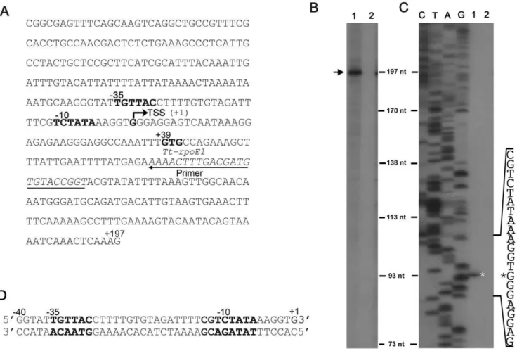
Many ECFsfactors are able to recognize their own promoter and thereby auto-regulate their own gene expression [14,21]. To test if Tt-RpoE1 can activate its own gene transcription, we used a PCR product containing 159-base pair (bp) (+39,+197) ofTt-rpoE1gene and a 218 bp upstream region as the template and analyzed the ability of Tt-RpoE1 to recognize it byin vitrorun-off transcription reconstitution assays (Fig. 2A). Indeed, with theE. colicore RNAP, Tt-RpoE1 could activate theTt-rpoE1promoterin vitro, resulting in a significant transcript (,197nt) (lane 1, Fig. 2B), indicating the
transcription start site is located about 39 bp upstream of the GTG start codon ofTt-rpoE1. In the control experiment when Tt-RpoE1 was omitted, there was no transcription product generated (lane 2,
Figure 1. The interaction betweenT. tengcongensisECFsfactor
Tt-RpoE1 and its putative anti-sfactor Tt-TolB.(A). Organization
Fig. 2B), indicating theT. tengcongenesissigma factor is required to initiate transcription from its own promoter.
To identify the recognition sequence in theTt-rpoE1promoter, the Tt-RpoE1-dependent start site was further confirmed by primer extension analysis with thein vitrotranscripts. As shown in Fig. 2C, the transcription start site (TSS) was located at the ‘‘G’’ exactly 39 bp upstream from the translational start codon. Based on the TSS, we deduced the location of the 235 region (235 TGTTAC 230) and 210 region (211 TCTATA 26), which are spaced apart by 18 bp (Fig. 2A). This putative promoter sequence was subjected to comprehensive interaction and mutagenesis analyses for further characterization of the recogni-tion determinant.
Interaction between Tt-RpoE1 and TheTt-rpoE1 Promoter in Fork-junction Structure
As the promoter sequence ofTt-rpoE1 was deduced, we then investigated how Tt-RpoE1 binds to its own promoter sequence as an auto-regulated ECF s factor. It has been previously demonstrated that free s70
could not bind to promoter DNA due to the inhibition of its subdomains1.1[22,23]. Because ECF sfactors lack this N-terminal subdomains1.1[24], we predicted
Tt-RpoE1 could bind its promoter sequence without core RNAP. To test this prediction, we conducted binding studies with an electrophoretic mobility shift assay (EMSA). First, the double-stranded probe (T+1/B+1) corresponding to240 to+1 bp of the promoter region was used (Fig. 2D), but only weak binding between the double-stranded promoter region and Tt-RpoE1 could be observed when the protein concentration was much high (25mM, data not shown).
Previous studies showed thats54and s70holoenzymes tightly bind to fork-junction promoter DNA [25], because this structure partially mimics the open state of the promoter DNA, which includes a duplex upstream of210 region and a single-stranded
210 region [26]. Therefore, we examined the interaction between Tt-RpoE1 and the fork-junction probe of the promoter DNA, which was obtained by ‘‘cutting back’’ the bottom strand from B+1 to B210 (Fig. 3, lane T+1/B210). The result was in striking contrast to the weak binding of duplex probe, there was a strong preference for Tt-RpoE1 to bind to the fork-junction probe. This finding is likely because the 210 region on the non-template strand became accessible when the template (bottom) strand was cut back. It was not surprising to observe only a weak interaction between Tt-RpoE1 and the non-template single-stranded DNA
Figure 2. Analysis of Tt-RpoE1 recognition of its own promoter sequence byin vitrotranscription and primer extension assays.(A). Sequence of thein vitrorun-off transcription which includes 218 bp upstream and 159 bp ofTt-rpoE1gene. (B).In vitrorun-off transcription of Tt-rpoE1template (A) byE. colicore RNAP with (lane 1) or without (lane 2) Tt-RpoE1. The transcription product is indicated with an arrow. (C). Mapping of the transcriptional start site (TSS) ofTt-rpoE1by primer extension, in which RNA was isolated fromin vitrotranscription reactions with (lane 1) or without (lane 2) Tt-RpoE1. The TSS is marked by an asterisk. Lanes C, T, A, and G are the DNA sequencing ladder corresponding to the primer extension results. The relevant sequence is shown at the side. TheTt-rpoE1promoter region (the deduced210 and235 regions), the TSS and the putative translation start codon (+39) are all indicated in (A). The position of the primer used for primer extension and DNA sequencing is underlined. (D). Sequence of the double-stranded parental probe for the following EMSA assay.
(ssTop, Fig. 3), which implied that the double-stranded 235 element is also important for Tt-RpoE1 binding to promoter DNA. Notably, a much weaker band formed by Tt-RpoE1 and the template strand (ssBot., Fig. 3) was also found at the same position. These results clearly indicated that Tt-RpoE1 could efficiently bind to theTt-rpoE1promoter, consistent with the result
of the run-off transcription assay, and showed thatTt-rpoE1gene would be auto-regulated inT. tengcongensis.
We also detected the interaction between the promoter and the reconstituted holoenzyme (formed by Tt-RpoE1 andE.coli core RNAP). It exhibited little difference in binding efficiency to that of Tt-RpoE1 alone (data not shown), implying that high concentra-tion of Tt-RpoE1 (5mM) could decrease the ‘‘activation effect’’ of core RNAP on the binding affinity [27]. Thus, we omitted the core RNAP in the following EMSA reaction. On the other hand, since
T. tengcongensisgrows at an optimum temperature of approximately 75uC, we also assayed the interaction between Tt-RpoE1 and the fork-junction promoter DNA at different temperatures from 25 to 80uC. Tt-RpoE1 exhibited a similar binding affinity to promoter DNA from 25 to 55uC, indicating that it functioned very well at a wide range of temperatures (data not shown). However, when performed at 60 to 80uC, the fork-junction promoter DNA partially melted (data not shown), which is not favorable for studying the interaction between Tt-RpoE1 and the promoter DNA. Therefore, in order to mimic the Tt-RpoE1/promoter interaction in an open complex of transcription initiationin vivo, we performed the EMSA experiment at 25uC to investigate the interaction between Tt-RpoE1 and the fork-junction promoter DNA in the following experiments.
Effect of Tt-TolB as an Anti-sigma Factor on the Activity of Tt-RpoE1
To investigate if the proposed anti-s factor Tt-TolB would affect the interaction between Tt-RpoE1 and its promoter, we added equimolar concentrations of TolB or its C-terminal Tt-TolB-C (N-terminal of Tt-TolB is not used as it is too short to be purified), respectively, to the EMSA reaction with Tt-RpoE1, using the fork-junction promoter DNA (T+1/B210) as a template. As shown in Fig. 4A, when Tt-TolB was added into the EMSA reaction system, a supershifted complex larger than the RpoE1/promoter complex was formed. However, adding Tt-TolB-C into the EMSA reaction system did not lead to the formation of a larger complex, and neither Tt-TolB nor Tt-TolB-C alone could bind to the fork-junction promoter DNA. The results suggested that the larger complex was formed by a direct interaction between Tt-RpoE1 and Tt-TolB (Fig. 4A), and the interaction was mediated by the N-terminal domain of Tt-TolB, consistent with Y2H result in Fig. 1C. We also tested if Tt-TolB affected the transcription of Tt-RpoE1 in vitro. Equimolar concentration of Tt-TolB to Tt-RpoE1 was added into the transcription system. In lane 1 of Fig. 4B, Tt-TolB was added into reaction at the same time with Tt-RpoE1, and in lane 2, Tt-TolB was added with NTP together, after Tt-RpoE1, promoter DNA and RNAP were incubated together for short time (see materials and methods). The transcription products decreased in both of them, which indicated that Tt-TolB could inhibit the transcription of Tt-RpoE1 by interaction with it. The product in lane 1 was less than that in lane 2, which might be due to Tt-TolB competing for Tt-RpoE1 with RNAP. When Tt-TolB was added at the same time with RNAP and Tt-RpoE1, it decreased the RNAP binding to Tt-RpoE1 more than Tt-TolB added into system later. Together with Y2H results, these results confirmed that Tt-TolB was the anti-sfactor of Tt-RpoE1 and that it interacted with the ECFsfactor Tt-RpoE1 via direct interaction.
Determination of the235 and210 Regions in the Tt-RpoE1-recognized Promoter
To experimentally determine the sequences of the promoter recognized by RpoE1, we analyzed the interaction between
Tt-Figure 3. The interaction between Tt-RpoE1 and different promoter DNA structures. The structure of parental probe is provided at the top. EMSA results of 5mM Tt-RpoE1 protein binding with single-stranded (ssTop, ssBot.) or fork-junction structure promoter DNA(T+1/B210); the vertical line indicates the terminal base-pair on the strands used in fork-junction probe. Free probe (T+1/B210) was loaded as a negative control. The arrows indicate complexes formed by Tt-RpoE1 and the different probes.
RpoE1 and different fork-junction promoter probes with trunca-tions in the putative 210 or 235 regions. For the 235 region using T+1/B210 as the parental probe, we cut back the double strands from240 to different positions (marked by vertical lines, Fig. 5A; for the sequences, see Table S1). Notably, the 5 bp truncation from240 to236 (D-5) had little effect on the binding strength between the promoter and Tt-RpoE1, but further truncation from235 to232 (D-9) resulted in a strong decrease in binding affinity. Tt-RpoE1 binding was almost abolished when the 235 region was removed (D-12) (Fig. 5A). This result suggested that the removal of 4 bp from 235 to 232 of the
235 region eliminated determinants of recognition. Therefore, the 4-bp sequence was very important for promoter recognition of Tt-RpoE1, and likely the 210 region could not be recognized and bound by Tt-RpoE1 without the235 region.
For the210 region using T+1/B+1 as the parental probe, the top strand (T+1) was left intact and the bottom strand was cut back from+1 to different positions (Fig. 5B). As shown in Fig. 5B, the binding strength became stronger with more non-template sequences of the 210 element exposed. The binding was the strongest with the nucleotide 211T in the non-template strand exposed (lane T+1/B212), but at position B213, the binding affinity decreased significantly (lane T+1/B213, Fig. 5B). These results indicated that the212 position remaining base-paired was required for Tt-RpoE1 binding, which might be the similar situation of the interaction between s54 and its promoter [25]. These data also confirmed our initial prediction of the235 and
210 regions.
Identification of the Specific Recognition Determinants in the Tt-RpoE1-recognized Promoter
Although the truncation results of the235 and 210 regions gave clues to the location of promoter recognition by Tt-RpoE1,
the contribution of each nucleotide to recognition was still unresolved. To precisely define the conserved nucleotides needed for recognition by Tt-RpoE1, we performed scanning mutagenesis by nucleotide substitutions between G-C and A-T in the235 and
210 regions.
For the235 region using D25/210 as the parental probe, the nucleotides were substituted in top and bottom strands simulta-neously. The EMSA results were shown in Fig. 6A. Among the seven nucleotide substitutions (from235 to229), substitution of nucleotide 230C to T abolished Tt-RpoE1 binding, and substitutions at positions 234, 233 and 231 also significantly decreased the binding, whereas substitution of 229C to T had little effect on the binding affinity. Not surprisingly, double, triple and quadruple substitutions severely affected the binding affinity, likely due to cumulative effects. The result of four substitutions (from235 to232) was consistent with the truncation result of D-9. Interestingly, the double substitutions232TA231 to GG led to a more significant decrease than the other two double mutations or even the 4-bp substitutions from235 to 232, which suggest that 232TA231 dinucleotide together play a key role in the interaction between Tt-RpoE1 and promoter DNA. Both the results of substitutions at230C and232TA231 were consistent with abolition of binding in the 4-bp substitutions from 232 to
229. Thus, we concluded that the235 element recognized by Tt-RpoE1 contained the following sequence: tGTTAC (with impor-tant nucleotides capitalized).
For the210 region using the strongest binding structure (D25/
212) as the parental probe, scanning mutations were made from
211 to26 on the top strand, while the213 (C/G) and212(G/ C) base pairs were substituted in both top and bottom strands. The EMSA results were shown in Fig. 6B. Single substitutions at212G to T,211T to C, and 210C to T almost abolished Tt-RpoE1 binding, whereas substitutions at position213 and each nucleo-tide in 29TATA26 had little effect on the binding affinity. In
Figure 4. The effect of putative anti-sfactor Tt-TolB on the activity of Tt-RpoE1.(A). The effect of TolB on the interaction between
Tt-RpoE1 and fork-junction structure promoter DNA (T+1/B210). The indicated proteins were added (+) in an EMSA reaction at a concentration of 5mM. The solid arrow indicates the supershifted complex formed by Tt-RpoE1, Tt-TolB, and the promoter, and the open arrow indicates the complex formed by Tt-RpoE1 and the promoter. (B). The effect of Tt-TolB onin vitrotranscription of Tt-RpoE1. Lane 1. Tt-TolB was added into the transcription system at the same time with Tt-RpoE1. Lane 2. Tt-TolB was added into the transcription system after Tt-RpoE1,E.colicore RNAP and promoter DNA being incubated (see materials and methods). Lane C, thein vitrotranscription system without Tt-TolB. The solid arrow indicates the products of transcription.
addition, the double substitution of213CG212 to TT and triple substitution of 213C212G210C to TTT also significantly decreased the binding, which further suggested that 212 GTC
210 was the determinant of Tt-RpoE1 recognition. In contrast, double substitution of29TA28 to CC and quadruple substitution of 29TATA26 to CCCC had little effect on binding affinity, indicating that the TATA region (from29 to26) downstream of GTC may not contribute to the recognition. Based on these scanning mutagenesis results, we propose that the core sequence recognized by Tt-RpoE1 at the210 region was determined to be
212GTC210.
We also carried outin vitro transcription assays to test if those important nucleotides determined by EMSA would affect the transcription activity of Tt-RpoE1. Since the structure of the complex ofE. colisE
4and its235 element has been determined
[28], which provided some clues for our results in235 element determinant, here, we only took a subset of the mutations at the
210 region of the promoter DNA as templates to detect their effect on transcription activity of Tt-RpoE1. For those promoters substituted from213 to29, it was clear that the substitutions at
212GTC210 decreased transcription significantly (Fig. 6C), which confirmed the EMSA results that the212GTC210 was indeed the recognition determinants at the210 region. For the substitution at 29T, it led to a slight increase of transcription, which was consistent to the EMSA results (Fig. 6B). Interestingly, substitution at213C also decreased the transcription, which was
Figure 5. The effects of truncation in the235 and210 regions on the interaction between Tt-RpoE1 and promoter DNA.(A). EMSA results of truncation in the235 region. The structure of parental probe is provided at the top, and vertical lines indicate truncated positions in the double-stranded region. Both the top and bottom strands were truncated from240 to different positions indicated at the left of the fork-junction probe. The 39terminus of top strand was kept at+1, and the 59terminus of the bottom strand was kept at210. (B). EMSA results of truncations in the210 region. The structure of parental probe is provided at the top. The T+1 was the top strand for all the probes, and the bottom strand was truncated from B+1 to different positions as indicated on the right of the bottom strand. The dots denote the terminal bases in the bottom strands in fork-junction probes. The protein concentration was 5mM in all of the following experiments.
different from the EMSA results, where binding was similar to the wild type (wt) promoter. The observed data suggested substitution at213C may affect the interaction between RNAP and promoter DNA.
Based on the results of EMSA and transcription analysis, the determinant sequence (235 and210 regions) for the promoter recognized by Tt-RpoE1 could be identified as 59
tGTTACN16CGTC 39.
Identification of Residues in Tt-RpoE1 Potentially Involved in Recognition of the210 Region
The studies above showed that Tt-RpoE1 recognized a specific
210 element (213CGTC210) which is distinct from that recognized bys70(TATAAT) [29]. To assess the importance of particular amino acid residues for Tt-RpoE1-specific promoter recognition in the210 region, we employed alanine substitution mutagenesis to the Region 2.4 of Tt-RpoE1. For s70 family, Region 2 has been implicated in recognition of 210 regions [4,30]. Selection of amino acid residues for substitution was based on sequence alignments among group IV ECFsfactors (Fig. 7A). We substituted three residues (D66, Y67, R69) in the conserved motif ‘‘DXXR’’ based on the sequence alignment shown in Fig. 7A. Of the three alanine substitution mutations, only D66A strongly decreased the binding affinity to the wt promoter (Fig. 7B); while the binding affinity of Y67 and R69A remained (data not shown). We then tested whether this alanine mutation could suppress the promoter defects caused by base changes at
212GTC210 in the promoter, as it has been shown that213C to T had no effect on the Tt-RpoE1-promoter interaction (Fig. 6B). Interestingly, D66A could cure the defect caused by change at
212G position, but not at the211T and210C (Fig. 7B). Thus, D66 might contribute to the recognition of the 212G of the promoter.
Discussion
In this work, we addressed the function of Tt-RpoE1, one of seven ECFsfactors annotated in the genome ofT. tengcongensis. Y2H and EMSA results showed that Tt-TolB, the cognate downstream gene product of RpoE1, interacted with Tt-RpoE1 via its N-terminal domain. Tt-TolB also inhibited the transcription of RpoE1. These results demonstrated that Tt-TolB (TTE0322) was the anti-sigma factor of Tt-RpoE1. While TTE0322 was originally annotated as Tt-TolB for containing a conserved domain of TolB, a periplasmic component of the Tol biopolymer transport system [31], we now update the function of Tt-TolB to be an anti-sigma factor of Tt-RpoE1. Combined with these findings and that Tt-RpoE1 recognized its own promoter and initiated transcription, we confirmed Tt-RpoE1 functionally as an ECFsfactor.
Being an auto-regulated ECFs factor, the ECFs factor Tt-RpoE1 was first subjected to investigate the interaction with its promoter. Different froms70, ECFsfactor could bind to double-strand promoter DNA, but it preferred to bind fork-junction structure promoter (Fig. 3). With such structure, we identified the
specific promoter sequence recognized by Tt-RpoE1 with scanning mutations, which was further confirmed by in vitro
transcription assays. The determinant sequence in the Tt-RpoE1-recognized promoter was identified as 59tGTTACN16CGTC39,
which was similar some of the predictions by Staron and coworkers for promoters recognized by RpoE-like (ECF02) s
factors [19].
For the235 region of the Tt-RpoE1 promoter, we found that
234G,233T,230C and 232TA231 were functionally impor-tant for recognition by the ECF s factor Tt-RpoE1 (Fig. 6A). Substitutions at those positions significantly decreased the binding affinity of Tt-RpoE1. This finding was supported by the structural analysis of the complex ofE. colisE4and its235 element. In that
complex, specific protein-DNA base interactions occurred only at three positions of its 7 bp235 element GGAACTT (underlined):
235G,234G, and231C, which were specifically recognized by residues R176, S172 and R171 ofE. colisE, respectively [28]. We proposed that234G and 230 C of235 element (tGTTAC) of Tt-RpoE1 promoter played the same roles as235G,234G, and
231C in theE. colisEpromoter, serving as the key nucleotides to form strong hydrogen bonds or van der Waals interactions with Tt-RpoE1. For the233T, 232TA231, they may be similar to the ‘‘AA’’ motif in the235 element inE.coli sEpromoter [28], which plays an essential structure role in the sE4/promoter
interaction. Thus substitution at any one of those nucleotides would disrupt the structure, and affected the Tt-RpoE1/promoter interaction.
For the 210 region, scanning mutagenesis of Tt-RpoE1-recognized promoter indicated that the four nucleotides CGTC (from213 to210) are functionally important. Mutations at these bases resulted in loss of Tt-RpoE1 binding affinity and decreasing the transcription activity. However, the ‘‘TATA’’ box downstream of the CGTC motif did not seem to contribute to the interaction between Tt-RpoE1 and its promoter. Taking one of the substitutions 29T to G as example, we have not detected any effect in the EMSA and in vitro transcription assay (Fig. 6B, C), indicating that the ‘‘TATA’’ box does not contribute to the recognition of Tt-RpoE1 promoter by Tt-RpoE1. This kind of
210 motif has been found in the 210 regions recognized by several other ECFsfactors, such as PvdS ofP. aeruginosa, CarQ of
Myxococcus xanthus,sCofMycobacterium tuberculosis[32],sx,swand
sMinB. subtilis[1,15]. Thus, we proposed that the ‘‘CGTC’’ in the 210 region is a common feature of many promoters recognized by ECF s factors, especially for those RpoE-like (ECF02)sfactors [19].
We have also identified residues in Tt-RpoE1 contributing to base-specific interactions in the promoter by site-directed muta-genesis employing the same strategy as Koo and his colleagues in their studies [33]. Specifically, loss of the residue interacting with a particular base may suppress the deleterious effects of promoter mutants only at the interacting position(s). Interestingly, mutations at the D66 residue of the conserved motif ‘‘DXXR’’ had strong effect on the Tt-RpoE1/promoter interaction (Fig. 7B). D66A decreased the binding affinity and rescued the defect caused by substitution at212G. In another ECF s factor, PvdS from P. aeruginosa, the results also suggested that the ‘‘D’’ residue participated in discriminating210 region contacts [32]. EMSA results showed that 212 position (213CGTC210, underlined) kept in base-pair formation was required for recognition. While it is not clear howsfactor recognizes the sequence-specific duplex
210 element [30], here we identified for the first time that the residue ‘‘D’’ of DXXR motif in ECF s factor recognizes the duplex210 element (212G/C).
Figure 6. The effect of scanning substitutions in the235 and 210 regions on the activity of Tt-RpoE1.(A & B). EMSA results of substitutions in the235 (A) and210 (B) regions. The D25/210 or D25/212 fork-junction structure of promoter DNA was used as the parental probe respectively. The substitutions were made both on the top and opposite positions of the bottom strands in the duplex part as indicated. (C). The effect of a subset of substitutions in the210 region onin vitrotranscription of Tt-RpoE1.
Notably, we demonstrated that the GC-rich motif in the210 region recognized by ECFsfactors is significantly different from the consensus sequence (TATAAT) recognized by group I factor
s70
. This is consistent with their different functions in bacteria. The group Isfactors contain conserved melting residues (F427, Y430, W433 and W434) [5], which makess70tolerate a great deal of promoter sequence diversity when directing the transcription of thousands of housekeeping genes. Whereas only one melting residue corresponding to ‘‘W’’ (Fig. 7A, marked by ‘‘*’’) exists in ECF s factors. Most recent studies suggest that weak melting capacity of ECFsfactors is consistent with their function acting as local regulators, which are confined to direct the transcription of a more restricted set of promoters in adverse environments [11,34]. Thus, there is a balance between melting capacity of as factor and its promoter specificity. Here, we suggest that recognition of the specific ‘‘CGTC’’ motif in 210 region of Tt-RpoE1-recognized promoter is an important strategy employed by ECF
s factor to strengthen the stringency of its promoter, which enables ECF s factors respond to environmental stresses in a focused way by regulating a tightly defined regulon [34]. On the other hand, Koo and his colleagues also found that a GC-rich extended210 motif played important roles in the recognition of group III s factors s28
of E.coli, they proposed that GC-rich promoters may avoid their transcription by the housekeepingss [33]. It should be the same case for the Tt-RpoE1-recognized promoter. Similar to ECF sigma factor, this GC-rich motif was also recognized by a ‘‘DXXR’’ motif ofs28
[33]. Thus, it might be proposed that a distinct210 element and a ‘‘DXXR’’ motif are the general strategy used by alternative s factor-dependent regulons to function in the bacterial world, although more structural details for these interactions remain to be investigated in the future.
Figure 7. The effects of single amino acid substitution in Region 2.4 of Tt-RpoE1 on the interaction between Tt-RpoE1 and promoter DNA.(A). Alignment of the amino acid sequence of Regions 2.4 in group IV ECFsfactors. The numbers at each end of the sequence indicate the amino acid position. The substitutions in Tt-RpoE1 used in this study are shown. The asterisk indicates the conserved amino acids of the melting residues ofs70. Species abbreviations and GenBank accession numbers of their proteins are as follows:Thermoanaerobacter tengcongensis (T.tc), Tt-RpoE1 (NP_622011.1);Bacillus subtilis(B.sub), SigX (NP_390191.2), SigW (NP_388054.1);Pseudomonas aeruginosa(P.aer), PvdS (NP_251116.1); Myxococcus Xanthus(M.xan), CarQ (YP_632266.1). The recognition between D66 and212G is indicated by arrow. (B). The effects of single amino acid substitution in Region 2.4 of Tt-RpoE1 on the interaction between Tt-RpoE1 and wild-type (D25/212) or mutated promoter DNA. Tt-RpoE1 D66A uniquely suppressed single nucleotide changes at position212G.
doi:10.1371/journal.pone.0040885.g007
Table 1.Plasmids used in this study.
Plasmids Description Sources or references
pET228a Kanr, expression vector with His-tag coding sequence Novagen
pET223b Ampr, expression vector with His-tag coding sequence Novagen
P28Tt-RpoE1 pET228a derivative for expression of the Tt-RpoE1 this work
P28Tt-TolB pET228a derivative for expression of the Tt-TolB this work
P28Tt-TolB-C pET228a derivative for expression of the C-terminal domain of Tt-TolB this work
pGBKT7(BD) Yeast two-hybrid DNA-binding domain vector clontech
pGADT7(AD) Yeast two-hybrid activation domain vector clontech
BD-Tt-RpoE1 pGBKT7 derivative for expression of the Tt-RpoE1 this work
AD-Tt-TolB pGADT7 derivative for expression of the Tt-TolB this work
AD-Tt-TolBN pGADT7 derivative for expression of the N-terminal domain of Tt-TolB this work
AD-Tt-TolBC pGADT7 derivative for expression of the C-terminal domain of Tt-TolB this work
AD- Permase pGADT7 derivative for expression of the Permase this work
AD- PtsB pGADT7 derivative for expression of the PtsB this work
Materials and Methods
Bacteria, Plasmids and Oligonucleotides
T. tengcongensis MB4T was routinely grown in modified MB
medium at 75uC without shaking [18].E. coliDH5awas used as a host for the cloning experiments, and E. coli BL21DE3 (lysS) (Novagen, UK) for overproduction of the recombinant proteins. Both E. coli strains were grown in LB medium containing the appropriate antibiotic, ampicillin (Amp, 100mg/ml) or kanamycin
sulfate (km, 50mg/ml) if necessary. The plasmids and partial
oligonucleotides used in this study were described in Tables 1 and 2, respectively.
DNA Manipulations
TheTt-rpoE1(TTE0323) andTt-tolB(TTE0322) coding regions were amplified by PCR from genomic DNA with primers P1/P3 and P9/P10 (Table 2), respectively. Similarly, the DNA fragment encoding the carboxy-terminal portion (residues 62–645) of
Tt-TolB protein was obtained by PCR amplification with primers P8/P10 (Table 2). The PCR products were digested and inserted into the corresponding sites of pET28a (Novagen, UK) to generate the expression plasmids RpoE1, TolB and p28Tt-TolB-C respectively. The plasmid p28Tt-RpoE1 was used as a template for the following mutagenesis. Derivatives of Tt-RpoE1 mutated at different residues were amplified with the primers listed in Table 2. The PCR-amplified sequences were verified by DNA sequencing for all of these constructs.
Yeast Two-hybrid – Assay
Yeast two-hybrid analysis was carried out using the Matchmak-er system 3 (Clontech, Palo Alto, CA, USA) according to the manufacturer’s protocol. Genes encoding Tt-RpoE1 (TTE0323) and the other three proteins (TTE0320-0322) including Tt-tolB
were amplified by PCR from T. tengcongensisgenomic DNA (for primer sequences, see Table 2). The PCR products were digested with appropriate restriction enzymes and cloned into both
Table 2.Partial oligonucleotides used in this study.
Names Sequences(59to 39)* Purposes
P1 GCGAATTCAGCTTTATTGAATTTTATGAG P1/P2:BD-Tt-RpoE1
P2 TCGGATCCTCATCCCTCCAAACATTT
P3 TCCTCGAGTCATCCCTCCAAACATTT P1/P3: p28Tt-RpoE1
P4 ATGAATTCCGGCGAGTTTCAGCAAGT P4/P2: pPTt-RopE1-T
P5 AGGAATTCGACGAAAAGAGAATAGAG P5/P6: AD-Tt-TolB
P6 TTGGATCCGTATTAAAACCTGCCCTT
P7 CTCTCGAGTATCTTTTTCCATCTGTT P5/P7: AD-Tt-TolBN
P8 AGGAATTCCAAGATAATTTAATAACA P8/P6: AD-Tt-TolBC
P9 ACGAATTCATGGACGAAAAGAGAATAGA P9/P10: p28Tt-TolB
P10 CTCTCGAGGTATTAAAACCTGCCCTT P8/P10: p28Tt-TolB-C
P11 TGGAATTCAGTACTAGCTCTTTGATTTT P11/P12: AD-Permase
P12 TGGGATCCGGCCTGCCTTAGTTGATG
P13 CGGAATTCGTGGAAATAGAGCTTAAAAA P13/P14: AD-PtsB
P14 CGGGATCCTCACTTTATCATCTCCTTTA P4/P15 for transcription template
P15 CTTTGAGTTTGATTTTAC
P16 ACCGGTACACATCGTCAAAGTTTT For primer extension
P17 CCGAAATACTGTGACAGCCTATTACAGAATGAGGA P17/18: D66A of p28Tt-RpoE1
P18 TCCTCATTCTGTAATAGGCTGTCACAGTATTTCGG
P39 TTTTGTGTAGATTTTTGTCTATAAAGGTGGGAGGAG P39/40:mutation at213C of transcription
template
P40 CTCCTCCCACCTTTATAGACAAAAATCTACACAAAA
P41 TTTGTGTAGATTTTCTTCTATAAAGGTGGGAGGAGT P41/42:mutation at212G of transcription
template
P42 ACTCCTCCCACCTTTATAGAAGAAAATCTACACAAA
P43 TTGTGTAGATTTTCGCCTATAAAGGTGGGAGGAGTC P43/44:mutation at211T of transcription
template
P44 GACTCCTCCCACCTTTATAGGCGAAAATCTACACAA
P45 TGTGTAGATTTTCGTTTATAAAGGTGGGAGGAGTCA P45/46:mutation at210C of transcription
template
P46 TGACTCCTCCCACCTTTATAAACGAAAATCTACACA
P47 GTGTAGATTTTCGTCGATAAAGGTGGGAGGAGTCAA P47/48:mutation at29T of transcription
template
P48 TTGACTCCTCCCACCTTTATCGACGAAAATCTACAC
pGADT7 and pGBKT7 to generate the AD (active domain) and BD (binding domain) fusion plasmids, respectively. Protein– protein interactions were carried out as described previously [35].
Transcription and Primer Extension Assays
Run-off transcriptionin vitroassays were performed as described previously by Huang et al with minor modifications [21]. The template used for transcription was amplified with primers P4/ P15, the mutated templates were derived from it by PCR with primers listed in Table 2, and the mutated templates were equimolar concentration in the in vitro transcription system. Typical reaction mixtures (25ml) contained 1mg template DNA, 2.5 pmol ofE.colicore RNAP (Epicentre, USA), 50 to 60 pmol of ECFsfactor Tt-RpoE1 in transcription buffer (40 mM Tris-HCl [pH 7.5], 10 mM MgCl2, 150 mM KCl, 10 mM DTT, 0.01% TritonX2100) with 0.8 mM ATP, GTP, CTP and 5mCi [a-32
P]-UTP. DNA and RNAP were preincubated at 4uC for 30 min and 37uC for 8 min to allow promoter binding. Nucleotide triphos-phates (NTPs) were then added, and transcription proceeded for another 8 min. The RNA transcripts were extracted with phenol-chloroform and precipitated with ethanol. The pellet was resuspended in 10ml of urea stop solution, heated to 95uC for 3 min, and separated by 7 M urea–6% polyacrylamide gel electrophoresis and autoradiography. For assay to detect the effect of Tt-TolB on the in vitro transcription of RpoE1, TolB was added into the reaction system at the same time with Tt-RpoE1 or after Tt-Tt-RpoE1 and RNAP incubated for 38 min with the same concentration of Tt-RpoE1 (5mM).
For primer extension assays, the RNA samples were obtained from the transcription reaction with or without ECFsfactor Tt-RpoE1 as described above except that [a-32P]-UTP was substituted with UTP. The primer P16 (Table 2) was labeled at the 59-end with [c-32P]-ATP, and was used for both DNA sequencing and primer extension as described previously [36].
Expression and Purification of Recombinant Proteins
To overproduce the His-tagged proteins Tt-RpoE1, Tt-TolB and Tt-TolB-C,E. coliBL21DE3 (lysS) harboring plasmid p28Tt-RpoE1, p28Tt-TolB and p28Tt-TolB-C were cultivated in LB to
an optimal density at 600 nm of 0.6 at 37uC and induced with 0.3 mM isopropyl b-D-1-thiogalactopyranoside (IPTG) at 24uC overnight. Then the proteins were extracted and purified as described previously [35]. All the purified proteins were analyzed by SDS-PAGE and the protein concentrations were determined by using the BCATMprotein concentration assay kit (PIERCE).
Electrophoretic Mobility Shift Assay (EMSA)
Double-stranded or fork-junction probes were obtained by annealing reaction containing equimolar concentration of two oligonucleotides (Table S1) in 50 mM Tris-HCl (pH8.0), 10 mM MgCl2, 50 mM NaCl and 1 mM EDTA, with the top strands
labeled at the 59-end with [c-32P] ATP. The 20ml-standard binding reaction contained: 50 mM Tris-HCl (pH 8.0), 10 mM MgAc2,
30 mM NaCl, 1 mM DTT, 5% glycerol, 30mg/ml BSA, 0.5 mM EDTA, 1mg poly (dI-dC), 20 fmol labeled DNA probe and the indicated amounts of appropriate proteins. After incubation at 25uC for 20 min, samples were immediately loaded on native 5% polyacrylamide gel (mono/bis, 80:1) in 0.56TBE buffer and
electrophoresis at 150 V for 2 h. Gels were dried and exposed to Biomax radiographic film (Kodak) for autoradiography.
Supporting Information
Table S1 Nucleotide sequences for electrophoretic mobility shift assay.
(DOC)
Acknowledgments
We thank Professor Shiladitya DasSarma for critical reading and valuable comments on an earlier version of this manuscript.
Author Contributions
Conceived and designed the experiments: J. Liu HX. Performed the experiments: J. Liu J. Li ZW HP JZ. Analyzed the data: J. Liu HX. Contributed reagents/materials/analysis tools: J. Liu J. Li ZW HX. Wrote the paper: J. Liu HX.
References
1. Helmann JD (2002) The extracytoplasmic function (ECF) sigma factors. Adv Microb Physiol46: 47–110.
2. Malhotra A, Severinova E, Darst SA (1996) Crystal structure of a sigma 70 subunit fragment fromE. coliRNA polymerase.cell87: 127–136.
3. Murakami KS, Darst SA (2003) Bacterial RNA polymerases: the wholo story.
Curr Opin Struct Biol13: 31–39.
4. Wosten MM (1998) Eubacterial sigma-factors.FEMS Microbiol Rev22: 127–150. 5. Lonetto M, Gribskov M, Gross CA (1992) The sigma 70 family: sequence
conservation and evolutionary relationships.J Bacteriol174: 3843–3849. 6. Ades SE (2004) Control of the alternative sigma factor sigmaE inEscherichia coli.
Curr Opin Microbiol7: 157–162.
7. Campbell EA, Tupy JL, Gruber TM, Wang S, Sharp MM, et al. (2003) Crystal structure ofEscherichia colisigmaE with the cytoplasmic domain of its anti-sigma RseA.Mol Cell11: 1067–1078.
8. Schobel S, Zellmeier S, Schumann W, Wiegert T (2004) The Bacillus subtilis
sigmaW anti-sigma factor RsiW is degraded by intramembrane proteolysis through YluC.Mol Microbiol52: 1091–1105.
9. Missiakas D, Mayer MP, Lemaire M, Georgopoulos C, Raina S (1997) Modulation of theEscherichia colisigmaE (RpoE) heat-shock transcription-factor activity by the RseA, RseB and RseC proteins.Mol Microbiol24: 355–371. 10. Rhodius VA, Suh WC, Nonaka G, West J, Gross CA (2006) Conserved and
variable functions of the sigmaE stress response in related genomes.PLoS Biol4: e2.
11. Koo BM, Rhodius VA, Nonaka G, deHaseth PL, Gross CA (2009) Reduced capacity of alternative sigmas to melt promoters ensures stringent promoter recognition.Genes Dev23: 2426–2436.
12. Rhodius VA, Mutalik VK (2010) Predicting strength and function for promoters of theEscherichia colialternative sigma factor, sigmaE.Proc Natl Acad Sci U S A107: 2854–2859.
13. Cao M, Kobel PA, Morshedi MM, Wu MF, Paddon C, et al. (2002) Defining the
Bacillus subtilissigma(W) regulon: a comparative analysis of promoter consensus search, run-off transcription/macroarray analysis (ROMA), and transcriptional profiling approaches.J Mol Biol316: 443–457.
14. Huang X, Fredrick KL, Helmann JD (1998) Promoter recognition byBacillus subtilissigmaW: autoregulation and partial overlap with the sigmaX regulon.
J Bacteriol180:3765–3770.
15. Huang X, Gaballa A, Cao M, Helmann JD (1999) Identification of target promoters for theBacillus subtilisextracytoplasmic function sigma factor, sigma W.Mol Microbiol31: 361–371.
16. Huang X, Helmann JD (1998) Identification of target promoters for theBacillus subtilissigma X factor using a consensus-directed search.J Mol Biol279: 165– 173.
17. Xue Y, Xu Y, Liu Y, Ma Y, Zhou P (2001)Thermoanaerobacter tengcongensissp.nov., a novel anaerobic, saccharolytic, thermophilic bacterium isolated from a hot spring in Tengcong, China.Int J Syst Evol Microbiol51: 1335–1341.
18. Bao Q, Tian Y, Li W, Xu Z, Xuan Z, et al. (2002) A complete sequence of theT. tengcongensisgenome.Genome Res12: 689–700.
19. Staron A, Sofia HJ, Dietrich S, Ulrich LE, Liesegang H, et al. (2009) The third pillar of bacterial signal transduction: classification of the extracytoplasmic function (ECF) sigma factor protein family.Mol Microbiol74: 557–581. 20. Raivio TL, Silhavy TJ (2001) Periplasmic stress and ECF sigma factors.Annu Rev
Microbiol55: 591–624.
21. Huang X, Decatur A, Sorokin A, Helmann JD (1997) TheBacillus subtilis
sigma(X) protein is an extracytoplasmic function sigma factor contributing to survival at high temperature.J Bacteriol179: 2915–2921.
23. Dombroski AJ, Walter WA, Record MT, Jr, Siegele DA, Gross CA (1992) Polypeptides containing highly conserved regions of transcription initiation factor sigma 70 exhibit specificity of binding to promoter DNA.cell70: 501–512. 24. Brooks BE, Buchanan SK (2008) Signaling mechanisms for activation of extracytoplasmic function (ECF) sigma factors.Biochim Biophys Acta1778: 1930– 1945.
25. Guo Y, Gralla JD (1998) Promoter opening via a DNA fork junction binding activity.Proc Natl Acad Sci U S A95: 11655–11660.
26. Roberts CW, Roberts JW (1996) Base-specific recognition of the nontemplate strand of promoter DNA byE. coliRNA polymerase.cell86: 495–501. 27. Zenkin N, Kulbachinskiy A, Yuzenkova Y, Mustaev A, Bass I, et al. (2007)
Region 1.2 of the RNA polymerase sigma subunit controls recognition of the
210 promoter element.EMBO J26: 955–964.
28. Lane WJ, Darst SA (2006) The structural basis for promoter235 element recognition by the group IV sigma factors.PLoS Biol4: e269.
29. Lisser S, Margalit H (1993) Compilation ofE. colimRNA promoter sequences.
Nucleic Acids Res21: 1507–1516.
30. Feklistov A, Darst SA (2011) Structural basis for promoter210 element recognition by the bacterial RNA polymerase sigma subunit.cell147: 1257– 1269.
31. Lazzaroni JC, Dubuisson JF, Vianney A (2002) The Tol proteins ofEscherichia coliand their involvement in the translocation of group A colicins.Biochimie84: 391–397.
32. Wilson MJ, Lamont IL (2006) Mutational analysis of an extracytoplasmic-function sigma factor to investigate its interactions with RNA polymerase and DNA.J Bacteriol188: 1935–1942.
33. Koo BM, Rhodius VA, Campbell EA, Gross CA (2009) Mutational analysis of
Escherichia coli sigma28 and its target promoters reveals recognition of a composite210 region, comprised of an ‘extended210’ motif and a core210 element.Mol Microbiol72: 830–843.
34. Feklistov A, Darst SA (2009) Promoter recognition by bacterial alternative sigma factors: the price of high selectivity?Genes Dev23: 2371–2375.
35. Liu J, Pei H, Mei S, Li J, Zhou L, et al. (2008) Replication initiator DnaA interacts with an anti-terminator NusG inT. tengcongensis.Biochem Biophys Res Commun371: 573–577.