173
Jornal Brasileiro de Pneumologia 31(2) - Mar/Abr de 2005
Langerhans-cell histiocytosis: rapid resolution after
smoking cessation*
JOSÉ MIGUEL CHATKIN, VINICIUS D. SILVA, CARLOS C. FRITSCHER, JUSSARA FITERMAN, CLÁUDIA RECK
* Study carried out at the Hospital São Lucas of the Pontifícia Universidade Católica of Rio Grande do Sul, Porto Alegre, Brazil Correspondence to: Jose Miguel Chatkin - Av.Ipiranga 6690 - 3º andar - CEP 90610-000, Porto Alegre, RS, Brazil. Fax: 55 51 3320-3316. E-mail: [email protected]
Submitted: 20 April 2004. Accepted, after review: 19 August 2004.
We describe a case of pulmonary Langerhans cell histiocytosis with a close temporal relationship between smoking cessation and radiological improvement. High-resolution computed tomography revealed multiple small nodules located in the upper and middle lobes of both lungs. Microscopy of these lesions showed histiocytic infiltration that reacted strongly to staining for S100 protein. The histiocytes resembling Langerhans cells showed strong reactivity for S100 protein. Smoking cessation was recommended and the patient complied. Chest X-ray and computed tomography performed 6 and 24 months later revealed almost complete resolution of the radiographic abnormalities. Despite the possibility that this evolution was attributable to spontaneous remission, in this case, the lesions did, in fact, disappear rapidly after smoking cessation.
J Bras Pneumol 2005; 31 (2): 173-6.
Key words: Histiocytosis. Langerhans-Cell. Tabaco use cessation.
Case Report
INTRODUCTION
Pulmonary Langerhans cell histiocytosis is a disease of unknown etiology, characterized by the presence in the lungs of destructive granulomatous lesions, which, during their active phase, contain a number of Langerhans cells(1).
Cases of sole pulmonary involvement were initially described by Farinacci et al.(2) Since then, this situation
has been reported in a number of series(3-5). Pulmonary
Langerhans cell histiocytosis is usually seen in patients in their 30s or 40s(1,6,7), and it is uncommon to find
isolated pulmonary involvement in children(1,8,9).
The principal recognized risk factor is smoking. Most patients are smokers at the time of diagnosis (3,6,9-12). Only 3 of the 87 patients with pulmonary
Langerhans cell histiocytosis in a study conducted at the Mayo Clinic series were nonsmokers(9). It has
been confirmed through a case control study that smoking is an significant risk factor for development of the disease(13), and that there is a relationship with
the nicotine load(9). There have also been reports of
174
Chatkin, JM, et al.
Langerhans-cell histiocytosis: rapid resolution after smoking cessation
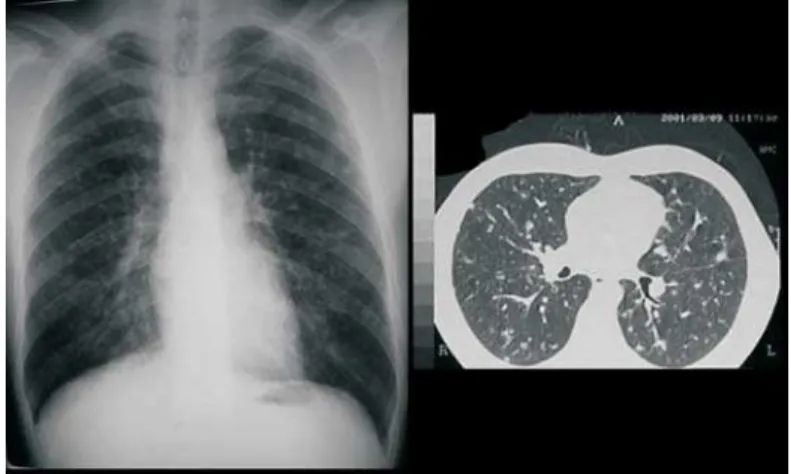
Figure 1. Chest X-ray showing multiple focal nodules distributed throughout both lungs, high-resolution computed tomography confirming the presence of multiple small nodules, without cavitations or cysts
The beneficial effect of smoking cessation on the evolution of the disease has previously been presented in some studies(15-17), although it has not
been formally established in prospective controlled studies. However, the lack of a correlation between smoking and extrapulmonary histiocytosis and the fact that pulmonary Langerhans cell histiocytosis is found in only a small percentage of smokers lead us to believe that there are probably other factors to be considered in the genesis and evolution of the disease(1,7).
The objective of this study was to report a case of pulmonary Langerhans cell histiocytosis with rapid resolution (clinical and radiological i m p r o v e m e n t ) a f t e r s m o k i n g c e s s a t i o n , contributing to the hypothesis that smoking is a risk factor and affects the evolution of this disease.
CASE REPORT
A previously healthy, 41-year-old male sought medical attention at the pulmonology outpatient clinic of the of the Pontifícia Universidade Católica of Rio Grande do Sul (Rio Grande do Sul Pontifical Catholic University) Hospital São Lucas, in the city of Porto Alegre (state of Rio Grande do Sul), for assessment of dry cough and progressive dyspnea persisting for approximately three months, with no other symptoms. He had been a smoker for 20 pack years, having started using tobacco at the age of 20. There was no history of exposure to dust or chemical products. He reported an episode of spontaneous pneumothorax at the age of 31.
Upon physical examination, the patient presented good general condition, and the only abnormality was the presence of occasional cracking rales in the medial thirds of both lungs. He presented no digital clubbing, masses or organomegaly.
Results of laboratory testing, which included standard urinalysis, complete blood count, erythrocyte sedimentation, creatinine, glucose, bilirubin, total proteins and albumin, globulin, calcium, electrolytes, aspartate aminotransferase, alanine aminotransferase and alkaline phosphatase, were all normal. Blood gas analysis while the patient was on room air showed arterial oxygen tension at 97 mmHg and arterial oxygen tension at 38 mmHg. The chest X-ray showed multiple focal nodules in both lungs. High-resolution computed tomography confirmed the presence of these small nodules, the largest of which measured 10 mm in
diameter. The lesions were located predominantly in the upper lobes and, to a lesser extent, throughout the medial pulmonary fields, sparing the bases and the costophrenic angles. There were no cysts or cavitations within these small nodules. No enlarged lymph nodes were detected (Figure 1).
The spirometric parameters were considered normal, including the carbon monoxide diffusion. The bone scintigraphy with Tc99m was also normal.
An open lung biopsy was performed, the macroscopic exam of which revealed multiple nodules interspersed with normal tissue. The histological assessment showed many peribronchial granulomas composed of histiocytes, lymphocytes and eosinophils. The histiocytic infiltration also affected the septal fibrous tissue. The histiocytes presented elongated, irregular and folded nuclei, w i t h d e l i c a t e n u c l e a r m e m b r a n e s a n d inconspicuous nucleoli. The cytoplasm was abundant and eosinophilic (Figure 2). The histiocytes stained positive for S100 protein.
175
Jornal Brasileiro de Pneumologia 31(2) - Mar/Abr de 2005
Figure 2. Material from the pulmonary biopsy, showing a mixed population of folded and elongated histiocytes with inconspicuous nucleoli, pale cytoplasm and barely visible cellular membrane, as well as a larger macrophage in the upper left quadrant (H&E; x500)
Figure 3. Chest X-ray and tomography obtained within 24 months after smoking cessation, showing significant resolution of the lesions
confirmed the favorable evolution and the tendency was maintained up to the last control tomography, performed at 24 months after smoking cessation (Figure 3).
DISCUSSION
Langerhans cell histiocytosis, formerly known as bone eosinophilic granuloma and later as histiocytosis X(18), was classified in 1997 by the
Reclassification Working Group of the Histiocyte Society(19), which categorized the histiocytic disorders
into two groups: one of malignant behavior and another of varied biological behavior. Each category is further subdivided into dendritic or macrophagic,
according to the cellular lineage. Langerhans cell histiocytosis was classified as a nonmalignant dendritic cell-related disorder. Langerhans cells differ from dendritic cells in that the former stain for S100 protein and present a strong expression of DC1a antigen on the cell surface(6).
Vassalo et al.(20) speculated that pulmonary
Langerhans cell histiocytosis represents a polyclonal reactive process induced by antigens present in cigarette smoke. This hypothesis is reinforced by the peribronchial lesional distribution, possibly following antigen inhalation(6). However, since only
a small percentage of smokers develop this condition, there must be other factors involved(7).
Pulmonary Langerhans cell histiocytosis differs from systemic histiocytosis because the latter seems to be the result of a monoclonal proliferation of these cells, similar to neoplasia(5,6).
The case reported here was classified as a definitive diagnosis according to the European Histiocyte Society Writing Group since it stained positive for S100 protein(18).
The high-resolution computed tomography findings most frequently described are pulmonary nodules with cystic alterations distributed between the central and peripheral portions, usually sparing the lung bases. The lesions evolve from firm nodules to cavitations, to cysts with thick walls and finally to cysts with thin walls. Some lesions might regress, but, upon reaching the cystic form, they tend to remain stable or increase in size. This progression is generally parallel to the deterioration of the pulmonary function(21).
No cystic lesions were detected in the X-rays of this patient, probably due to the short time elapsed between the onset of the symptoms and the diagnosis. That might also explain the marked resolution of the lesions following smoking cessation(10).
It is not uncommon for pulmonary function tests to be found normal or near normal in cases of pulmonary Langerhans cell histiocytosis with advanced radiological lesions(7). The most frequent
alteration is reduced diffusing capacity for carbon monoxide, which was reported in 70% to 100% of the published series(5,7). Blood gas analysis is
typically normal, as was observed in this patient. Although smoking probably predisposes to the disease, it is not a predictive factor for progression(9).
176
Chatkin, JM, et al.
Langerhans-cell histiocytosis: rapid resolution after smoking cessation
functional alterations since the establishment of the condition, are indicative of worse prognosis. However, none of these are sufficient to establish a true prognosis for a specific case(7).
Remission may occur spontaneously, even in patients who continue smoking, which makes it difficult to assess the role of smoking cessation in the evolution of the disease, although a few cases of clinical-radiological improvement following smoking cessation have been reported(15-17). In
1999, Mogulkoc conducted a review of the literature and identified only five cases in which improvement was attributable to smoking cessation(15).
In the present case, there was a clear t e m p o r a l r e l a t i o n s h i p b e t w e e n s m o k i n g cessation and clinical-radiological improvement, confirmed by serial assessments. We observed o b j e c t i v e l y q u a n t i f i a b l e r a d i o l o g i c a l improvement within approximately three months after smoking cessation, which leads us to believe that the present case contributes to the hypothesis that smoking cessation promotes the favorable evolution of the disease.
Patients with pulmonary Langerhans cell histiocytosis require long-term follow-up treatment, clinical as well as radiological and functional. Patients must be encouraged to remain smoke-free since, as has been demonstrated, smoking may influence the prognosis of the disease.
REFERENCES
1. S u n d a r K M , G o s s e l i n M V, C h u n g H L , C a h i l l B C . Pulmonary Langerhans cell histiocytosis: emerging concepts in pathobiology, radiology, and clinical evolution of disease. Chest 2003;123:1673-83. 2. Farinacci CJ, Jeffrey HC, Lackey RW. Eosinophilic
granuloma of the lung. US Armed Forces Med J 1951;2:1085-91.
3. Bernstrand C; Cederlund K; Sandstedt B; Ashstrom L; Lundell M; Dahlquist G; Henter JI. Pulmonary abnormalities at long-term follow up of patients with Langerhans' cell histiocytosis. Med Pediatr Oncol 2001;36:459-68. 4. H a r a r i S , C o m e l A . P u l m o n a r y L a n g e r h a n s C e l l
Histiocytosis. Sarcoidosis Vasc Diffuse Lung Dis 2001;18:253-62.
5. Vassallo R, Ryu JH, Schroeder DR, Decker PA, Limper AW. Clinical outcomes of pulmonary Langerhans' cell histiocytosis in adults. N Engl J Med 2002; 346: 484-90. 6. Ryu JH, Colby TV, Hartman T, Vassallo R. Smoking-related intersticial lung diseases: a concise review. Eur Respir J 2001;17:122-32.
7. Tazi A, Soler P, Hance AJ. Adult pulmonary Langerhans' cell histiocytosis. Thorax 2000;55:405-16.
8. Chatkin JM, Bastos JC, Stein RT, Gaiger AM. Sole p u l m o n a r y i n v o l v e m e n t b y L a n g e r h a n s ' c e l l histiocytosis in a child. Eur Respir J 1993;6:1226-8. 9. Horwath DM, Gilchrist GS, Mullan BP, Wiseman GA,
E d m o n s o n J H , S c h o m b e r g P J . L a n g e r h a n s c e l l histiocytosis: diagnosis, natural history, management and outcome. Cancer 1999;85:2278-90.
1 0 . Delobbe A, Durieu J, Duhamel A, Wallaert B, Group d'etude en pathologie interstitiale de la Societe de Pathologie Thoracique du Nord. Determinants of survival in pulmonary Langerhans' cell granulomatosis (histiocytosis X). Eur Respir J 1996;9:2002-6. 11. Ha SY, Helms P, Fletcher M, Broadbent V, Pritchard J. Lung
involvement of Langerhans cell histiocytosis: prevalence, clinical features and outcome. Pediatrics 1992;89:466-9. 1 2 . Vassallo R, Ryu JH, Colby TV, Hartman T, Limper AW.
Pulmonary Langerhans's cell histiocytosis. N Engl J Med 2000;342:1969-78.
1 3 . Hance AJ, Basset F, Saumon G. Smokingand interstitial lung disease: the effect of cigarette smoking on the incidence of pulmonary histiocytosis X and sarcoidosis. Ann NY Acad Sci 1986;465:643-56.
14. Etienne B, Bertocchi M, Gamondes JP. Relapsing pulmonary Langerhans cell histiocytosis after lung transplantation. Am J Respir Crit Care Med 1998;157:288-91.
1 5 . Mogulkoc N, Veral A, Bishop PW, Bayindir U, Pickering AC, Egan JJ. Pulmonar Langerhans's cell histiocytosis: radiologic resolution following smoking cessation. Chest 1999;115:1452-5.
1 6 . Morimoto T, Matsumara T, Kitaichi M. Rapid remission of pulmonary eosinophilic granuloma in a young male patient after cessation of smoking. Nikhon Kokyuki Gakkai Zasshi 1999;37:140-5.
17. von Essen S, West W, Sitorius M, Rennard SI. Complete resolution of roentgenographic changes in a patient with pulmonary histiocytosis X. Chest 1990;98:765-67. 1 8 . Chu T, D'Angio GJ, Favara BE. Histiocytosis syndromes
in children. Lancet 1987;i:208-09.
1 9 . Favara BE, Feller AC, with members of the WHO Committee on Histiocytic /Reticulum Cell Profileration. Contempory classification of histiocytic disorders. Med Pediatr Oncol 1997;29:157-66.