Objective: To report a case of late‑onset self‑healing Langerhans cell histiocytosis.
Case description: A 4½‑month‑old female patient presenting with an eythematopurpuric eruption underwent a skin biopsy for histopathology and was irst diagnosed with isolated cutaneous Langerhans cell histiocytosis. Her lesions regressed within a few months and she was retrospectively diagnosed with late‑onset self‑healing Langerhans cell histiocytosis after being without skin or systemic involvement in a follow‑up four years later.
Comments: Self‑healing Langerhans cell histiocytosis, which is characterized by clonal proliferation of Langerhans cells and presents with cutaneous lesions, is a rare self‑limited variant of histiocytosis and can only be diagnosed retrospectively, after the patient remains free from systemic involvement for several years. Although it presents at birth or during the neonatal period, only a few cases of its late‑onset type regarding the age of onset have been reported. Purpuric lesions that appear after the neonatal period serve as a clue for late‑onset self‑healing Langerhans cell histiocytosis and the patients should be monitored regularly for systemic involvement if the diagnosis is confirmed by a cutaneous biopsy.
Keywords: Infant; Histiocytosis, Langerhans‑cell; Self‑healing.
Objetivo: Relatar um caso de histiocitose de células de Langerhans autolimitada e de início tardio.
Descrição do caso: Paciente com 4 meses e meio de idade do sexo feminino, apresentando uma erupção cutânea eritematosa purpúrea, foi submetida a uma biópsia de pele, sendo diagnosticada com histiocitose de células de Langerhans cutânea isolada. As lesões regrediram em poucos meses e ela foi diagnosticada, retrospectivamente, com histiocitose de células de Langerhans autolimitada e de início tardio, após não apresentar nenhum envolvimento cutâneo ou sistêmico durante um seguimento de quatro anos.
Comentários: A histiocitose de células de Langerhans autolimitada caracteriza‑se pela proliferação clonal das células de Langerhans e apresenta-se com lesões cutâneas, sendo uma variante autolimitada rara de histiocitose. A doença só pode ser diagnosticada de forma retrospectiva, após o paciente não apresentar nenhum envolvimento sistêmico durante vários anos. Embora existam casos de manifestações ao nascimento ou durante o período neonatal, apenas alguns casos de histiocitose de células de Langerhans de idade tardia foram relatados. Lesões purpúreas que aparecem após o período neonatal podem sugerir histiocitose de células de Langerhans autolimitada e de início tardio. Uma vez conirmado o diagnóstico por biópsia cutânea, tais pacientes devem ser acompanhados regularmente, pois pode haver comprometimento sistêmico.
Palavras-chave: Criança; Histiocitose de células de Langerhans; Cura espontânea.
ABSTRACT
RESUMO
*Corresponding author. E‑mail: [email protected] (F.S. Afsar). aAtaturk Research and Training Hospital, Izmir, Turkey.
bDr. Behcet Uz Children’s Hospital, Izmir, Turkey.
cKatip Celebi University, School of Medicine, Izmir, Turkey.
Received on May 27, 2016; accepted on October 2, 2016; available online on March 08, 2017.
LATE‑ONSET SELF‑HEALING LANGERHANS
CELL HISTIOCYTOSIS: REPORT
OF A VERY RARE ENTITY
Histiocitose de células de Langerhans autolimitada
e de início tardio: relato de uma entidade raríssima
Fatma Sule Afsar
a,*, Malik Ergin
b, Gulcihan Ozek
b,
INTRODUCTİON
Langerhans cell histiocytosis (LCH) is a generic term that identiies several clinical cases characterized by the prolifera‑ tion of distinctive cells that are S100 and CD1a positive and contain Birbeck granules in their cytoplasm.1,2 “Self‑healing”
Langerhans cell histiocytosis (SHLCH) is a rare, self‑limited variant of LCH that presents cutaneous lesions at birth or in the neonatal period with the absence of systemic manifes‑ tations and spontaneous resolution.3 Here we report a late‑
‑onset type of SHLCH, which was irst diagnosed as isolated cutaneous LCH.
CASE DESCRİPTİON
A 4½‑month‑old female patient presented to the pediatric der‑ matology clinic with an erythematopurpuric eruption on her torso. he parents reported that the the lesions had been pre‑ sent since she was 3‑months‑old. he patient was born at term after an uncomplicated pregnancy and was otherwise healthy with normal development for her age. Her physical examina‑ tion was within normal limits and dermatologic examination revealed tiny erythematopurpuric papules, some of which were crusted, scattered over the torso (Fig. 1).
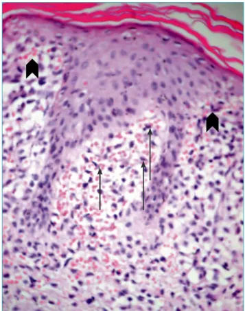
An incisional biopsy was taken from one of the papules on the torso with clinical diferential diagnoses of LCH and congenital leukemia cutis. he skin biopsy revealed a dense iniltrate of neoplastic cells in papillary dermis with sparse epidermal iniltration (Fig. 2). In an immunhistochemical
Figure 1 Erythematopurpuric and crusted papules on the torso
analysis, the neoplastic cells were positive for S100 and CD1a and negative for mast cell tryptase, CD117, and myelope‑ roxidase (Fig. 3).
Figure 3 (A) Focal immunohistochemical staining with
CD1a in Langerhans cells (arrows) (immunoperoxidase;
original magnification, X400). (B) Diffuse and dense positive staining with S100 protein in the
Langerhans cells (arrows) (immunoperoxidase; original magniication, X400).
A B
Figure 2 Dense iniltrate of neoplastic cells in papillary
In a laboratory investigation, hemoglobin was 10.5 g/dL, total leukocyte count was 6,300/mm3, and platelet count was
285,000/mm3. Liver enzymes, renal function tests, serum
chemistry, and urinanalysis were all within normal limits. Chest X‑ray and skeletal radiographs did not show abnorma‑ lities. Ultrasonography of the abdomen and cranial magnetic resonance imaging were normal.
he patient was irst diagnosed with isolated cutaneous LCH and no treatment was given to her. Within a few mon‑ ths, her lesions showed signs of regression. he patient is still being followed‑up with and she is doing well. She has been without skin or systemic symptoms for four years, and now retrospectively evaluated as late‑onset SHLCH.
DİSCUSSİON
LCH represents a group of rare histiocytic disorders that are characterized by tissue iniltration with dendritic cells typically seen in infants and children. hree to four cases per million occur annually in children under 15 years of age with a peak incidence in infants aged one to two years old.4,5 he classi‑
ication of histiocytic disorders are proposed by the World Health Organization as Class I (Langerhans cell histiocytosis), Class II (Histiocytosis of mononuclear phagocytes other than Langerhans cells, Familial and reactive hemophagocytic lym‑ phohistiocytosis, Sinus histiocytosis with massive lymphade‑ nopathy, Rosai‑Dorfman disease, Juvenile xhantogranuloma, reticulohistiocytoma) and Class III (Malignant histiocytic disor‑ ders, Acute monocytic leukemia, malignant histiocytosis, True histiocytic lymphoma).6
he etiology of LCH is unknown, but neoplasia, immu‑ nostimulation, and dendritic cell disorders have been implica‑ ted in its pathogenesis.7 A common progenitor dendritic cell
is hypothesized to give rise to Langerhans cells (LC) residing in the epidermis with dermal dendritic cells in the dermal and hypodermal areas.8
Congenital self‑healing Langerhans cell histiocyto‑ sis (CSHLCH), also known as Hashimato Pritzker disease, is a rare, benign variant of histiocytosis. It is characterized by disseminated papules, vesicles, or nodules, occasionally with scaling, sometimes urticarial or hemangioma like. Afected infants are otherwise healthy and skin lesions tend to invo‑ lute spontaneously within weeks to months.9 he diagnosis of
LCH is based on histopathology, which is indistinguishable for all forms of LCH revealing a proliferation of CD1a and S100 protein‑positive cells.10
After LCH is diagnosed, a thorough evaluation should be performed to rule out systemic involvement. he most com‑ mon organs involved are the skin, liver, lymph nodes, bone
marrow, spleen, and the skeletal system. A physical examination for lymphadenopathy, and an abdominal ultrasound for hepa‑ tosplenomegaly should be performed. A skeletal survey would reveal lesions within the skull or large bones. Urine osmalility should be checked to screen for diabetes insipidus.10
Neoplastic disorders to consider in a newborn with papu‑ lovesicles are congenital leukemia, LCH, and neuroblastoma.11
Characteristic histopathology and absence of other system invol‑ vement permit diferentiation of benign forms of LCH. Because of the potential for recurrence in the skin or systematically, it has been suggested that the diagnosis of CSHLCH be made retrospectively, after a patient has remained free from systemic involvement for several years.12 About 100 cases of CSHLCH
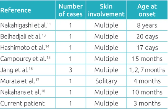
have been reported in the literature and we inally evaluated our case as CSHLCH after a four year follow‑up. Regarding the age of onset, self‑healing LCH is divided into the common type, which presents at birth or during the neonatal period, namely CSHLCH, and a late onset type, which presents after the neonatal period as observed in our case. Only a few cases of late onset SHLCH have been reported (Table 1).11,13‑18
CSHLCH generally carries a good prognosis. Its true inci‑ dence may be underestimated since spontaneous resolution often occurs before assessment by a dermatologist.19 CSHLCH
patients with multisystem involvement may also show sponta‑ neous regression.20 he self‑regressing character of CSHLCH
has been explained by the tumor cells of CSHLCH which eventually become apoptotic upon terminal maturation during the natural course of LC activation.19 However, there are no
deinitive clinical or histopathologic indings that reliably pre‑ dict the long‑term behavior of skin‑only LCH in neonates; therefore, it is recommended that all patients be monitored at regular intervals throughout childhood with noninvasive monitoring.10,21 In neonates and young infants, cutaneous
involvement is also the most common presentation of non‑self
Reference Number of cases
Skin involvement
Age at onset
Nakahigashi et al.11 1 Multiple 8 years
Belhadjali et al.13 1 Multiple 20 days
Hashimoto et al.14 1 Multiple 17 days
Campourcy et al.15 1 Multiple 15 months
Jang et al.16 3 Multiple 1, 2, 7 months
Murata et al.17 1 Solitary 4 months
Nakahara et al.18 1 Multiple 10 months
Current patient 1 Multiple 3 months
REFERENCES
1. Larralde M, Rositto A, Giardelli M, Gatti CF, Santos‑Muñoz A. Congenital self‑healing Langerhans cell histiocytosis: the need for a long term follow‑up. Int J Dermatol. 2003;42:245‑6.
2. Walia M, Paul P, Mishra S, Mehta R. Congenital Langerhans cell histiocytosis: the self‑healing variety. J Pediatr Hematol Oncol. 2004;26:398‑402.
3. Orle J, Mósca AM, Sousa MA, Lima CM, Adriano AR, Rezende PM. Congenital self healing reticulohistiocytosis in a newborn (Hashimoto‑Pritzker). An Bras Dermatol. 2011;86:785‑8.
4. Broekaert SM, Metzler G, Burgdorf W, Röcken M, Schaller M. Multisystem Langerhans cell histiocytosis: successful treatment with thalidomide. Am J Clin Dermatol. 2007;8:311‑4.
5. Shahidi‑Dadras M, Saeedi M, Shakoei S, Ayatollahi A. Langerhans cell histiocytosis: an uncommon presentation, successfully treated by thalidomide. Indian J Dermatol Venereol Leprol. 2011;77:587‑90.
6. Harris NL, Jafe ES, Diebold J, Flandrin G, Muller-Hermelink HK, Vardiman J, et al. World Health Organization classiication of neoplastic diseases of the hematopoietic and lymphoid tissues: report of the Clinical Advisory Committee meeting‑Airlie House, Virginia, November 1997. J Clin Oncol. 1999;17:3835‑49.
7. Brazzola P, Schiller P, Kühne T. Congenital self‑healing langerhans cell histiocytosis with atrophic recovery of the
skin: clinical correlation of an immunologic phenomenon. J Pediatr Hematol Oncol. 2003;25:270‑3.
8. Zwerdling T, Konia T, Silverstein M. Congenital, single system, single site, Langerhans cell histiocytosis: a new case, observations from the literature, and management considerations. Pediatr Dermatol. 2009;26:121‑6.
9. Popadic S, Brasanac D, Arsov B, Nikolic M. Congenital self-healing histiocytosis presenting as blueberry muin baby: a case report and literature review. Indian J Dermatol Venereol Leprol. 2012;78:407.
10. Mohr MR, Sholtzow MN, Bevan HE, Fisher RG, Williams JV. Exploring the diferential diagnosis of hemorrhagic vesicopustules in a newborn. Pediatrics. 2011;127:e226‑30.
11. Nakahigashi K, Ohta M, Sakai R, Sugimoto Y, Ikoma Y, Horiguchi Y. Late‑onset self‑healing reticulohistiocytosis: pediatric case of Hashimoto‑Pritzker type Langerhans cell histiocytosis. J Dermatol. 2007;34:205‑9.
12. Sankilampi U, Huikko‑Tarvainen S, Kärjä V, Pirinen E, Naukkarinen A, Hollmén A. Congenital Langerhans cell histiocytosis mimicking a “blueberry muin baby”. J Pediatr Hematol Oncol. 2008;30:245‑8.
13. Belhadjali H, Mohamed M, Mahmoudi H, Youssef M, Moussa A, Chouchane S, et al. Self-healing Langerhans cell histiocytosis (Hashimoto‑Pritzker disease): two Tunisian cases. Acta Dermatovenereol Alp Pannonica Adriat. 2008;17:188‑92.
regressive Langerhans cell histiocytosis (NSRLCH).22 Patients
with systemic involvement may have a mortality rate as high as 20%. Also, it has been reported that of patients with LCH who initially presented with skin‑only involvement at birth, 50% of cases had lesions that did not self‑heal and later pro‑ gressed to multisystem disease requiring treatment with syste‑ mic chemotherapy.21
However, the total mortality rate in infants initially diagno‑ sed with CSHLCH in the literature is approximately 3%. he relatively substantial mortality rate in CSHLCH is noteworthy because CSHLCH has historically been considered a benign con‑ dition.23 While cutaneous involvement is observed in only 10%
cases of children with single system LCH, the 53% incidence of cutaneous involvement is signiicantly higher in children with the multisystem disease.21 In the absence of systemic involvement,
regular physical examinations for at least two years with repeti‑ tion of blood work every six months is a valid approach in the long term management of patients with CSHLCH.24
here is no speciic treatment for CSHLCH. Following the clinical picture and awaiting spontaneous regression is recom‑ mended. If the lesions persist, topical corticosteroids or topical nitrogen mustard may be efective. In cases of systemic recur‑ rence, chemotherapy with vinblastine or etoposide, with or
without corticosteroid is recommended.3 he early recognition
of CSHLCH may spare children from redundant and poten‑ tially toxic systemic treatment.9 he most common sequela of
CSHLCH is post‑inlammatory hyper‑ or hypopigmentation.23
he course of LCH varies, from spontaneous resolution to a progressive multisystem disorder with organ dysfunction and potential life‑threatening complications. Diagnosis of LCH is often diicult and may be delayed because of its rarity and espe‑ cially so if it occurs with unusual presentation. A high index of suspicion and awareness of characteristic cytological features of LCH and its diferential diagnoses is necessary.25 he late
onset type of CSHLCH that appears after the neonatal period is a very rare and retrospective diagnosis. In the case of purpu‑ ric lesions, which serve as a clue for histiocytosis, the diagnosis should be conirmed by a cutaneous biopsy with immunohis‑ tochemical staining and the patients should be monitored at regular intervals to rule out systemic involvement.
Funding
his study did not receive funding.
Conflict of interests
© 2017 Sociedade de Pediatria de São Paulo. Published by Zeppelini Publishers. This is an open access article under the CC BY license (http://creativecommons.org/licenses/by/4.0/).
14. Hashimoto K, Griffin D, Kohsbaki M. Self‑healing reticulohistiocytosis: a clinical, histologic, and ultrastructural study of the fourth case in the literature. Cancer. 1982;49:331‑7.
15. Campourcy M, Moreau‑Cabarrot A, Gorguet B, Samalens G, Daste B, Eclache F, et al. Acquired regressive cutaneous non‑Langerhans‑cell histiocytosis in an infant. Ann Dermatol Venereol. 1997;124:167‑70.
16. Jang KA, Ahn SJ, Choi JH, Sung KJ, Moon KC, Koh JK. Histiocytic disorders with spontaneous regression in infancy. Pediatr Dermatol. 2000;17:364‑8.
17. Murata S, Yoshida Y, Adachi K, Morita E, Yamamoto O. Solitary, late‑onset, self‑healing Langerhans cell histiocytosis. Acta Derm Venereol. 2011;91:103‑4.
18. Nakahara T, Kido‑Nakahara M, Itoh E, Furue M. Late‑onset self‑healing Langerhans cell histiocytosis in a patient with atopic dermatitis. J Dermatol. 2014;41:450‑1.
19. Zanuncio VV, Carvalho LR, Guedes AC, Silva CM, Gontijo B. Case for diagnosis. Hashimoto‑Pritzker disease. An Bras Dermatol.2013;88:1001‑3.
20. Chunharas A, Pabunruang W, Hongeng S. Congenital self‑healing Langerhans cell histiocytosis with pulmonary involvement: spontaneous regression. J Med Assoc Thai. 2002;85 Suppl 4:S1309‑13.
21. Lau L, Krafchik B, Trebo MM, Weitzman S. Cutaneous Langerhans cell histiocytosis in children under one year. Pediatr Blood Cancer. 2006;46:66‑71.
22. Battistella M , Fraitag S, Teillac DH, Brousse N, Prost Y, Bodemer C. Neonatal and early infantile cutaneous Langerhans cell histiocytosis: comparison of self‑regressive and non‑self‑regressive forms. Arch Dermatol. 2010;146:149‑56.
23. Larsen L, Merin MR, Konia T, Armstrong AW. Congenital self‑healing reticulohistiocytosis: concern for a poor prognosis. Dermatol Online J. 2012;18:2.
24. Zunino‑Goutorbe C, Eschard C, Durlach A, Bernard P. Congenital solitary histiocytoma: a variant of Hashimoto‑Pritzker histiocytosis. A retrospective study of 8 cases. Dermatology. 2008;216:118‑24.