Key words:
Prostate cancer; Testosterone; Hypogonadism; Prognosis
Int Braz J Urol. 2013; 39: 173-81
__________________ Submitted for publication: August 08, 2012
__________________ Accepted after revision: November 30, 2012
Purpose: A growing body of evidence suggests that low testosterone can be an indepen-dent predictor of adverse clinicopathological features and worse prognosis in prostate cancer (PCa) patients. However, this association is still incompletely understood and the results are divisive. The aim of this study was to analyze testosterone as a predictor of aggressive disease in subjects with clinically localized PCa.
Materials and Methods: A cohort was conducted including the patients submitted to radical prostatectomy in our institution during a period of four years. The patients had clinically localized disease and their total testosterone (TT) was routinely measured pre-operatively in the morning before surgery. They were stratified in groups with low (< 300 ng/dL) and normal TT (≥ 300 ng/dL). Tumor aggressiveness was inferred based on preoperative PSA levels, pathological Gleason score (lower, equal or greater than 7), TNM stage and surgical margins status.
Results: After analyzing 164 patients we found a significant association between mean preoperative TT and extraprostatic disease (379 for pT3 vs. 421 ng/for pT2 - p < 0.001, AUC > 0.99). Conversely, men with high Gleason score had similar mean TT compared to those with lower scores. Preoperative low TT (defined as TT < 300 ng/dL) could not be statistically correlated with either preoperative PSA levels, pathological Gleason score, extraprostatic extension, positive surgical margins or seminal vesicles involvement.
Conclusions: This study indicates that testosterone may be a useful predictive tool once pathological extraprostatic extension was somewhat signaled by lower TT levels preope-ratively. However, it does not consolidate a clear association between aggressive tumor biology and hypogonadism.
INTRODUCTION
Prostate cancer is a biologically heteroge-neous disease and both indolent and aggressive tumors are found in clinical practice (1). Defining in which group a patient fits is critical for selecting the adequate treatment. In fact, there has been ex-tensive research in this area and three major prog-nostic factors were universally established, namely
the clinical TNM stage of the disease, preoperative levels of PSA, and degree of tumor differentiation as expressed by the Gleason score (1).
Testosterone is a hormone necessary for the development of the prostate and has been conside-red for more than 70 years an inductor of prolife-ration of normal and cancerous cells (2). This con-cept was introduced by Huggins’ landmark study demonstrating that androgen deprivation caused
Study of testosterone as a predictor of tumor
aggressiveness in patients with prostate cancer
_______________________________________________
Pedro Henrique Oliveira Cabral, Marcelo Wassano Iwamoto, Victor Silvestre Soares Fanni, Luciano
da Rocha Barros, Sandro Nassar Cardoso, Luiz Figueiredo Mello, Sidney Glina
Ipiranga Hospital (PHCO, LRB, SNC, LFM, SG), Department of Urology and Brazilian Institute of Cancer Control - IBCC (MWI, VSSF), São Paulo, Brazil
ABSTRACT
ARTICLE
INFO
tumoral regression in men with metastatic (but not localized) prostate cancer (3). Interestingly, when analyzing the failure cases, the author found that those with small testes at the time of castration had a poor prognosis, the first description of a more ominous cancer arising in men with low testos-terone. Surprisingly, it was not until recently that preoperative testosterone has been investigated as a new marker to identify aggressive disease among men with non-metastatic cancers (4).
While many controversies and uncertain-ties regarding the correlation between testosterone and the aggressiveness of non-metastatic PCa per-sist (5), an increasing body of evidence demons-trates not only an association between low total testosterone (TT) and pathologically advanced di-sease (6-8), but also with more undifferentiated tumors (9-11) and worse prognosis (12).
In addition, the usefulness of testosterone as a prognostic factor for clinically localized PCa in the Brazilian population has yet to be determi-ned. To our best knowledge, there´s only one pre-vious retrospective Brazilian survey of 64 patients that failed to validate TT as a predictor of either pathological stage or Gleason score (13).
The aim of this study was to evaluate pros-pectively the association between serum TT and clinicopathological features (preoperative PSA, Gleason score, pathological stage and surgical margins status) in patients submitted to radical retropubic prostatectomy (RRP) for the treatment of clinically localized PCa.
MATERIALS AND METHODS
We analyzed a prospective cohort of 164 patients submitted to open RRP and bilateral obtu-ratory lymphadenectomy for the treatment of cli-nically organ-confined PCa. None of the patients received any type of neoadjuvant therapy or had previous testosterone replacement therapy. We excluded those on medications that could induce testosterone levels decrease, such as glucocorticoi-ds, loop diuretics, cimetidine, digoxin, neuroleptic drugs, opiates, cannabinoids and others. The sur-geries were performed by the team of urologists according to the technique previously described by Walsh (14), at the Department of Urology of the
Ipiranga Hospital (Brazil), from April 2005 to May 2009. Nerve-sparing was pursued in all the proce-dures, except when it was judged to compromise oncological principles, in those cases in which the-re was an induration palpable in the lateral pelvic fascia after the endopelvic fascia was opened or when the neurovascular bundle seemed to be fixed to the prostate at the time it was being released.
The diagnosis of PCa was done by trans-rectal ultrasound-directed biopsy of a minimum of 12 fragments. The indications for biopsy were PSA > 4 ng/dL or suspect digital rectal examination.
Total testosterone was determined by a sin-gle sample of venous blood using a commercially available radioimmunoassay collected in the mor-ning of the day before surgery. Two groups were devised: one with normal TT (≥ 300 ng/dL) and other with low TT (< 300 ng/dL). This threshold to delineate the low TT group was adopted because it is recommended by the American Society of Cli-nical Endocrinologists to indicate hypogonadism depending on symptoms and widely used in pre-vious studies on testosterone and PCa (15).
The pathological staging of the surgical specimens was based on the 1997 TNM classifi-cation (AJCC/UICC). The surgical specimens were assessed for Gleason score, tumor volume, extra-capsular extension, seminal vesicle invasion and lymph node involvement. Organ-confined tumors (pT2) included those tumors without extracapsu-lar extension or seminal vesicles invasion. Locally advanced tumors (pT3-T4) included those with extracapsular extension (pT3a) or seminal vesicle invasion (pT3b). According to the Gleason score, patients were divided into low (Gleason < 7), in-termediate (Gleason = 7) and high-grade disease (Gleason ≥ 8).
Collected data was allocated in an electro-nic spreadsheet and statistical analysis was accom-plished by a statistician using the Mann-Whitney and Kruskall-Wallis tests for comparing the means of continuous numeric variables, and the likelihood ratio test to analyze proportions of categorized va-riables (groups with low and normal testosterone). Results were considered significant when p < 0.05.
RESULTS
Of the 164 patients included, the mean age, PSA and TT levels were 63.6 years (range: 44-76 years), 9.35 ng/mL and 400.4 ng/dL (range: 92-1050 ng/dL), respectively. Forty-seven patients (28.6%) had low TT. Figure-1 shows the distribu-tion of TT levels in the populadistribu-tion.
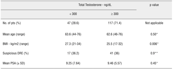
PSA levels, age or suspicious digital rectal examination did not differ significantly between the groups, but hypogonadal men had higher BMIs (Table-1).
One hundred twenty patients (73.2%) had stage organ-confined disease and 44 (26.8%) were pT3. Mean TT was 421 ng/dL for pT2 and 379 ng/dL for pT3 tumors (Table-2). This difference was
sta-Figure 1 - Total testosterone levels in the study population.
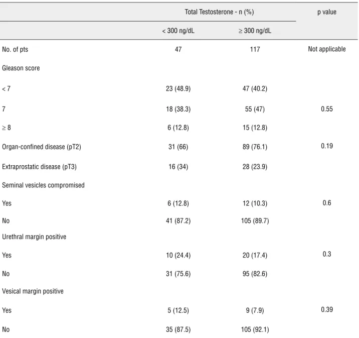
tistically significant, with an area under the curve (AUC) > 0.99. In the categorized analysis, the rate of extraprostatic disease was higher in the hypogo-nadic group: 34% vs. 23.9%, but without statistical significance (Table-3).
Involvement of seminal vesicles was noted in 14 (8.5%) and positive surgical margins in 44 pa-tients (26.8%). The occurrence of these events was
comparable in both groups (Table-3). There wasn´t any case of lymph node involvement or T4 tumors.
In regard to tumor differentiation, 70 (42.7%) patients had Gleason < 7, 73 (44.5%) Gle-ason = 7 and 21 (12.8%) GleGle-ason ≥ 8. The mean levels of TT were statistically equivalent in each one of these groups: 400.6, 432.2 and 365.8 ng/dL, respectively (Table-2). In the categorized analysis,
NUMBER OF P
ATIENTS
TESTOSTERONE LEVEL ng/dL
<150 150-200 201-250 251-300 301-350 351-400 400-450 450-500 >500 50
45
40
35
30
25
20
15
10
5
Gleason scores were also similar in groups with low and normal testosterone (Table-3).
DISCUSSION
The selection of the adequate method of treatment in oncology relies greatly on the
ba-lance between the aggressiveness of the disease and the benefits and morbidity of the therapy. This is particularly valid for PCa, a malignancy that is frequently indolent and which treatment (regardless of the method chosen) may be both deleterious and unnecessary. For better patient selection, D´Amico and others have stratified risk
Table 1 - Baseline clinical characteristics stratified by total serum testosterone.
Total Testosterone - ng/dL p value
< 300 ≥ 300
No. of pts (%) 47 (28.6) 117 (71.4) Not applicable
Mean age (range) 63.6 (44-76) 62.6 (46-76) 0.50*
BMI - kg/m2 (range) 27.3 (21-34) 25.5 (17-32) 0.006*
Suspicious DRE (%) 17 (36.2) 41 (36) 0.9**
Mean PSA (± SD) 9.25 (7.64) 9.46 (5.57) 0.45*
* Mann-Whitney test. ** Likelihood ratio test
DRE: digital rectal examination.
BMI: body mass index.
Table 2 - Mean total testosterone levels according to the pathological outcomes.
Pathological feature No. Pts (%) Mean testosterone level - ng/dL (± SD) p value
Organ-confined disease (pT2) p(pT2)(p(pT2) 120 (73.2) 421.6 (± 173)
Extraprostatic disease (pT3) 44 (26.8) 379.1 (± 178) < 0.001*
Gleason < 7 70 (42.7) 400.6 (± 172) 0.4 **
Gleason = 7 73 (44.5) 432.2 (± 183)
Gleason ≥ 8 21 (12.8) 365.8 (± 153)
Total 164 (100) 410.2 (± 175)
Table 3 - Gleason score and pathological features stratified by total serum testosterone level.
Total Testosterone - n (%) p value
< 300 ng/dL ≥ 300 ng/dL
No. of pts 47 117 Not applicable
Gleason score
< 7 23 (48.9) 47 (40.2)
7 18 (38.3) 55 (47) 0.55
≥ 8 6 (12.8) 15 (12.8)
Organ-confined disease (pT2) 31 (66) 89 (76.1) 0.19
Extraprostatic disease (pT3) 16 (34) 28 (23.9)
Seminal vesicles compromised
Yes 6 (12.8) 12 (10.3) 0.6
No 41 (87.2) 105 (89.7)
Urethral margin positive
Yes 10 (24.4) 20 (17.4) 0.3
No 31 (75.6) 95 (82.6)
Vesical margin positive
Yes 5 (12.5) 9 (7.9) 0.39
No 35 (87.5) 105 (92.1)
Likelihood ratio test
groups considering only three major prognostic markers: clinical stage, Gleason score and PSA levels (1). Despite universally accepted these cri-teria are not flawless and urologists are still limi-ted in their ability to predict pathological tumor stage in a reliable manner (4,5). Understanding other determinants of disease aggressiveness may be extremely helpful in selecting appropriate the-rapy for individual patients and advances in the
comprehension of other prognostic factors such as cancer density in biopsy, third Gleason grade, genetic mutations, tumor characteristics on MRI and, more recently, testosterone have been made (12,16).
is still incompletely understood and divisive, as we depict in Table-4. While there is evidence that cancers in a low testosterone environment tend to be more aggressive (6-10,12,18,19), many groups failed to demonstrate this association (1,20-22).
Approximately one third of our patients had TT deficiency, accordingly to surveys that also noted an increased incidence of biochemical hypogonadism in PCa patients compared to the general population (15). Again, this scenario is not unequivocal and a recent trial noted a rate of 15% of hypogonadism, which is comparable to the populational prevalence (7).
We adopted as primary endpoints the pa-thological features (Gleason score, stage, surgical margins status) as determined by the analysis of the surgical specimen because it´s the most relia-ble manner to determine the actual status of dise-ase and biopsy frequently understages the tumor (16). In our view, this avoids confusing and con-flicting results of others who relied exclusively on clinical staging and non-standardized biopsies.
The major finding of this survey was the significant difference in the mean preoperative TT levels when there was non-organ confined di-sease (421 vs. 379 ng/dL). This association was
Table 4 - Synthesis of the principal studies on the relationship between clinically localized prostate cancer and tumor aggressiveness.
Clinicopathological features associated with low testosterone
No. of cases
Design Gleason TNM stage PSA Surgical margins
Recurrence
Hoffman (11) (2000) 57 Retrospective Yes*** No No NA NA
Schatz (13) (2001) 156 Retrospective Yes NA Yes NA NA
Massengill (10) (2003) # * 879 Retrospective No Yes No No No
Teloken (16) (2005) 64 Retrospective No No No Yes NA
Isom-Batz (12) (2005) #* 326 Retrospective No Yes No NA No
Imamoto (30) (2005) * 82 Retrospective No Yes No NA Yes
Yamamoto (14) (2007)* 272 Retrospective No No No No No
Lane (17) (2008) 455 Prospective Yes No No No No
Pierorazio (19) (2010) 781 Retrospective No No NA NA Yes
Xylinas (8) (2010)** 107 Retrospective Yes Yes No No No
Botto (7) (2011) 431 Prospective Yes No Yes Yes NA
Salonia (21) (2011) 673 Prospective No## No### No No NA
Isbarn (9) (2009) --- Review Uncertain Uncertain Uncertain NA No
very strong, with an AUC > 0.99. Curiously, when patients were divided in groups of low and nor-mal TT, the rate of pT3 disease was 11% higher in the hypogonadic group, but still not statistically significant. The reasons for this are unknown to the authors. Possibly, this difference may become significant with an inclusion of a higher number of patients. Another pertinent explanation addres-ses the TT cut-off level of 300 ng/dL adopted by us and other authors. Clearly, while a threshold of 300 ng/dL may be adequate to hypogonadism diagnosis according to consensus definition of endocrinology and urology societies (15), it may be inappropriate to predict tumor aggressiveness. The relatively high mean TT values we found in the groups (421 and 379 ng/dL) support this idea by themselves. Hoffman also reported a mean TT of 490 and 390 ng/dL when Gleason was < 8 or ≥ 8 respectively (9), levels similar to ours and to Imamoto et al., who also correlated lower mean TT with locally advanced PCa (18).
This ability to predict extraprostatic exten-sion in prostatectomy specimens is important be-cause it´s a proven indicator of aggressive disease, determining greater likelihood of clinical progres-sion, greater risk of a positive surgical margin and poorer long-term cancer control (5). Massengill et al. were the first to demonstrate, in a retrospective cohort, results similar to ours less than ten years ago (6). In that study, there was a higher likelihood of non-organ confined disease (pT3–T4) as TT decreased, but testosterone was collected “at the discretion of the treating physician”, potentially imparting a selection bias. The only previous stu-dy in a Brazilian population is retrospective and analyzed retrospectively 64 patients after RRP, with the only statistically significant association found between low TT and positive surgical mar-gins, which in our experience was not more fre-quent in the men with TT < 300 ng/dL (13).
We failed to demonstrate that Gleason score or preoperative PSA levels are influen-ced by preoperative TT levels, like some groups (12,13,18,23) and in contrast to others (9,10,20). In our opinion, this seems somewhat logical because dihydrotestosterone (the most biologically active prostatic androgen) concentration in prostate cells does not reflect the concentration of total
testos-terone (24). Notably, when DHT was inhibited by finasteride or dutasteride in PCPT (25) and REDU-CE (26) trials, a higher proportion of high grade tumors was detected.
Some of the most important outcomes in oncologic treatment are disease recurrence and actual clinical progression. Their relationship with testosterone lacks confirmation (20). Interestingly, there are studies demonstrating a correlation with Gleason score (20) and pathological staging (6,10) but not with PSA recurrence or clinical progression (18,20). In 2007, Yamamoto et al. demonstrated that preoperative TT was an independent predictor of biochemical recurrence, but paradoxically it did not correlate with any pathologic features (Glea-son score, pathologic stage, surgical margins). The authors state that the reason of these discrepancies is unclear (12). In a well-conducted prospective study, Lane et al. concluded that low pretreatment TT was associated with Gleason pattern 4-5 cancer at prostatectomy, but not with pathological stage or disease progression thereafter (20). They affirm that “at present, routine measurement of TT in men treated by prostatectomy does not appear to be of any clinical value”. An argument can be done, ho-wever, because this study used TT, which is not the most biologically active form. In this regard, Hoffman et al. showed that free testosterone corre-lated with mean percent of biopsies demonstrated cancer (47% vs. 28%, p = 0.018) and also with pa-thological stage while TT did not (9).
The greater strength of our study was its prospective design, allowing routine morning tes-tosterone measurement before surgery during a 4 years period and the formation of a cohort of men representative of the reality in which PCa is tre-ated in Brazil, including both high and low-risk disease. To our knowledge, this is also the first prospective study to address testosterone as a pre-dictor of aggressive disease in Brazilian men with clinically localized PCa. Validation of a prognos-tic factor in a different population is important because prostate cancer may be genetically and clinically diverse in different populations (30).
The limitations of our study include the absence of central pathological review and una-vailability of data on long-term post-operative follow-up and survival. Body mass index was lo-wer in the hypogonadic group (a finding shared by others (7)) and we did not control the groups for ethnicity because it´s particularly complex to dis-criminate race in the Brazilian population, that´s multiracial and heterogeneous. Free and bioavai-lable testosterone (considered more biologically active forms) were not determined. Furthermore, a single dosage of TT in the day before surgery could imply on an incorrect value, once the stress of preoperative period could modify testosterone levels on an individual fashion (15).
CONCLUSIONS
Preoperative TT was associated with ex-traprostatic disease and may become a useful tool to improve our ability to recognize more advanced carcinomas. This correlation was not validated for other variables indicative of tumor aggressiveness and is not unequivocally consolidated in the lite-rature. Nonetheless, the concept that testosterone and other androgens have a permissive role and promote the development of PCa seems to be in-correct and an oversimplification in view of the current evidences in the field.
ABBREVIATIONS
BMI: Body mass index
DRE: Digital rectal examination NA: Not analyzed
PCa: Prostate cancer
PSA: Prostate Specific Antigen TT: Total testosterone
CONFLICT OF INTEREST
None declared.
REFERENCES
1. Partin AW, Kattan MW, Subong EN, Walsh PC, Wojno KJ, Oesterling JE, et al.: Combination of prostate-specific anti-gen, clinical stage, and Gleason score to predict pathologi-cal stage of lopathologi-calized prostate cancer. A multi-institutional update. JAMA. 1997; 277: 1445-51. Erratum in: JAMA 1997; 278: 118.
2. Morgentaler A: Testosterone deficiency and prostate can-cer: emerging recognition of an important and troubling relationship. Eur Urol. 2007; 52: 623-5.
3. Huggins C, Hodges CV: Studies on prostatic cancer. I. The effect of castration, of estrogen and androgen injection on serum phosphatases in metastatic carcinoma of the pros-tate. Cancer Res. 1941; 22: 293-7.
4. Morgentaler A: Turning conventional wisdom upside-down: low serum testosterone and high-risk prostate cancer. Can-cer. 2011; 117: 3885-8.
5. Isbarn H, Pinthus JH, Marks LS, Montorsi F, Morales A, Morgentaler A, et al.: Testosterone and prostate cancer: re-visiting old paradigms. Eur Urol. 2009; 56: 48-56. 6. Massengill JC, Sun L, Moul JW, Wu H, McLeod DG,
Am-ling C, et al.: Pretreatment total testosterone level predicts pathological stage in patients with localized prostate can-cer treated with radical prostatectomy. J Urol. 2003; 169: 1670-5.
7. Botto H, Neuzillet Y, Lebret T, Camparo P, Molinie V, Rayn-aud JP: High incidence of predominant Gleason pattern 4 localized prostate cancer is associated with low serum tes-tosterone. J Urol. 2011; 186: 1400-5.
8. Xylinas E, Ploussard G, Durand X, Fabre A, Salomon L, Al-lory Y, et al.: Low pretreatment total testosterone (< 3 ng/ mL) predicts extraprostatic disease in prostatectomy spec-imens from patients with preoperative localized prostate cancer. BJU Int. 2011; 107: 1400-3.
9. Hoffman MA, DeWolf WC, Morgentaler A: Is low serum free testosterone a marker for high grade prostate cancer? J Urol. 2000; 163: 824-7.
11. Schatzl G, Madersbacher S, Thurridl T, Waldmüller J, Kramer G, Haitel A, et al.: High-grade prostate cancer is associated with low serum testosterone levels. Prostate. 2001; 47: 52-8.
12. Yamamoto S, Yonese J, Kawakami S, Ohkubo Y, Tatokoro M, Komai Y, et al.: Preoperative serum testosterone level as an independent predictor of treatment failure following radical prostatectomy. Eur Urol. 2007; 52: 696-701. 13. Teloken C, Da Ros CT, Caraver F, Weber FA, Cavalheiro AP,
Graziottin TM: Low serum testosterone levels are associ-ated with positive surgical margins in radical retropubic prostatectomy: hypogonadism represents bad prognosis in prostate cancer. J Urol. 2005; 174: 2178-80.
14. Walsh PC: Radical prostatectomy in locally confined pros-tatic carcinoma. Prog Clin Biol Res. 1990; 359: 199-207; discussion 223-9.
15. Wang C, Nieschlag E, Swerdloff R, Behre HM, Hellstrom WJ, Gooren LJ, et al.: ISA, ISSAM, EAU, EAA and ASA rec-ommendations: investigation, treatment and monitoring of late-onset hypogonadism in males. Int J Impot Res. 2009; 21: 1-8.
16. Freedland SJ, Presti JC Jr, Terris MK, Kane CJ, Aronson WJ, Dorey F, et al.: Improved clinical staging system com-bining biopsy laterality and TNM stage for men with T1c and T2 prostate cancer: results from the SEARCH database. J Urol. 2003; 169: 2129-35.
17. Chodak GW, Vogelzang NJ, Caplan RJ, Soloway M, Smith JÁ: Independent prognostic factors in patients with meta-static (stage D2) prostate cancer. The Zoladex Study Group. JAMA. 1991; 265: 618-21.
18. Imamoto T, Suzuki H, Fukasawa S, Shimbo M, Inahara M, Komiya A, et al.: Pretreatment serum testosterone level as a predictive factor of pathological stage in localized pros-tate cancer patients treated with radical prospros-tatectomy. Eur Urol. 2005; 47: 308-12.
19. Pierorazio PM, Ferrucci L, Kettermann A, Longo DL, Met-ter EJ, CarMet-ter HB: Serum testosMet-terone is associated with aggressive prostate cancer in older men: results from the Baltimore Longitudinal Study of Aging. BJU Int. 2010; 105: 824-9.
20. Lane BR, Stephenson AJ, Magi-Galluzzi C, Lakin MM, Klein EA: Low testosterone and risk of biochemical recurrence and poorly differentiated prostate cancer at radical prosta-tectomy. Urology. 2008; 72: 1240-5.
21. Salonia A, Gallina A, Briganti A, Abdollah F, Suardi N, Capi-tanio U, et a.: Preoperative hypogonadism is not an inde-pendent predictor of high-risk disease in patients undergo-ing radical prostatectomy. Cancer. 2011; 117: 3953-62. 22. Salonia A, Gallina A, Briganti A, Suardi N, Capitanio U,
Ab-dollah F, et al.: Circulating estradiol, but not testosterone, is a significant predictor of high-grade prostate cancer in patients undergoing radical prostatectomy. Cancer. 2011; 117: 5029-38.
23. Monda JM, Myers RP, Bostwick DG, Oesterling JE: The correlation between serum prostate-specific antigen and prostate cancer is not influenced by the serum testosterone concentration. Urology. 1995; 46: 62-4.
24. Marks LS: Words of wisdom. Re: Endogenous sex hor-mones and prostate cancer: a collaborative analysis of 18 prospective studies. Eur Urol. 2009; 55: 750-1.
25. Thompson IM, Goodman PJ, Tangen CM, Lucia MS, Miller GJ, Ford LG, et al.: The influence of finasteride on the de-velopment of prostate cancer. N Engl J Med. 2003; 349: 215-24.
26. Andriole G, Bostwick D, Brawley O, Gomella L, Marberger M, Tindall D, et al.: Chemoprevention of prostate cancer in men at high risk: rationale and design of the reduction by dutasteride of prostate cancer events (REDUCE) trial. J Urol. 2004; 172: 1314-7
27. Miller LR, Partin AW, Chan DW, Bruzek DJ, Dobs AS, Ep-stein JI, et al.: Influence of radical prostatectomy on serum hormone levels. J Urol. 1998; 160: 449-53.
28. Zhang PL, Rosen S, Veeramachaneni R, Kao J, DeWolf WC, Bubley G: Association between prostate cancer and serum testosterone levels. Prostate. 2002; 53: 179-82.
29. Imamoto T, Suzuki H, Yano M, Kawamura K, Kamiya N, Ara-ki K, et al.: Does presence of prostate cancer affect serum testosterone levels in clinically localized prostate cancer patients? Prostate Cancer Prostatic Dis. 2009; 12: 78-82. 30. Lima MM Jr, Oliveira MN, Granja F, Trindade AC, De
Cas-tro Santos LE, Ward LS: Lack of association of GSTT1, GSTM1, GSTO1, GSTP1 and CYP1A1 polymorphisms for susceptibility and outcome in Brazilian prostate cancer pa-tients. Folia Biol (Praha). 2008; 54: 102-8.
_____________________
Correspondence address: