Malnutrition enhances cardiovascular responses to
chemoreflex activation in awake rats
Arlete Rita Penitente
a, Luciano Gonçalves Fernandes
a, Leonardo Máximo Cardoso
a,
Marcelo Eustáquio Silva
b, Maria Lúcia Pedrosa
a, Antônio Leite Silva
a,
Andréa Siqueira Haibara
c, Márcio Flávio Dutra Moraes
c,
Deoclécio Alves Chianca Júnior
a,⁎
a
Department of Biological Sciences, ICEB/NUPEB, Federal University of Ouro Preto, 35400-000, Ouro Preto, MG, Brazil
b
Department of Foods, Nutrition School, Federal University of Ouro Preto, 35400-000, Ouro Preto, MG, Brazil
c
Department of Physiology and Biophysics, Institute of Biological Sciences, Federal University of Minas Gerais, 31270-901, Belo Horizonte, MG, Brazil
Received 23 March 2007; accepted 6 July 2007
Abstract
Several studies in the literature suggest that low-protein intake is associated with increases in sympathetic efferent activity and cardiovascular disease. Among the possible mechanisms, changes in the neurotransmission of cardiovascular reflexes have been implicated. Therefore, the present study comprised the evaluation of chemoreflex responsiveness in rats subjected to a low-protein diet during the 35 days after weaning. As a result, we observed that malnourished rats presented higher levels of baseline mean arterial pressure and heart rate and exhibited a mild increase in the pressor response to chemoreflex activation. They also exhibited a massive bradycardic response to chemoreflex activation. Interestingly, bilateral ligature of the carotid body arteries further increased baseline mean arterial pressure and heart rate in malnourished animals. The data suggest severe autonomic imbalance and/or change in the central interplay between neural and cardiovascular mechanisms.
© 2007 Elsevier Inc. All rights reserved.
Keywords:Arterial pressure; Chemoreflex; Albumin; Sympathetic activity; Low-protein diet
Introduction
Cardiovascular diseases are the most frequent causes of morbidity and mortality in the world. In the last few decades, scientific research has yielded great advances in cardiovascular disease diagnosis and therapy, however the need to better understand the pathophysiological mechanisms of cardiovas-cular diseases still exists. Regulation of the cardiovascardiovas-cular system invariably involves neural and hormonal systems, such as the sympathetic nervous system (SNS) and renin– angioten-sin system (RAS), which play central roles in cardiovascular
regulation in both health and disease. SNS involvement in the pathogenesis of hypertension, coronary artery disease or heart failure has been well-established (Sinski et al., 2006). Several studies have shown that reduced protein intake leads to changes in cardiovascular homeostasis, which affects peripheral vascular resistance, renin secretion, renal hemodynamics (reducing the renal blood flow and glomerular filtration rate) and central neurotransmission of cardiovascular reflexes pathways (Barker et al., 1990, 1993; Benabe et al., 1993a,b; Benabe and Martinez-Maldonado, 1993, 1998; Langley-Evans et al., 1996, 2003; Plagemann et al., 2000; Woods and Raju, 2001). Indeed, studies performed in our laboratory have shown that animals submitted to our malnutrition model (reduction of 60% in the dietary protein) are characterized by increased levels of baseline mean arterial pressure and suggest increased sympathetic efferent activity directed to the heart relative to normal diet-fed rats (Oliveira et al., 2004; Tropia et al., 2001). These studies also ⁎Corresponding author. Universidade Federal de Ouro Preto-UFOP,
Labor-atório de Fisiologia Cardiovascular, Depto. Cien. Biol. ICEB/UFOP/NUPEB, Ouro Preto, 35400-000, MG, Brazil. Tel.: +55 31 3559 1721; fax: +55 31 3559 1680.
E-mail address:[email protected](D.A. Chianca).
suggest that malnutrition affects cardiovascular homeostasis resulting in hypertension in this experimental model. Similar results were observed in humans (Sawaya et al., 2003) suggesting that the animal malnutrition represents a reliable model to study cardiovascular alterations following malnutri-tion states in humans. Nevertheless, mechanisms involved in the development of hypertensive states in malnourished animals should be explored further. A growing number of studies have suggested that the enhancement of sympathetic efferent activity could represent a risk factor for cardiovascular disease (Hawkins et al., 2000; Irigoyen et al., 2005; Young et al., 1985). We hypothesize that autonomic imbalance due to the low-protein diet could affect the sympathetic output to the heart and vessels and possibly contribute to the development of hypertension.
The chemoreflex is one of the most important cardiovascular reflexes involved in the maintenance of cardiovascular homeostasis. Activation of the chemoreflex response by cytotoxic or hypoxic hypoxia activates sympathoexcitatory and a parasympathoexcitatory efferent pathways resulting in a pressor and bradycardic responses, respectively (Barros et al., 2002). The activation of the chemoreflex response depends on the sensory mechanisms of glomic cells (Franchini and Krieger, 1992; Gonzalez et al., 1995; Marshall, 1994). Therefore, potassium cyanide (KCN) is an excellent tool to assess the chemoreflex response. The chemoreflex neural pathways are involved in the generation of hypertension, since chronic chemoreflex activation and increase in sympathetic efferent activity could lead to a sustained rise in mean arterial pressure (Fletcher, 2000, 2001; Fletcher et al., 2002; Tahawi et al., 2001). The present study aimed to evaluate the chemoreflex response in malnourished animals to address whether this nutritional condition contributes to disruptions in the autonomic control of the cardiovascular system driven by the chemoreflex pathway.
Materials and methods
Animals
Male Fischer rats (180–210 g) from the Experimental
Nutrition Laboratory of the Nutrition School were used in this study. The animals were kept in individual cages and fed with regular or low-protein diet and filtered waterad libitum. They were maintained in a climate controlled area (24 °C) on a 12-hour dark–light cycle of 12 h. All the experimental procedures are in accordance with the Brazilian Council for Animal Experimentation(COBEA).
Diets
To induce malnutrition, rats were fed with a normal or low-protein content diet manufactured at the Experimental Nutrition Laboratory of the Nutrition School. The regular protein diet contained 15% protein while the low-protein diet contained 6% protein. The diets were isocaloric (422 kcal/100 g of diet) and the salts and vitamins were at similar concentrations in both diets.
Malnutrition protocol
Two female per male Fischer rats (four months old) were maintained in plastic cages (47 × 33 × 15 cm) for mating. After 10 days, the animals were separated and kept in individual cages. During pregnancy and weaning periods, the females received regular rat chow and filtered waterad libitum. After the puppies were born, they were handled randomly, keeping eight puppies per mother, and the weaning period was set to 28 days. After the weaning period, the male rats were separated in individual cages and divided into four groups according to diet and ligature surgery: 1) control intact (n= 8); 2) control ligated (n= 8); 3) malnourished intact (n= 8) and 4) malnour-ished ligated (n= 8). The animals were maintained on these diet protocols for 35 days and carotid body artery ligation was conducted 1 day prior to the experiments. The experiments were conducted on the 36th day after weaning.
Blood measurements
Blood samples were collected and subsequent measurements of biochemical parameters (glucose, total proteins and albumin concentration) were performed before and after carotid body artery ligature. The blood samples were centrifuged at 4000 rpm for 10 min and the serum was kept in sterile centrifuge tubes and stored at−20 °C until the colorimetric analysis. For glucose measurements, the blood was centrifuged for 10 min at 2000 rpm. All measurements were performed using an automated system (Wiener Lab, Germany).
Catheterization of the femoral and artery vein
One day prior to the cardiovascular recordings, animals received polyethylene catheters into the femoral artery (for cardiovascular measurements) and vein (for systemic drug administration) under tribromoethanol (2.5%, Merck, Darm-stadt, Germany) anesthesia. The catheters were tunneled through the subcutaneous and exteriorized on the back of the neck. The animals were maintained in individual cages in the experimental room until the next day to recover from anesthesia and adapt to the experimental room.
Ligature of the carotid body artery
We performed carotid body artery ligation to promote the degeneration of chemosensitive cells in the carotid body resulting in impairment of chemoreflex activation on two groups (normal protein diet fed and one low-protein diet fed) 1 day prior to experimentation, as described byFranchini and Krieger (1992).
Cardiovascular measurements
digital acquisition system (PowerLab/400, ADInstruments, Australia) in a 200-Hz sample frequency. Mean arterial pressure and heart rate were derived on-line from the pulsatile arterial pressure using Chart4 software (ADInstruments, Australia).
Chemoreflex activation
After a 30-minute stabilization period, the arterial chemore-flex was activated by intravenous administration of KCN in doses ranging from 5 to 40μg/kg. The pressor and bradycardic responses were recorded and a dose–response curve to KCN
injection was established. To verify that the baroreflex was not affected by the carotid body ligature, we performed injections of phenylephrine (PHE—1.5μg/kg) and Sodium Nitroprusside (SNP—0.5μg/kg).
Statistical analysis
The data were expressed as mean ± mean standard error. The statistical test used was one-way ANOVA for repeated measures followed by Tukey post-test or Student's t-test, where appropriate. The significance level was fixed at 5%.
Experimental protocol
Evaluation of the cardiovascular responses to chemoreflex in normal and malnourished rats
We evaluated cardiovascular responses (hypertension and bradycardic responses) to chemoreflex activation in normal protein diet or low-protein-diet-fed animals by the intravenous administration of KCN. This protocol allowed us to evaluate the chemoreflex function in normal and malnourished animals.
Results
Effects of the malnutrition on biochemical parameters
The malnutrition (n= 4) protocol significantly reduced the measured biochemical parameters (glucose, total protein and albumin) in comparison to normal diet-fed rats (n= 4). We also
observed that ligation of the carotid body artery had no additional effects on these biochemical parameters in normal or low-protein-diet animals. Furthermore, we observed that the malnutrition protocol reduced body weight in low-protein-diet-fed animals (n= 18) in comparison to normal diet rats [(n= 18) (Table 1)].
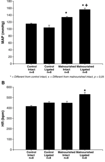
Effect of the malnutrition on the basal mean arterial pressure (MAP) and heart rate (HR) before and after the bilateral ligature of the carotid body artery
The basal levels of MAP (panel A,Fig. 1) and HR (panel B,
Fig. 1) were evaluated in malnourished and control animals, before and after the bilateral ligature of the carotid body artery. We observed that the malnourished intact group exhibited higher levels of baseline mean arterial pressure and heart rate relative to the control intact group (134 ± 2 mm Hg;n= 8vs
115 ± 2 mm Hg;n= 8 and 448 ± 16 bpm;n= 8vs 416 ± 8 bpm;
n= 8, respectively) and the bilateral ligature surgery increased
Table 1
Biochemical parameters of glucose (mg/dL), total protein content (g/dL), albumin (g/dL) and body weight (g) in control (intacts and ligated) and malnourished (intact and ligated) animals
Control intact
Control ligated
Malnourished intact
Malnourished ligated
Reference values Body
weight (g)
204 ± 3 – 68 ± 1⁎ – –
Glucose (mg/dL)
119.2 ± 15.1 115.7 ± 10.10
95.0 ± 7.10⁎ 88.0 ± 3.90⁎ 50–135
Total protein (g/dL)
7.1 ± 0.02 7.0 ± 0.36
6.0 ± 0.90⁎ 5.7 ± 0.04⁎ 4.7–8.2
Albumin (g/dL)
4.0 ± 0.37 4.0 ± 0.37
2.6 ± 0.03⁎ 2.4 ± 0.03⁎ 2.7–5.1
⁎ Different from control group (pb0.05).
Fig. 1. A—Baseline mean arterial pressure in control (n= 8) and malnourished
animals (n= 8) before (intact) and after (ligated) bilateral ligation of the carotid body artery. B—Basal heart rate in control (n= 8) and malnourished animals
baseline mean arterial pressure and heart rate in the malnour-ished group relative to the control ligated group [(156 ± 4 mm Hg, n= 8vs 104 ± 7 mm Hg, n= 8 and 531 ± 19 bpm, n= 8vs
450 ± 11 bpm, n= 8, respectively) (F= 3.466 and p= 0.023 for heart rate comparisons; F= 27.668 and pb0.001 for arterial pressure comparisons)].
Cardiovascular responses to chemoreflex activation in malnourished and control animals, before and after the bilateral ligature of the carotid body arteries
Fig. 2 represents changes in arterial pressure and heart rate due to chemoreflex activation by the intravenous injection of several doses of KCN (from 5 to 40 μg/kg) in malnourished (gray columns) and control (black columns) animals. The malnourished group exhibited an enhanced pressor response at doses of 5, 10 and 15μg/kg (10 ± 2 mm Hg,n= 8; 16 ± 1 mm Hg,
n= 8 and 21 ± 2 mm Hg,n= 8, respectively;p= 0.034;pb0.001 andp= 0.002, respectively) in comparison to the control group (6 ± 1 mm Hg,n= 8; 8 ± 1 mm Hg,n= 8 and 14 ± 2 mm Hg,n= 8, respectively). The doses of 20 and 40 μg/kg elicited pressor responses in malnourished (26 ± 1 mm Hg,n= 8 and 33 ± 2 mm Hg,n= 8, respectively;p= 0.138 andp= 0.585 respectively) and control (23 ± 2 mm Hg, n= 8 and 31 ± 2 mm Hg, n= 8, respectively) animals that were not different between the groups. Also, the bradycardic responses to chemoreflex activation by the
injection of 5, 10, 15, 20 and 40μg/kg were significantly greater in malnourished rats (−45 ± 7 bpm, n= 8; −70 ± 9 bpm, n= 8;
−86 ± 10 bpm,n= 8;−118 ± 11 bpm,n= 8 and−150 ± 13 bpm,
n= 8, respectively; pb0.001 for 5, 10, 15, 20 mg/kg and
p= 0.004 for 40 mg/kg) than in control animals (−8 ± 2 bpm,
n= 8; −20 ± 4 bpm, n= 8, −30 ± 5 bpm, n= 8; −59 ± 10 bpm,
n= 8 and −92 ± 13 bpm, n= 8). We also observed that chemoreflex activation after bilateral ligature of the carotid body arteries in malnourished and normal animals produced negligible effects on heart rate or arterial pressure indicating that ligature surgeries were effective (data not shown). It is important to point out that, in ligated malnourished and control animals, we tested the cardiovascular responses to baroreflex activation by PHE intravenous injection (1.5μg/kg) or deactivation by SNP intravenous injection (0.5 μg/kg) and we observed that both groups had intact functional baroreflex mechanisms indicating that bilateral ligature of the carotid body arteries did not affect baroreflex afferent pathways.
Discussion
Several studies have shown that the arterial chemoreflex plays an important role on cardiovascular homeostasis ( Gonza-lez et al., 1995; Haibara et al., 1999; Marshall, 1994). Chemoreflex activation is dependent on glomus cell activation since bilateral ligature of the carotid body arteries virtually abolished the cardiovascular responses to chemoreflex activa-tion in this and other studies (Barros et al., 2002; Franchini and Krieger, 1992, 1993; Haibara et al., 1995, 1999). It is important to note that, at least in the rat, carotid chemoreceptors seem to be more important than aortic ones for cardiovascular responses to chemoreflex activation and, therefore, to cardiovascular ho-meostasis (Sapru and Krieger, 1977). Evidence suggests that chronic chemoreceptor activation has been implicated in the development of hypertension: in rats subjected to chronic intermittent hypoxia resulting in increases in baseline mean arterial pressure and basal sympathetic efferent activity (Dick et al., 2007; Fletcher, 2000, 2001; Fletcher et al., 2002; Prabhakar et al., 2007; Tahawi et al., 2001). With respect to hypertension mechanisms, it is imperative that we understand the cardiovas-cular chemoreceptive mechanisms in health and disease since chronic increases in the sympathetic efferent activity constitute a risk for several cardiovascular diseases (Mancia, 1997; Sinski et al., 2006; Vasquez et al., 1997).
The present study examined cardiovascular changes ob-served in malnourished rats and the role of carotid chemoreflex on cardiovascular regulation in these animals. Initially, we observed that the animals maintained on a low-protein diet presented a body weight that was 67% lower than that observed in normal protein diet-fed animals. Also, we observed lower serum glucose, lower serum albumin and lower serum total protein levels. These results were completely in accordance with previous studies from our lab (Oliveira et al., 2004). These results confirm the development of malnutrition status and validate our animal model. In the present study, malnourished animals presented significant higher levels of baseline mean arterial pressure in comparison to the control animals. Interestingly, bilateral ligature
of the carotid body artery further increased baseline mean arterial pressure and basal heart rate in malnourished animals. The explanation for this observation could rely on the central interplay between neural pathways, the arterial baroreflex and carotid chemoreflex: Franchini and Krieger (Franchini and Krieger, 1992, 1993) observed that, in normotensive rats, bilateral ligature of the carotid body artery reduced the baseline mean arterial pressure, which disclosed the inhibitory influence of the chemoreflex response on the baroreflex pathways. However, malnourished animals often present an autonomic imbalance that lead to increased sympathetic and reduced parasympathetic efferent activity (Barker et al., 1993; Benabe et al., 1993a,b; Benabe and Martinez-Maldonado, 1993; Langley-Evans et al., 1996, 2003; Plagemann et al., 2000). For example, studies from our lab have shown that malnourished animals present impaired baroreflex function (Tropia et al., 2001). This central autonomic imbalance could lead to changes in the central interplay between the baroreflex and chemoreflex responses (Vasquez et al., 1997). Therefore, in malnourished animals, it seems that the carotid chemoreflex has a stimulatory effect on the baroceptive activity and the ligature of the carotid body arteries further increased the baseline mean arterial pressure and heart rate. Still, further investigations are required to prove these hypothesis and clarify the underlying mechanisms, but the data presented here is unique in that a model in which the removal of the chemoreflex response led to an increase in blood pressure. It should be observed that changes in the arterial pressure and heart rate observed in malnourished ligated animals were not dependent on afferent baroreflex denervation (as an artifact from the ligature surgery), since baroreflex activation with PHE or deactivation with SNP produced cardiovascular responses that were not changed after bilateral ligature of the carotid body arteries.
With respect to cardiovascular responses to chemoreflex response activation, the intravenous injection of KCN in doses that ranged from 5 to 40 μg/kg produced pressor and bradycardic responses that were dose-related and completely abolished by the bilateral ligature of the carotid body arteries. We observed that at all KCN doses, bradycardic responses were significantly increased in malnourished relative to control animals suggesting increased basal efferent sympathetic tonus in malnourished animals. Moreover, following KCN injection at doses of 5, 10 and 15μg/kg, we observed that malnourished animals exhibited significantly greater pressor responses when compared to those observed in control animals suggesting the enhancement of efferent sympathetic activity. Several possible mechanisms could be involved in these autonomic changes:
Efron and Barbul (1999)suggested that malnutrition could lead to reduced nitric oxide synthesis and therefore could account for increased sympathetic efferent activity; malnutrition can increase tissue and plasma levels of angiotensin, as well angiotensin II mRNA expression, and could be responsible for autonomic changes observed in malnourished animals (Benabe et al., 1993a,b; Benabe and Martinez-Maldonado, 1993; Tonkiss et al., 1998). Also, it is possible that malnutrition affects the neurotransmitter content in the chemoreflexes synapses and affects neurotransmission of the cardiovascular
responses to chemoreflex activation or increased central and/or peripheral chemosensitive mechanisms (Agarwal et al., 1981). Doses of 20 and 40 μg/kg failed to promote different pressor responses in malnourished and control animals. The latter effect is probably associated with maximal increases in sympathetic efferent activity or with a maximal constriction level of the vasculature in response to chemoreflex activation (Pimentel et al., 2003). The plateau observed in the pressor response to chemoreflex activation was also reported by others (Haibara et al., 1999).
The results of the present study raise important questions that should be addressed in further studies: 1) the identification of central and/or peripheral mechanisms involved in the increased sympathetic efferent activity observed in malnourished animals could reveal extremely important data that would help to understand the development of essential hypertension; 2) malnutrition is a health problem that affects individuals in several countries around the world and knowledge surrounding the potential risks for the cardiovascular system of malnour-ished individuals could suggest new medical treatments, as well as new prophylactic procedures.
In conclusion, the findings of the present study suggest that malnutrition status leads to an autonomic imbalance, which affects the neural pathways of the chemoreflex response. Moreover, this autonomic change alters the interplay between baro- and chemoreflex central mechanisms leading to increased sympathetic efferent activity and, therefore, serves as a risk factor and imposes deleterious effects on the cardiovascular system.
Acknowledgements
We thank Fundação de Amparo à Pesquisa do Estado de Minas Gerais (FAPEMIG) and Conselho Nacional de Desen-volvimento Científico e Tecnológico (CNPq) for their financial support of the present study.
References
Agarwal, K.N., Prasad, C., Taneja, V., 1981. Protein deprivation and the brain: effect on enzymes and free amino acids related to glutamate metabolism in rats. Annals of Nutrition & Metabolism 25, 228–233.
Barker, D.J., Bull, A.R., Osmond, C., Simmonds, S.J., 1990. Fetal and placental size and risk of hypertension in adult life. British Medical Journal 301, 259–262.
Barker, D.J., Gluckman, P.D., Godfrey, K.M., Harding, J.E., Owens, J.A., Robinson, J.S., 1993. Fetal nutrition and cardiovascular disease in adult life. Lancet 341, 938–941.
Barros, R.C., Bonagamba, L.G., Okamoto-Canesin, R., de Oliveira, M., Branco, L.G., Machado, B.H., 2002. Cardiovascular responses to chemoreflex acti-vation with potassium cyanide or hypoxic hypoxia in awake rats. Autonomic Neuroscience 97, 110–115.
Benabe, J.E., Martinez-Maldonado, M., 1993. Dietary modification of the renin angiotensin system. Seminars in Nephrology 13, 567–572.
Benabe, J.E., Martinez-Maldonado, M., 1998. The impact of malnutrition on kidney function. Mineral and Electrolyte Metabolism 24, 20–26.
Benabe, J.E., Fernandez-Repollet, E., Tapia, E., Luo, C., Martinez-Maldonado, M., 1993a. Angiotensin II and catecholamines interaction in short-term low protein feeding. Kidney International 44, 285–293.
Dick, T.E., Hsieh, Y.H., Wang, N., Prabhakar, N., 2007. Acute intermittent hypoxia increases both phrenic and sympathetic nerve activities in the rat. Experimental Physiology 92, 87–97.
Efron, D.T., Barbul, A., 1999. Arginine and nutrition in renal disease. Journal of Renal Nutrition 9, 142–144.
Fletcher, E.C., 2000. Hypertension in patients with sleep apnoea, a combined effect? Thorax 55, 726–728.
Fletcher, E.C., 2001. Invited review: physiological consequences of intermittent hypoxia: systemic blood pressure. Journal of Applied Physiology 90, 1600–1605.
Fletcher, E.C., Orolinova, N., Bader, M., 2002. Blood pressure response to chronic episodic hypoxia: the renin–angiotensin system. Journal of Applied
Physiology 92, 627–633.
Franchini, K.G., Krieger, E.M., 1992. Carotid chemoreceptors influence arterial pressure in intact and aortic-denervated rats. American Journal of Physiology 262, R677–R683.
Franchini, K.G., Krieger, E.M., 1993. Cardiovascular responses of conscious rats to carotid body chemoreceptor stimulation by intravenous KCN. Journal of the Autonomic Nervous System 42, 63–69.
Gonzalez, C., Lopez-Lopez, J.R., Obeso, A., Perez-Garcia, M.T., Rocher, A., 1995. Cellular mechanisms of oxygen chemoreception in the carotid body. Respiration Physiology 102, 137–147.
Haibara, A.S., Colombari, E., Chianca Jr., D.A., Bonagamba, L.G., Machado, B.H., 1995. NMDA receptors in NTS are involved in bradycardic but not in pressor response of chemoreflex. American Journal of Physiology 269, H1421–H1427.
Haibara, A.S., Bonagamba, L.G., Machado, B.H., 1999. Sympathoexcitatory neurotransmission of the chemoreflex in the NTS of awake rats. American Journal of Physiology 276, R69–R80.
Hawkins, P., Steyn, C., McGarrigle, H.H., Saito, T., Ozaki, T., Stratford, L.L., Noakes, D.E., Hanson, M.A., 2000. Effect of maternal nutrient restriction in early gestation on responses of the hypothalamic–pituitary–adrenal axis to
acute isocapnic hypoxaemia in late gestation fetal sheep. Experimental Physiology 85, 85–96.
Irigoyen, M.C., Paulini, J., Flores, L.J., Flues, K., Bertagnolli, M., Moreira, E.D., Consolim-Colombo, F., Bello-Klein, A., De Angelis, K., 2005. Exercise training improves baroreflex sensitivity associated with oxida-tive stress reduction in ovariectomized rats. Hypertension 46, 998–1003. Langley-Evans, S.C., Phillips, G.J., Benediktsson, R., Gardner, D.S., Edwards, C.R., Jackson, A.A., Seckl, J.R., 1996. Protein intake in pregnancy, placental glucocorticoid metabolism and the programming of hypertension in the rat. Placenta 17, 169–172.
Langley-Evans, S.C., Langley-Evans, A.J., Marchand, M.C., 2003. Nutritional programming of blood pressure and renal morphology. Archives of Physiology and Biochemistry 111, 8–16.
Mancia, G., 1997. Bjorn Folkow Award Lecture. The sympathetic nervous system in hypertension. Journal of Hypertension 15, 1553–1565.
Marshall, J.M., 1994. Peripheral chemoreceptors and cardiovascular regulation. Physiological Reviews 74, 543–594.
Oliveira, E.L., Cardoso, L.M., Pedrosa, M.L., Silva, M.E., Dun, N.J., Colombari, E., Moraes, M.F., Chianca Jr., D.A., 2004. A low protein diet causes an increase in the basal levels and variability of mean arterial pressure and heart rate in Fisher rats. Nutritional Neuroscience 7, 201–205.
Pimentel, F.F., Bonagamba, L.G., Machado, B.H., 2003. Pressor response to chemoreflex activation before and after microinjection of glycine into the NTS of awake rats. American Journal of Physiology. Regulatory, Integrative and Comparative Physiology 284, R1000–R1009.
Plagemann, A., Harder, T., Rake, A., Melchior, K., Rohde, W., Dorner, G., 2000. Hypothalamic nuclei are malformed in weanling offspring of low protein malnourished rat dams. Journal of Nutrition 130, 2582–2589.
Prabhakar, N.R., Dick, T.E., Nanduri, J., Kumar, G.K., 2007. Systemic, cellular and molecular analysis of chemoreflex-mediated sympathoexcitation by chronic intermittent hypoxia. Experimental Physiology 92, 39–44. Sapru, H.N., Krieger, A.J., 1977. Carotid and aortic chemoreceptor function in
the rat. Journal of Applied Physiology 42, 344–348.
Sawaya, A.L., Martins, P., Hoffman, D., Roberts, S.B., 2003. The link between childhood undernutrition and risk of chronic diseases in adulthood: a case study of Brazil. Nutrition Reviews 61, 168–175.
Sinski, M., Lewandowski, J., Abramczyk, P., Narkiewicz, K., Gaciong, Z., 2006. Why study sympathetic nervous system? Journal of Physiology and Pharmacology 57 (Suppl 11), 79–92.
Tahawi, Z., Orolinova, N., Joshua, I.G., Bader, M., Fletcher, E.C., 2001. Altered vascular reactivity in arterioles of chronic intermittent hypoxic rats. Journal of Applied Physiology 90, 2007–2013.
Tonkiss, J., Trzcinska, M., Galler, J.R., Ruiz-Opazo, N., Herrera, V.L., 1998. Prenatal malnutrition-induced changes in blood pressure: dissociation of stress and nonstress responses using radiotelemetry. Hypertension 32, 108–114.
Tropia, F.C., Cardoso, L.M., Pedrosa, M.L., Silva, M.E., Haibara, A.S., Moraes, M.F., Chianca Jr., D.A., 2001. Effects of low-protein diet on the baroreflex and Bezold–Jarisch reflex in conscious rats. Nutritional Neuroscience 4, 99–107.
Vasquez, E.C., Meyrelles, S.S., Mauad, H., Cabral, A.M., 1997. Neural reflex regulation of arterial pressure in pathophysiological conditions: interplay among the baroreflex, the cardiopulmonary reflexes and the chemoreflex. Brazilian Journal of Medical and Biological Research 30, 521–532.
Woods, S.E., Raju, U., 2001. Maternal smoking and the risk of congenital birth defects: a cohort study. Journal of the American Board of Family Practice 14, 330–334.