Blo ckade o f NK-1 re ce pto rs in the
late ral co m m issural nucle us tractus
so litarii o f awake rats had no e ffe ct
o n the cardio vascular re spo nse s to
che m o re fle x activatio n
Departamento de Fisiologia, Faculdade de Medicina de Ribeirão Preto, Universidade de São Paulo, Ribeirão Preto, SP, Brasil
C. Zhang, L.G.H. Bonagamba and B.H. Machado
Abstract
The neurotransmission of the chemoreflex in the nucleus tractus solitarii (NTS), particularly of the sympatho-excitatory component, is not completely understood. There is evidence that substance P may play a role in the neurotransmission of the chemoreflex in the NTS. Microinjection of substance P (50 pmol/50 nl, N = 12, and 5 nmol/50 nl, N = 8) into the commissural NTS of unanesthetized rats produced a significant increase in mean arterial pressure (101 ± 1 vs 108 ± 2 and 107 ± 3 vs 115 ± 4 mmHg, respectively) and no significant changes in heart rate (328 ± 11 vs 347 ± 15 and 332 ± 7 vs 349 ± 13 bpm, respectively) 2 min after microinjection. Previous treatment with WIN, an NK-1 receptor antagonist (2.5 nmol/50 nl), microinjected into the NTS of a specific group of rats, blocked the pressor (11 ± 5 vs 1 ± 2 mmHg) and tachycardic (31 ± 6 vs 4 ± 3 bpm) responses to substance P (50 pmol/50 nl, N = 5) observed 10 min after microinjec-tion. Bilateral microinjection of WIN into the lateral commissural NTS (N = 8) had no significant effect on the pressor (50 ± 4 vs 42 ± 6 mmHg) or bradycardic (-230 ± 16 vs -220 ± 36 bpm) responses to chemoreflex activation with potassium cyanide (iv). These data indi-cate that the activation of NK-1 receptors by substance P in the NTS produces an increase in baseline mean arterial pressure and heart rate. However, the data obtained with WIN suggest that substance P and NK-1 receptors do not play a major role in the neurotransmission of the chemoreflex in the lateral commissural NTS.
Co rre spo nde nce B.H. Machado
Departamento de Fisiologia FMRP, USP
Av. Bandeirantes, 3900 14049-900 Ribeirão Preto, SP Brasil
Fax: + 55-16-633-0017 E-mail: bhmachad@ fmrp.usp.br
Research supported by FAPESP, CNPq and PRO NEX. C. Zhang was the recipient of a CNPq postdoctoral fellowship (No. 150065/96-5).
Received January 21, 2000 Accepted August 7, 2000
Ke y wo rds
·Tachykinins
·Cardiovascular regulation
·Carotid chemoreceptors
·Autonomic regulation
·NK-1 receptors
·Substance P
Intro ductio n
Studies from our laboratory have evalu-ated the autonomic processing of the cardio-vascular reflexes in the nucleus tractus soli-tarii (NTS), with particular emphasis on the role of excitatory amino acid (EAA) recep-tors (1-6). Several studies have indicated a
chemoreflex in the NTS (11-14).
In a recent study (15) we observed that microinjection of EAA receptor antagonists (kynurenic acid; 6,7-dinitroquinoxaline-2,3-dione (DNQX) or a -methyl-4-carboxyphen-ylglycine (MCPG)) into the NTS produced only partial blockade of the pressor response to chemoreflex activation, indicating that the sympatho-excitatory component of this reflex was not exclusively mediated by L-glutamate and EAA receptors. Considering the possible involvement of substance P in the processing of the sympatho-excitatory component of the chemoreflex in the NTS (11-14), in the present study we evaluated the role of substance P and NK-1 receptors in the processing of the chemoreflex affer-ents in the lateral commissural NTS of awake rats. To achieve these goals we microin-jected substance P into the NTS of awake rats before and after local microinjection of WIN, an NK-1 receptor antagonist, and we also activated the chemoreflex before and after bilateral microinjection of an effective dose of WIN into the lateral commissural NTS of awake rats. A preliminary report of these data has been published as an abstract (16).
Mate rial and Me tho ds
Male Wistar rats weighing 280-300 g were used in the present study. Four days before the experiments rats under 2.5% tribromoethanol anesthesia (1 ml/100 g, ip) were placed in a stereotaxic apparatus (David Kopf, Tujunga, CA, USA) and the technique described by Michelini and Bonagamba (17) was used to implant bilateral guide cannulas in the direc-tion of the lateral commissural NTS (0.5 mm lateral to the midline and ~0.5 mm rostral to the calamus scriptorium) according to the co-ordinates of the Paxinos and Watson atlas (18). To implant each guide cannula we made a small window in the skull caudal to the lambda and introduced a 15-mm long stainless steel guide cannula (22 gauge; Small Parts, Miami Lakes, FL, USA) perpendicularly
through the window at the following coordi-nates: 0.5 mm lateral to the bregma, 14.00 mm caudal to the bregma and 7.9 mm below the skull surface at the bregma. The tip of the guide cannula was positioned ~1.0 mm above the dorsal surface of the brainstem. The guide cannula was fixed to the skull with methacry-late and watch screws and then closed with an occluder until the time of experimentation (2,5,17). The needle (33 gauge, Small Parts) used for microinjection into the NTS was 1.5 mm longer than the guide cannula and was connected by PE-10 tubing to a 1-µl syringe (Hamilton, Reno, NV, USA). The needles for microinjection of L-glutamate, used for func-tional identification of the NTS (5), substance P and WIN were carefully inserted into the guide cannula and manual injection was initi-ated 30 s later.
One day before the experiments, under 2.5% tribromoethanol anesthesia, a catheter (PE-10 connected to PE-50; Clay Adams, Parsippany, NJ, USA) was inserted into the abdominal aorta through the femoral artery for measurement of pulsatile arterial pres-sure (PAP), mean arterial prespres-sure (MAP) and heart rate (HR). A second catheter was inserted into the femoral vein for drug ad-ministration. Both catheters were tunneled and exteriorized through the back of the neck to be connected to a pressure trans-ducer under conscious freely moving condi-tions. PAP was measured with a pressure transducer (model CDX III; Cobe Laborato-ries, Lakewood, CO, USA) connected to a physiological recorder (Narcotrace 80; Narco Bio-Systems, Austin, TX, USA) and MAP was also evaluated using a Narco Universal gain coupler (type 7189). HR was quantified with a Narco biotachometer coupler (type 7302).
after bilateral microinjection of WIN (2.5 nmol/50 nl) into the lateral commissural NTS. At the end of each experiment 50 nl of Evans blue (2%) was microinjected into the same sites for histological analysis. The ani-mals were then submitted to intracardiac perfusion with saline followed by 10% buff-ered formalin under ether anesthesia. The brains were removed and stored in buffered formalin for 2 days, and serial coronal sections (15 µm) were cut and stained by the Nissl method. Only the rats in which the site of microinjection was located in the lateral com-missural NTS were used for data analysis.
The following drugs and solutions were used: saline (0.9% NaCl), L-glutamate (Sigma Chemical Co., St. Louis, MO, USA), tribromoethanol (Aldrich Chemical Company, Inc., Milwaukee, WI, USA), KCN (Sigma), substance P (RBI, Natick, MA, USA) and WIN 51.708 (NK-1 receptor antagonist, RBI). All values are reported as means ± SEM. Statistical analysis was performed using one-way ANOVA and the paired Student t-test. Differences were considered significant at the P<0.05 level.
Re sults
Change s in base line MAP and HR in re spo nse
to bilate ral micro inje ctio n o f substance P into
the NTS
Table 1 shows the basal values of MAP and HR before and 2 min after bilateral microinjection of two doses of substance P (50 pmol/50 nl and 5 nmol/50 nl) or saline into the NTS of three different groups of rats. There were significant increases in basal MAP 2 min after microinjection of 50 pmol/ 50 nl and 5 nmol/50 nl of substance P com-pared to control. Considering that these two doses of substance P produced a similar increase in MAP, it is possible that both are in the maximal dose range and for this rea-son we used the dose of 50 pmol/50 nl in the subsequent experiments.
Comparison of the changes in MAP and HR in response to two sequential bilateral microinjections of substance P (50 pmol/50 nl) into the NTS at 20-min intervals in a specific control group (N = 5) showed no significant changes in the pressor (9 ± 3 vs 6 ± 2 mmHg) or tachycardic responses (32 ± 12 vs 13 ± 9 bpm) to substance P microinjec-tion. These data show that repeated microin-jections of substance P into the NTS pro-duced no tachyphylactic effect.
Effe ct o f micro inje ctio n o f WIN o n the
cardio vascular re spo nse s to m icro inje ctio n
o f substance P into the NTS
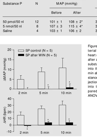
Figure 1 summarizes the changes in MAP and HR at 2, 5 and 10 min after unilateral
Table 1 - Basal mean arterial pressure (M AP) and heart rate (HR) before and 2 min after bilateral microinjection of substance P (50 pmol/50 nl and 5.0 nmol/50 nl) or saline into the NTS of three different groups of rats.
Values are reported as means ± SEM . * P<0.05 compared to control (paired t-test).
Substance P N M AP (mmHg) HR (bpm)
Before After Before After
50 pmol/50 nl 12 101 ± 1 108 ± 2* 328 ± 11 347 ± 15
5 nmol/50 nl 8 107 ± 3 115 ± 4* 332 ± 7 349 ± 13
Saline 4 103 ± 1 106 ± 2 314 ± 10 313 ± 9
Figure 1 - Changes in mean
ar-t erial pressure (DM AP) and
heart rate (DHR) 2, 5 and 10 min
after a control microinjection of substance P (SP, 50 pmol/50 nl) into the NTS and 2, 5 and 10 min after microinjection of sub-stance P preceded by microin-jection of WIN (2.5 nmol/50 nl) into the NTS. * P< 0.05 com-pared t o cont rol (one-w ay ANOVA).
D
M
A
P
(
m
m
H
g
)
20
15
10
5
0
D
H
R
(
b
p
m
)
40 30
0 -10 20 10
2 min 5 min 10 min
* *
*
* *
2 min 5 min 10 min
croinjection of substance P into the NTS at 2, 5 and 10 min. In a specific group of rats (N = 5) microinjection of a low dose of WIN (1 nmol/50 nl) into the NTS produced no block-ade in the cardiovascular responses to sub-stance P (50 pmol/50 nl; data not shown).
Effe ct o f bilate ral micro inje ctio n o f WIN into
the NTS o n the che mo re fle x
Figure 2 presents a typical tracing of one rat representative of the group showing that the effect of bilateral microinjection of WIN (2.5 nmol/50 nl) into the lateral NTS pro-duced no changes in the cardiovascular re-sponses to chemoreflex activation. The data summarized in Figure 3 show no significant changes in the pressor or bradycardic re-sponses to chemoreflex activation at 2, 10, 20 and 30 min after microinjection of WIN when compared to control responses.
Figure 4 is a photomicrograph of a coro-nal section of the brainstem of one rat repre-sentative of the group that received microin-jection of WIN and shows the bilateral sites of microinjection in the lateral aspect of the commissural NTS.
D iscussio n
The NTS is the site of termination of arterial baroreceptors, carotid chemorecep-tors (chemoreflex) and cardiopulmonary af-ferents (Bezold-Jarisch reflex) in the brain-stem and different subpopulations of post-synaptic neurons in the NTS are involved in the autonomic responses to the activation of these cardiovascular reflexes. In a recent study from our laboratory (15), we observed that microinjection of ionotropic receptor antagonists (kynurenic acid and DNQX) or a metabotropic receptor antagonist (MCPG) into the lateral commissural and medial NTS produced only partial blockade of the pres-sor response to chemoreflex activation, sug-gesting that the sympatho-excitatory compo-nent of this reflex is not exclusively
medi-Figure 2 - Typical tracing of a conscious rat representative of the group show ing the changes in heart rate (HR), pulsatile arte-rial pressure (PAP) and mean ar-t erial pressure (M AP) in re-sponse to chemoreflex activa-t ion w iactiva-t h poactiva-t assium cyanide
(KCN, 40 µg/rat, iv) before and 2,
10, 20 and 30 min after bilateral microinjection of WIN (2.5 nmol/ 50 nl) into the commissural NTS.
Figure 4 - Photomicrograph of a coronal section of the brainstem of one rat representative of the group that received bilateral mi-croinjection of WIN show ing the bilateral sites of microinjections in the lateral commissural NTS (Nissl, 120X).
H
R
(b
p
m
) 500
P
A
P
(m
m
H
g
)
300
100 150
50
150 50
M
A
P
(m
m
H
g
)
KCN control
WIN NTS 2 10 20 30 min
D
M
A
P
(
m
m
H
g
)
100
D
H
R
(
b
p
m
)
80
60
40
20
0
0 -50 -100 -150 -200
-350 -300 -250
Control (N = 5) After WIN (N = 5) Figure 3 - Changes in mean
arte-rial pressure (DM AP) and heart
rate (DHR) in response to
che-moreflex activation (KCN, 40 µg/
rat, iv) before (control) and 2, 10,
20 and 30 min after bilateral mi-croinjection of WIN (2.5 nmol/50 nl) into the NTS.
Control 2 10 20 30 min
mi-ated by EAA receptors (ionotropic and me-tabotropic). In view of these previous data and of evidence in the literature in favor of substance P as a neurotransmitter of the chemoreflex (13,14), in the present study we considered the possibility that substance P may be involved in the processing of the chemoreflex in the NTS, particularly in terms of the sympatho-excitatory component. For this reason we studied the possible role of substance P receptors (NK-1) in the NTS in the processing of the chemoreflex in unanes-thetized rats.
Substance P is a peptide belonging to the neurokinin family (20) and seems to play an important role as a neurotransmitter or neu-romodulator in primary baroreceptor and chemoreceptor afferent fibers, but the mechanisms of action of substance P in the brain with respect to the cardiovascular re-flexes in the NTS are not completely under-stood (12,21-25). It is important to note that microinjections of substance P into the NTS have produced variable effects on arterial pressure in different studies. Whereas some investigators failed to elicit cardiovascular responses to microinjection of substance P into the NTS (26,27), others reported that substance P induced hypotensive responses (22,23,25). The present study shows that bilateral microinjections of substance P into the lateral commissural NTS of unanesthe-tized rats produced a significant long-lasting increase (at least 10 min) in MAP and HR. On the other hand, the hypothesis that sub-stance P may be involved in baroreflex neu-rotransmission was raised in studies show-ing that low doses of substance P into the NTS elicit decreases in arterial pressure (11,25). However, at higher doses substance P produces long-lasting hypertension (24). The different cardiovascular responses to microinjection of substance P into the NTS may be related to the different experimental conditions used in each laboratory such as the anesthetic administered and the level of anesthesia. In the present study, we used
unanesthetized rats because in a previous study we observed that urethane or chlora-lose anesthesia produced a major effect on the cardiovascular responses to microinjec-tion of L-glutamate into the NTS (5).
In the present study, WIN, a selective, high-affinity, nonpeptide antagonist of NK-1 receptors, was used to test the hypothesis that substance P acting on these receptors could be playing a role in the neurotransmis-sion of the chemoreflex in the NTS. The effective dose of WIN to block NK-1 recep-tors was determined in experiments in which the long-lasting increase in MAP and HR produced by substance P was blocked by prior local administration of WIN. In these experiments we observed that substance P microinjected into the NTS produced sig-nificant cardiovascular changes which were entirely blocked by an NK-1 receptor an-tagonist. These data confirm findings of pre-vious studies showing that substance P and NK-1 receptors may play some role in car-diovascular regulation at the NTS level (11,25).
experi-ments were performed in awake rats. Anes-thetics significantly reduce the cardiovascu-lar responses to the chemoreflex and conse-quently the neurotransmission at the NTS level may not be the same under anesthesia (19; Haibara AS and Machado BH, unpub-lished data).
Bilateral microinjection of an NK-1 re-ceptor antagonist into the lateral commis-sural NTS, as shown in Figure 2, produced no changes in baseline MAP or HR, indicat-ing that these receptors do not play a tonic role in the processing of the baroreflex at the NTS level. However, whether or not NK-1 receptors in the NTS play a neuromodulatory role in the gain of the baroreflex is still matter for further investigation in awake rats.
Substance P has also been suggested to be one of the major neurotransmitters of the primary afferent barosensory fibers that ter-minate in the NTS (12,24). The study by Morilak et al. (24) using in vivo microdialy-sis showed that substance P can be released in the medial intermediate NTS during bilat-eral stimulation of the aortic depressor nerve in the rabbit, but Feldman (29) reported that blockade of NK-1 receptors in the NTS has no effect on the baroreflex in urethane-anes-thetized rats. There is also evidence showing that endogenous neurokinins, especially sub-stance P, act as mediators of stress responses in the brain (30). Therefore, considering this previous evidence and the fact that in the present study the microinjection of substance P produced a significant increase in MAP and HR, although the blockade of NK-1 receptors produced no effect on the chemore-flex, we may suggest that neurokinin recep-tors are present in the NTS and in some specific physiological situations like stress or defense reaction they can be activated to produce neuromodulation of the autonomic components of cardiovascular regulation, probably by facilitating the sympathetic
out-flow, considering that in the present study substance P microinjection produced a small but consistent increase in MAP and in some cases tachycardia.
With respect to the neurotransmission of the sympatho-excitatory component of the chemoreflex in the NTS, the present study indicates that substance P plays no major role in this processing and our previous study (15) demonstrated that excitatory amino ac-ids are not the sole system involved in this neurotransmission. Therefore, considering that substance P also plays no major role in this neurotransmission, further experiments are necessary to evaluate other potential neu-rotransmitters that may be involved in this processing. In this case, adenosine and puri-nergic receptors seem to be an important system to be evaluated in the neurotransmis-sion of the sympatho-excitatory component of the chemoreflex in the NTS of unanesthe-tized animals, especially due to the evidence reported by St. Lambert et al. (31) indicating that adenosine plays an important role in the neurotransmission of hypothalamic projec-tions to the NTS.
In conclusion, these results demonstrate that microinjection of substance P into the lateral commissural NTS produced an in-crease in MAP and HR, suggesting that this neurokinin may play some neuromodulatory role at the NTS level in some specific physi-ological situations. However, the blockade of NK-1 receptors with WIN had no effect on the cardiovascular responses to chemore-flex activation, indicating that substance P and NK-1 receptors are not involved in the neurotransmission of the chemoreflex at the level of the lateral commissural NTS.
Ackno wle dgm e nts
Re fe re nce s
1. Chianca-Jr DA & M achado BH (1996). M i-croinjection of NM DA antagonist into the NTS of conscious rats blocks the
Bezold-Jarisch reflex. Brain Research, 718:
185-188.
2. Colombari E, Bonagamba LGH & M acha-do BH (1994). M echanisms of pressor and bradycardic responses to L-glutamate mi-croinjected into the NTS of conscious rats.
Am erican Journal of Physiology, 266 (Regulatory, Integrative and Comparative Physiology, 35): R730-R738.
3. Colombari E, Bonagamba LGH & M acha-do BH (1997). NM DA receptor antagonist blocks bradycardic but not pressor re-sponse to L-glutamate microinjected into
the NTS of unanesthetized rats. Brain
Re-search, 749: 209-213.
4. Haibara AS, Colombari E, Chianca-Jr DA, Bonagamba LGH & M achado BH (1995). NM DA receptors in NTS are involved in bradycardic but not in pressor response
of chem oreflex. Am erican Journal of
Physiology, 269 (Heart and Circulatory Physiology, 38):H1421-H1427.
5. M achado BH & Bonagamba LGH (1992). M icroinjection of L-glutamate into the nucleus tractus solitarii increases arterial
pressure in conscious rats. Brain
Re-search, 576: 131-138.
6. M achado BH, M auad H, Chianca-Jr DA, Haibara AS & Colombari E (1997). Auto-nomic processing of the cardiovascular reflexes in the nucleus tractus solitarii.
Brazilian Journal of M edical and Biological Research, 30: 533-543.
7. Brew S, de Casto D, Housley GD & Sinclair JD (1990). The role of glutamate in neurotransmission of the hypoxic input to respiration through the nucleus tractus solitarius. In: Acker H, Trzebski A &
O’Regan D (Editors), Chemoreceptors and
Chemoreceptor Reflexes. Plenum Press, New York, 331-338.
8. Gordon FJ & Talman WT (1992). Role of excitatory amino acids and their receptors in bulbospinal control of cardiovascular function. In: Kunos G & Ciriello J (Editors),
Central Neural M echanisms in Cardiovas-cular Regulation. Birkhäuser, Boston, 209-225.
9. Le Galloudec E, M erahi N & Laguzzi R (1989). Cardiovascular changes induced by the local application of glutamate-re-lated drugs in the rat nucleus tractus
soli-tarii. Brain Research, 503: 322-325.
10. Talm an W T, Perrone M H & Reis DJ (1980). Evidence for L-glutamate as the
neurotransmitter of baroreceptor afferent
nerve fibers. Science, 209: 813-815.
11. Hall M E, M iley FB & Stew art JM (1989). Cardiovascular effects of substance P peptides in the nucleus of the solitary
tract. Brain Research, 497: 280-290.
12. Gillis RA, Helke CJ, Hamilton BL, Norman WP & Jacobow itz DM (1980). Evidence that substance P is a neurotransmitter of baro- and chemoreceptor afferents in
nucleus tractus solitarius. Brain Research,
181: 476-481.
13. Lindefors N, Yamamoto Y, Pantaleo T, Lagercrantz H, Brodin E & Ungerstedt U
(1986). In vivo release of substance P in
the nucleus tractus solitarii increases
dur-ing hypoxia. Neuroscience Letters, 69:
94-97.
14. Srinivasan M , Goiny M , Pant aleo T, Lagercrantz H, Brodin E, Runold M &
Yamamoto Y (1991). Enhanced in vivo
re-lease of substance P in the nucleus trac-tus solitarii during hypoxia in the rabbit:
role of peripheral input. Brain Research,
546: 211-216.
15. Haibara AS, Bonagamba LGH & M achado BH (1999). Sympathoexcitatory neuro-transmission of the chemoreflex in the
NTS of aw ake rats. American Journal of
Physiology, 275 (Regulatory, Integrative and Comparative Physiology, 44): R69-R80.
16. Zhang CH, Bonagamba LGH & M achado BH (1997). Substance P and neurotrans-mission of the baro- and chemoreflex in the nucleus tractus solitarii of
unanesthe-tized rats. Journal of the Autonomic
Ner-vous System, 65: 86 (Abstract). 17. M ichelini LC & Bonagamba LGH (1988).
Baroreceptor reflex modulation by vaso-pressin microinjected into the nucleus
tractus solitarii of conscious rats.
Hyper-tension, 11 (Suppl I): 75-79.
18. Paxinos G & Watson C (1996). The Rat
Brain in Stereotaxic Coordinates. Academ-ic Press, New York, NY.
19. Franchini KG & Krieger EM (1993). Cardio-vascular responses of conscious rats to carotid body chemoreceptor stimulation
by intravenous KCN. Journal of the
Auto-nomic Nervous System, 42: 63-70. 20. Guard S & Watson SP (1991). Tachykinin
receptor types: Classification and
mem-brane signalling m echanism s.
Neuro-chemistry International, 18: 149-165. 21. Gallagher PJ, Paxinos G & White SW
(1985). The role of substance P in arterial
chemoreflex control of ventilation.
Jour-nal of the Autonomic Nervous System, 12: 195-210.
22. Haeusler B & Osterw alder R (1980). Evi-dence suggesting a transmitter or neuro-modulatory role for substance P at the first synapse of the baroreceptor reflex.
Naunyn-Schm iedeberg’ s Archives of Pharmacology, 314: 111-121.
23. Kubo T & Kihara M (1987). Blood pressure modulation by substance P in the rat
nucleus solitarius. Brain Research, 413:
379-383.
24. M orilak DA, M orris M & Chalm ers J (1988). Release of substance P in the
nucleus tractus solitarius measured by in
vivo microdialysis: response to
stimula-tion of the aortic depressor nerves in
rab-bit. Neuroscience Letters, 94: 131-137.
25. Nagashim a A, Takano Y, Tat eishi K, M atsuoka Y, Hamaoka T & Kamiya H (1989). Cardiovascular roles of tachykinin peptides in the nucleus tractus solitarii of
rats. Brain Research, 487: 392-396.
26. Spencer SE & Talman WT (1986). Central modulation of gastric pressure by sub-stance P: A comparison w ith glutamate
and acetylcholine. Brain Research, 385:
371-374.
27. Talman WT & Reis DJ (1981). Baroreflex actions of substance P microinjected into the nucleus tractus solitarii in rat: A
con-sequence of local distortion. Brain
Re-search, 220: 402-407.
28. Bond SM , Cervero F & M cQueen DS (1982). Influence of neonatally adminis-t ered capsaicin on barorecepadminis-t or and chemoreceptor reflexes in the adult rat.
British Journal of Pharmacology, 77: 517-524.
29. Feldman PD (1995). Neurokinin-1 recep-tor mediation of the vasodepressor ef-fects of substance P in the nucleus of the
tractus solitarius. Journal of
Pharmacolo-gy and Experimental Therapeutics, 273: 617-623.
30. Culman J, Klee S, Ohlendorf C & Unger T (1997). Effect of tachykinin receptor inhi-bition in the brain on cardiovascular and
behavioral responses to stress. Journal of
Pharmacology and Experimental Thera-peutics, 280: 238-246.
31. St Lambert JH, Dashw ood M R & Spyer KM (1996). Role of brainstem adenosine A1 receptors in the cardiovascular re-sponse to hypothalamic defense area
stimulation in the anaesthetized rat.