Margarida Cristina Camacho Pestana Correia
DOCTORATE IN BIOLOGICAL SCIENCES
September| 2014
Nematicidal activity of
Solanum nigrum
and
S. sisymbriifolium
extracts against the
root-lesion nematode
Pratylenchus goodeyi
and its effects on infection and gene expression
Nematicidal activity of
Solanum nigrum
and
S. sisymbriifolium
extracts against the
root-lesion nematode
Pratylenchus goodeyi
and its effects on infection and gene expression
DOCTORAL THESIS
Margarida Cristina Camacho Pestana Correia
DOCTORATE IN BIOLOGICAL SCIENCESSUPERVISOR
Maria Manuela Câmara de GouveiaCO-SUPERVISOR
“In short, if all the matter in the universe except the nematodes were swept away,
Acknowledgements
I would like to start by sharing my deepest thanks with my supervisors. To Professor Manuela Gouveia a heartfelt thank you for having accepted to supervise my project and for sharing her knowledge with me particularly with in respect to molecular analysis. Thank you for all the support, direction, friendship and comprehension; I will be always indepted to you. To Professor Isabel Abrantes for the vast knowledge in Nematology, your lessons were invaluable, without your guidance and teaching
I wouldn’t have been able to finish this work.
To Professor Nereida Cordeiro and her collaborators, thanks for all your help and valuable assistance in the chemical part of this thesis.
To Universidade da Madeira for accepting this thesis.
To Secretaria Regional do Ambiente e Recursos Naturais, Direção Regional de Agricultura e Desenvolvimento Regional, Direção de Serviços de Laboratórios Agroalimentares for making the development of this work possible.
To Laboratório de Genética Humana da Universidade da Madeira for allowing me the use of their equipment.
To Direção Regional do Ordenamento do Território e Ambiente for all the help and assistance.
To all my colleagues and friends of Laboratório de Qualidade Agrícola da Madeira especially Adelaide, Adriano, Alda, Décio, Délia, Duarte, Fátima, Miguel, and Rubina for the support, help and encouragement.
To my laboratory partners in Centro de Ciências da Vida Diana, Lucilia, Filipa and Mónica. Without you everything would have been more difficult. I leave here my sincere appreciation for all the support.
To my partners of the five o’clock tea, thanks for the knowledge and the brainstorming
that we’ve shared with each other.
I
Contents
Acknowledgments
List of tables……...………...………...…………IV
List of figures………...………..………...V
List of abbreviations………...………....….…..VII
Outline of this thesis……….….………VIII
Abstract ...1
Resumo ...2
Chapter 1 ...3
1. General Introduction...4
1.1 The banana culture ...4
1.2 The root-lesion nematodes ...5
1.3 Plant nematode control ...7
1.3.1 The nematicide potential of plants ...8
1.4 Parasitism and genes from plant-parasitic nematodes ...10
Chapter 2...13
2. Material and Methods ...14
2.1 Biological material ...14
2.1.1 Pratylenchus goodeyi isolates ...14
2.1.2 Nematode multiplication...14
2.1.3 Plant material...14
2.2 Chemicals ...15
2.3 Plant extractions ...15
2.3.1 Solanum aqueous extractions...15
2.3.2 Solanum sequential extraction...16
2.4 Chemical analysis ...16
2.4.1 Samples preparation and selection ...16
2.4.2 Gas chromatography-mass spectrometry (GC-MS) ...16
2.4.3 Ultra-high performance liquid chromatography (UHPLC) ...17
2.4.4 Mass spectrometry...17
2.4.5 HPLC quantification ...17
2.5 Biological assays ...18
2.5.1 Nematicidal activity ...18
2.5.2 Mobility and attraction ...19
2.5.3 Infection ...19
2.6 Molecular analysis ...20
2.6.1 Biological samples ...20
2.6.2 RNA extraction and cDNA synthesis ...20
2.6.3 cDNA amplification and cloning ...20
2.6.5 RT-PCR analysis...23
2.6.6 Expression analysis ...23
2.7Statistical analysis ...23
Chapter 3...25
3. Nematicidal activity of Solanum sisymbriifolium and S. nigrum extracts against the root-lesion nematode Pratylenchus goodeyi...26
3.1 Abstract...26
3.2 Introduction ...26
3.3 Results ...27
3.3.1 Fractions of S. sisymbriifolium and S. nigrum dry material ...27
3.3.2 P. goodeyi mortality and mobility in S. sisymbriifolium and S. nigrum extracts ...28
3.4 Discussion ...29
Chapter 4 ...31
4. In vitro evaluation of nematicidal properties of Solanum sisymbriifolium and S. nigrum extracts on Pratylenchus goodeyi ...32
4.1 Abstract ...32
4.2 Introduction ...32
4.3 Results ...34
4.3.1 Aqueous extracts ...34
4.3.2 Sequential extracts ...34
4.3.3 Nematicidal activity of aqueous extracts ...35
4.3.4 Nematicidal activity of sequential extracts ...37
4.4 Discussion ...40
Chapter 5 ...43
5. Comparative analysis of lipophilic and phenolic compounds from Solanum nigrum and S. sisymbriifolium ...44
5.1 Abstract ...44
5.2 Introduction ...45
5.3 Results ...46
5.3.1 Lipophilic composition ...46
5.3.2 Phenolic composition ...50
5.4 Discussion ...54
Chapter 6...57
6. Effect of chemical stress imposed by Solanum nigrum in calreticulin and beta-1,4- endoglucanase genes and in infectivity of Pratylenchus goodeyi ...58
6.1 Abstract ...58
6.2 Introduction ...58
6.3 Results ...60
6.3.1 Cloning of calreticulin gene ...60
6.3.2 Characteristics of the calreticulin protein ...62
III
6.3.4 Characteristics of the beta-1,4-endoglucanase protein...64
6.3.5 Expression of Pg-CRT and Pg-ENG ...66
6.3.6 Biological assays ...67
6.4 Discussion ...68
Chapter 7...71
7. Molecular cloning and characterization of cDNA encoding a Translocon-Associated Protein (TRAP δ) from the root-lesion nematode Pratylenchus goodeyi ...72
7.1 Abstract ...72
7.2 Introduction ...72
7.3 Results ...74
7.3.1 Cloning and sequence analysis of Pg-TRAP cDNA ...74
7.3.2 Protein Pg-TRAPδ characterization ...75
7.3.3 Gene expression analysis ...76
7.3.4 Expression pattern of Pg-TRAP in response to putative nematicides ...77
7.4 Discussion ...78
Chapter 8 ...81
8. Discussion and Conclusions ...82
8.1 General Discussion ...82
8.2 Conclusions ...86
List of tables
Table 1.1 Root-lesion nematodes genes and proteins related to infection and parasitism
with known function. ...11
Table 2.1 Calibration data used for the UHPLC–UV quantification of phenolic
compounds in Solanumnigrum and S. sisymbriifolium acetone and water extracts. ...18
Table 2.2 Primers used in this study for PCR amplification, RACE, RT-PCR, and
expression analysis. ...21
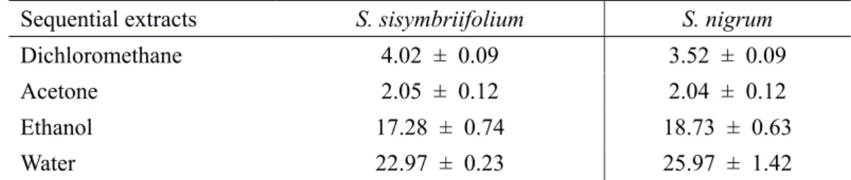
Table 4.1 Extractives (% of dry matter) obtained from Solanum sisymbriifolium and S. nigrum blended in hot and cold water or by reflux. Results are the mean of five replicates (mean ± SD). ...34
Table 4.2 Extractives (% of dry matter) obtained from Solanum sisymbriifolium and S. nigrum dry plants using sequential extraction. Results are the mean of five replicates (mean ± SD). ...35 Table 5.1 Lipophilic composition of Solanum nigrum and S. sisymbriifolium before (BH) and after (AH) hydrolysis expressed in mg/Kg of dry matter (DM). ...48
V
List of figures
Figure 1.1 Morphological characters of P. goodeyi. ...6
Figure 1.2 Drawing representing infection by the root-lesion nematode P. goodeyi.
(Adapted from Jones & Fosu-Nyarko 2014). ...7
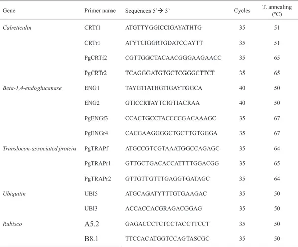
Figure 3.1 Percentage of extractives from dry S. sisymbriifolium and S. nigrum plants obtained from solvents sequence: dichloromethane (DMC), acetone (Acet), ethanol
(EtOH) and water. ...28
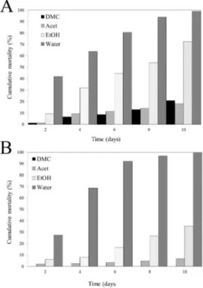
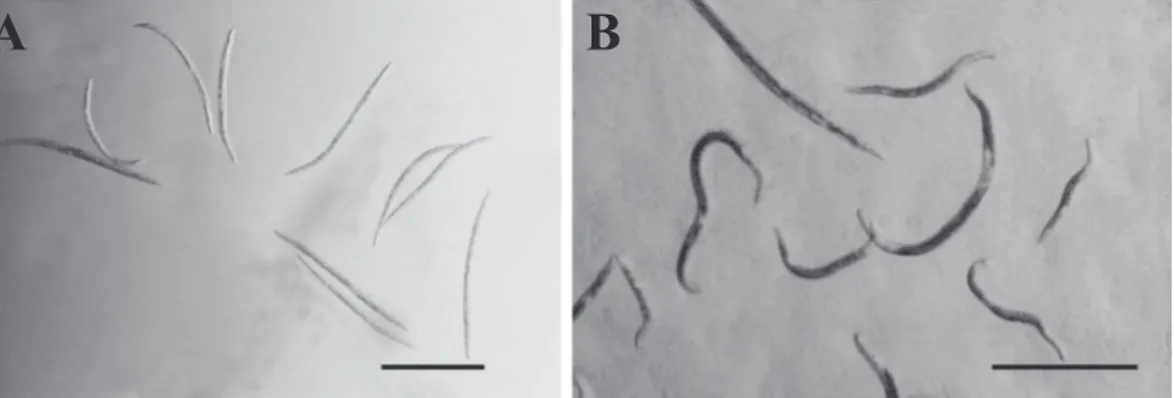
Figure 3.2 Mobility of P. goodeyi in dichloromethane (A), acetone (B), ethanol (C) and
water extracts (D) obtained from S. nigrum plant sequential extraction. ...28 Figure 3.3 P. goodeyi cumulative mortality for 10 days in S. sisymbriifolium (A) and S.
nigrum (B) solvents sequence extracts. ...29 Figure 4.1 Cumulative mortality of P. goodeyi in cold, hot and reflux aqueous extracts at
10 mg/mL concentration from fresh (A) and dry (B). ...35 Figure 4.2 Nematode activity after two days in aqueous extracts from fresh S. nigrum at
10 mg/mL plant concentration (A) and in water as control (B). ...36
Figure 4.3Cumulative mortality of P. goodeyi in cold, hot and reflux aqueous extracts, at
10 mg/mL concentration, from fresh (A) and dry (B) S. nigrum. ...36 Figure 4.4 Cumulative mortality of P. goodeyi in dichloromethane (DMC), acetone
(Acet), ethanol (EtOH) and water extracts, at 10 mg/mL concentration, from S.
sisymbriifolium (A) and S.nigrum (B). ...38 Figure 4.5Morphological changes of P. goodeyi in S. nigrum acetone extract. Separation between cuticle and internal content of the body (A, B and C). Condensation, rigidity and
necrosis of tissues (D). ...39
Figure 4.6 Mortality of P. goodeyi over 24 h incubation in S. nigrum acetone extract (10
mg/mL). ...39 Figure 5.1 GC-MS chromatogram of the derivatized dichloromethane extract from the S.
nigrum before alkaline hydrolysis (BH). ...47
Figure 5.2 Major families of lipophilic components identified by GC-MS in the Solanum
dichloromethane extracts. Fatty acids FA, long-chain aliphatic alcohols LCAA, before hydrolysis BH, after hydrolysis AH. ...47
Figure 5.3 Chemical structures of the main lipophilic and phenolic compounds identified
in S. nigrum and S. sisymbriifolium dichloromethane, acetone and water extracts. ...49
Figure 5.4 UHPLC-UV chromatograms of the acetone extract from S. nigrum (A) and S sisymbriifolium (B) at 340 nm. Water extract from S. nigrum (C)and S. sisymbriifolium (D) at 280 nm. ...51
Figure 6.1 Full-length cDNA and deduced amino acid sequence of P. goodeyicalreticulin
(GeneBank: KF993343). ...61 Figure 6.2 Comparison of deduced amino acid sequence of P. goodeyi calreticulin with
Figure 6.3 Beta-1,4-endoglucanase of P. goodeyi full-length cDNA and deduced amino acid sequence (GenBank: KM005101). ...64
Figure 6.4 Protein alignment of beta-1,4-endoglucanases from R. similis, P. penetrans, H.
glycines, R. reniformis and P. goodeyi ...65 Figure 6.5 Transcripts of the calreticulin, beta-1,4-endoglucanase and ubiquitin by specific
RT-PCR analysis from the cDNA samples of P. goodeyi (Pg), banana roots infected
with P. goodeyi (RPg) and uninfected banana roots from in vitro culture (Rv) and from plants grown in a pot with sterilized soil (Rp). ...66
Figure 6.6 Time-course expression levels of P. goodeyi: calreticulin, beta-1,4-endoglucanase and ubiquitin under chemical stress. Relative abundance of transcripts by semi-quantitative RT-PCR (A). Relative amount of calreticulin, beta-1,4-endoglucanase
normalized by ubiquitin transcripts (B). ...66 Figure 6.7 Nematode attraction to root of banana plants (Rv) in Pluronic gel (A). Data are the mean values ± SE of three independent experiments. Attraction after 2h (B), 4h
(C) and 6 h (D). ...67
Figure 6.8 Infection of banana root with P. goodeyi: J2 (A); females (B), males (C) and
the infectivity reduction (D) after exposure for 0, 6, 12 and 18 h to the acetone extract of
Solanum nigrum. ...68 Figure 7.1 Complete coding sequence and deduced amino acid sequence of P. goodeyi
translocon-associated protein (Pg-TRAPδ) (KF359552). ...75 Figure 7.2 Comparison of deduced amino acid sequence of P. goodeyi
translocon-associated protein (Pg-TRAPδ) (KF359552) with similar protein sequences from
nematodes listed in the GenBank. ...76
Figure 7.3 Transcripts of the TRAPδ, rubisco and ubiquitin by specific RT-PCR analysis
from the cDNA samples of P. goodeyi (Pg), banana roots infected with P. goodeyi (RPg), banana roots from in vitro culture (Rv) and roots from banana plants grown in a pot with sterilized soil (Rp). ...76
Figure 7.4 Mobility and morphological changes of P. goodeyi. ...77
Figure 7.5 Semi-quantitative RT-PCR analysis of Pg-TRAPδ expression during exposure for 0, 6, 12 and 18 h to the acetone extract of S. nigrum (A). Relative abundance of
VII
List of abbreviations
Acet, acetone AH, after hydrolysis BH, before hydrolysis
BLAST, Basic Local Alignment Search Tool CBS, Center for Biological sequence analysis
CRT, calreticulin
DMC, dichloromethane
EBI, European Bioinformatics Institute ENG, beta-1,4-endoglucanase
ER, endoplasmatic reticulum
EtOH, etanol
FA, fatty acids
GC-MS, chromatography-mass spectrometry
LCAA, long-chain aliphatic alcohols;
NCBI, National Center for Biotechnology Information
ORF, open reading frame Pg, Pratylenchus goodeyi
RACE, Rapid amplification of cDNA ends
Rp, uninfected banana roots from plants grown in a pot
RPg, banana roots infected with P. goodeyi RT retention time
Rv, uninfected banana roots from in vitro culture SMART, Simple Modular Architecture Research Tool
SN, Solanum nigrum
SS, Solanum sisymbriifolium
TMS, trimethylsilyl ethers and esters
TRAPδ,translocon associated protein delta subunit UTR, untranslated region
Outline of this thesis
Since nematicides used to control plant-parasitic nematodes are not effective enough to justify its application, besides being toxic to environment and animal
life, search for plant extracts that could be efficient to control nematodes was the
starting point of the present study. The main purpose of this doctoral thesis was to evaluate the nematicidal potential of two Solanum species (S. nigrum L. and S.
sisymbriifolium Lam.) against the plant-parasitic nematode Pratylenchus goodeyi
Sher & Allen 1953, an important parasite of banana roots in Madeira Island. Within this subject mobility and mortality was assessed and the possible effect analysed at behavioural and molecular level using effector genes related to parasitism or stress. In Chapter 1 a general introduction to the subject is given with the background on the main issues of the thesis. Chapter 2 describes all materials and methods used in this study. The nematicide properties of S. nigrum and S. sisymbriifolium as control agents against P. goodeyi were investigated through sequential extracts obtained from dried plants, and are reported in Chapter 3. In Chapter 4 the in vitro evaluation of the nematicide potential from S.nigrum and S. sisymbriifolium against P. goodeyi was made using sequential and aqueous extracts obtained from dried and fresh plants. The chemical profile concerning lipophilic and phenolic compounds present in S.nigrum and S. sisymbriifolium is given in Chapter 5. In the following chapters the molecular characterization and cloning of calreticulin,beta-1,4-endoglucanase
(Chapter 6) and translocon associated protein delta subunit (Chapter 7) genes from
1
Abstract
The control of Pratylenchus goodeyi a common nematode parasite of
banana crop in Madeira Island can benefit from searching for natural
nematicides through plants extracts. With this aim we submitted Solanum nigrum and S. sisymbriifolium driedplants to a sequential extraction in the solvent sequence of dichloromethane, acetone, ethanol and water, and to an aqueous extraction of the fresh and dried plants. Analyses with the extracts at several concentrations were used to assess mobility and mortality on P.
goodeyi. Results showed that the water extract and aqueous extracts from both plants at a concentration of 10 mg/mL affected nematode mobility and caused mortality but the acetone extract from S. nigrum was the most
efficient, causing 100% mortality whereas dichloromethane had no effect on P. goodeyi. Determination of the lipophilic and phenolic compounds present in the two most effective Solanum extracts (acetone and water) and in dichloromethane extract revealed that some of these compounds had nematicidal activity. S. nigrum acetone extract (10 mg/mL) was used to
find out the nematicidal potential following the effect at gene expression
level and nematode behaviour. Genes coding for calreticulin and beta-1,4-endoglucanase related to parasitism and translocon-associated protein
putatively connected to stress were obtained and its relative expression assessed in nematodes exposed to the extract. Results revealed that expression of Pg-CRT decreased showing to influence the infection, Pg-ENG remained steady and Pg-TRAPδ was induced over time exposure. Biological assays showed that P. goodeyi mobility and ability to infect the banana roots were affected as a decrease in the number of nematodes that reached the roots was obtained with the increased exposure time to the extract being implicated in the infection success. The information obtained from this thesis showed that S. nigrum has potential to be used for the development of a new control strategy against plant-parasitic nematodes.
Resumo
O controlo do nemátode Pratylenchus goodeyi parasita da bananeira, comum
na Ilha da Madeira, pode beneficiar com a utilização de extratos naturais
obtidos das plantas. Com este intuito duas plantas de Solanum (S. nigrum e
S. sisymbriifolium)foram submetidas a uma extração sequencial utilizando a sequência de solventes: diclorometano; acetona; etanol e água e, ainda, a uma extração aquosa das plantas frescas ou secas. Foram efectuadas análises destes extratos, em várias concentrações, de modo a testar o efeito sobre a mobilidade e mortalidade de P. goodeyi. Os resultados mostraram que o extrato em água da sequência e os extratos aquosos das duas plantas na concentração de 10 mg/mL afetaram a mobilidade e causaram mortalidade do nemátode. O extrato de acetona de S. nigrum foi o mais eficaz causando 100% de mortalidade mas o extrato de diclorometano não afetou nem a mobilidade de P. goodeyi nem causou mortalidade. A determinação dos compostos lipofílicos e fenólicos presentes no extrato de diclorometano e nos extratos de acetona e água das plantas de Solanum revelou a presença de compostos com atividade nematicida conhecida. O extrato de acetona de S. nigrum (10 mg/mL) foi usado para avaliar o efeito nematicida ao nível da expressão de genes e no comportamento do nematode. Os genes
calreticulina e beta-1,4-endoglucanase associados com o parasitismo e
translocon-associated protein relacionado com o stress foramisolados e a expressão relativa avaliada em nematodes expostos ao extrato de acetona. Os resultados revelaram que a expressão de Pg-CRT diminuiu mostrando
que pode influenciar a infeção e a de Pg-ENG manteve-se constante. Mas, a expressão de Pg-TRAPδ aumentou com o tempo de exposição. Ensaios biológicos posteriores demostraram que a mobilidade e a capacidade de
P. goodeyi infetar a raiz de bananeira foram afetadas porque o número de nematodes que conseguiu atingir e penetrar na raiz diminuiu com a
exposição ao extrato, influenciando o sucesso da infeção. Os resultados
desta investigação indicaram que S. nigrum tem potencial para ser usada
como uma nova estratégia de controlo dos nemátodes fitoparasitas.
Chapter 1
1. General Introduction
The search for environmentally-friendly alternatives to control plant-parasitic nematodes that contribute to reduce the dependence on chemical nematicides is highly important not only to banana culture in Madeira agriculture, but also to other cultures worldwide. Recently, the demand for plants with nematicidal properties or that can be antagonistic to nematodes used as a soil amendment increased
significantly (Ntalli & Caboni 2012)
Previous research (Pestana 2007) revealed that Solanum nigrum L. and S.
sisymbriifolium Lam. plants are not hosts of Pratylenchus goodeyi Sher & Allen 1953. Besides that, its incorporation in the soil improved banana plantations, regarding to plant growth and reduction on the populations of root-lesion nematode which were affected due to the release of exudates with nematostatic or nematicides properties. Based on the hypothesis that these species have phytochemical compounds which
influenced P. goodeyi populations we decided to investigate what effects they might cause on the nematodes and if theycan be used to control plant-parasitic nematodes.
1.1 The banana culture
Banana plant was introduced in Madeira Island between the 16th and 17th centuries, maybe as a botanic curiosity (Silva & Meneses 1978). But, the real expansion and importance of banana culture for the development of agriculture in Madeira only occurred in the beginning of the 20th century. Because this culture is relatively easy to maintain, undemanding and conducted outdoors, it quickly spread on the Island assuming a great importance for the local economy (Ribeiro & Silva 1998).
Banana plantations from the species Musa acuminata Colla are carried out mainly in the southern part of the Island from sea level up to 300-400 m altitude and involve many human resources. This is the most important permanent crop with an estimated production of 16174 t, of which 12700 t was exported in 2013 (DREM
2014). Nevertheless, this culture has faced great difficulties due to external constraints
related to the production of banana worldwide and to internal limitations such as
the rough orography of the Island, the small size and difficult access to cultivated private properties. These factors contributed to the abandonment of cultivated fields
and consequently to the quick decline of this culture. In order to continue the renewal
5 production, balanced to the consumer and safer. With these goals in mind, since 1996 the Government Services are fully committed to support the development and production of organic banana. The differences in the organic production are related to fertilization level and control of pests and diseases through the adoption of plant health preventive strategies such as installation of a drip irrigation system, weed control and promotion of biodiversity (Silva & Guerreiro 2010).
In Madeira Island some serious damages in banana plantations are caused by insects and mites like Thrips exilicornis Hood and Tetranichus urticae Koch which attack the fruit and feed on their skin, thus reducing fruit quality. The banana-weevil
Cosmopolites sordidus Germar is another severe problem. The larvae bore into the plant underground stem, weakening the plant and its root system, causing its death in cases of heavy infestation (Aguiar 1999). The Panama disease or banana wilt caused by the fungus Fusarium oxysporum f. sp. cubense (E. F. Sm) Snyder and Hans. is also very common, although restricted to Funchal (Rodrigues & Sardinha 1999). Nematodes from the genera Helicotylenchus, Pratylenchus, Rotylenchulus and
Meloidogyne are widespread, although prevailing the first two; mixtures of species
from these genera often occur originating severe problems in banana plantations as toppling, decreasing the production and fruit quality (Pestana & Cravo 1999).
1.2 The root-lesion nematodes
The root-lesion nematodes Pratylenchus spp. are obligate root endoparasites of many crop plants, distributed worldwide are among those with the greatest impact on crops (Jones et al. 2013). There is a wide range of host plants for the most important root-lesion nematode species: P. coffeae (Zimmerman 1898) Filipjev & Schuurmans Stekhoven 1941; P. neglectus (Rensch 1924) Filipjev & Schuurmans Stekhoven 1941; P. penetrans (Cobb 1917) Filipjev & Schuurmans Stekhoven 1941;
P. thornei Sher & Allen 1953 and P. vulnus Allen & Jensen 1951. However, among the root-lesion nematodes, associated with banana culture, the species P. goodeyi and P. coffeae are recognized as damaging pathogens, the former being the most harmful to banana (Gowen & Quénérhervé 1990). P. goodeyi was initially detected in banana roots in Grenada (Cobb 1919) and later in Canary Islands, (De Guiran & Vilardebo 1962), then in Kenya, Tanzania, Cameroon, Greece, and Madeira Island (Gichure & Ondieki 1977; Walker et al. 1983; Gowen & Quénérhervé 1990; Waudo
et al. 1990; Sakwe & Geraert 1994; Vovlas et al. 1994; Prasad et al. 1995; Troccoli
et al. 1996).
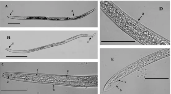
long and a diameter of 20 µm, an annulated lip region, a strong stylet with large basal knobs and an oesophagus that overlaps the intestine ventrally. Females of this species have a posterior vulva position at 73-75% and the males with paired slender spicules and usually with the bursa enveloping the tail (Fig. 1.1). The tail is conoid with a small irregular peg which is a distinguishing feature of this species (Machon
& Hunt 1985; Loof 1991). Nonetheless, the identification of P. goodeyi based on
morphological characters is complex because they are difficult to detect and also due to a high intraspecific variability. Furthermore, biochemical and molecular
characters are not well established to this species (De Waele & Elsen 2002) and the
identification is still being done on the basis of morphological characters.
Figure 1.1 Morphological characters of P. goodeyi. Female (A) and male (B). Stylet with large basal knobs and oesophagus (C), vulva position in female (D), spicules and bursa enveloping the male tail (E). Vulva a, bursa b, spicules c, stylet e, median bulb f and oesophageal glands of the oesophagus g and excretory pore h. Scale bar (A and B) 80 µm; (C, D and E) 45 µm.
P. goodeyi can be found in roots, rhizomes, tubers and in the host pseudostem. After penetrating the roots, they multiply rapidly reaching 1000 to 3500 specimens per gram of root. All life stages are considered to be infective, capable to enter and leave the root tissues. The life cycle is completed within the root in 24-30 days at 24-25 ºC, thus several generations may develop during one growing season (Gowen & Quénérhervé 1990). P. goodeyi are also found in the rhizosphere where they can survive for some time and search for new roots to infect (Machon & Hunt
1985). Females produce eggs and the first moult occurs in the egg as a first-stage
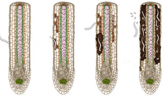
7 and their functions become severely impaired. Roots infected by P. goodeyi have small brownish-red elongated lesions that tend to enlarge and coalesce, causing an extensive root necrosis (Gowen & Quénérhervé 1990; Loof 1991) (Fig. 1.2).
Figure 1.2 Drawing representing infection by the root-lesion nematode P. goodeyi. Nematode penetration, migration, feeding and root lesion development (Adapted from Jones & Fosu-Nyarko 2014).
Symptoms of damage are identical to those observed with other plant-parasitic nematodes as Helicotylenchus multicinctus, P. coffeae or Radopholus similis and may include: stunting of plants; lengthening of the vegetative cycle; reduction in leaf size and number; reduction in bunch weight, decrease in the plantation productive life and toppling. The interactions between P. goodeyi and other nematodes as
Helicotylenchus,Rotylenchulus and Meloidogyne or P. goodeyi and other organisms as F. oxysporum or C. sordidus are common in Madeira potentiating symptoms and affecting severely the banana production (Pestana & Cravo 1999).
1.3 Plant nematode control
The management of nematode populations has relied on the use, by farmers all over the World, of expensive and toxic fumigant products (chemical nematicides) such as 1,3-dichloropropene, aldicarb, dazomet, emamectin, oxamyl, fenamiphos and metam-sodium among others (Whitehead 2002). These products are harmful to the environment, animals and humans and some of them have active substances which may affect hormones, causing a decrease in fertility, mutagenesis and carcinogenesis. Moreover, its use does not mean a suppression of nematodes and the cost of using them repeatedly is not economically viable for a sustainable agriculture. Nevertheless, chemical treatments are still used for the management of the plant-parasitic nematodes, maintaining the populations at low levels despite all the intrinsic hazards. In fact, the wide distribution and widespread application of chemicals on economically important crops made chemical nematicides detectable in ecosystems, aquifers and in other water systems of most agricultural areas (Ibrahim et al. 2006). These facts have led to the prohibition and restriction of application of such products in many countries. Legislation in European Union is continually being actualized in order to prevent and to prohibit the use of some chemical nematicides as aldicarb and 1,3-dichloropropene (EC Directive 2007/619/ EC).
Further measures can be applied to control nematodes but they are effective only when integrated with one or more practices such as: i) organic correction that
stimulates the proliferation of beneficial microorganisms or release toxins which
accelerate the decomposition of soil promoting natural equilibrium (Badra el al. 1979; Bradow 1991); ii) biological control using natural enemies including fungi, bacteria and predacious nematodes; iii) culture rotations; iv) trap crops and v) aqueous plant extracts.
1.3.1 The nematicide potential of plants
Many studies have reported that plants produce compounds that can kill or keep away harmful organisms destroying their life cycles and acting as an attractive or repellent (Ibrahim et al. 2006). These compounds can be simply organized in two major groups, one containing primary metabolites as carbohydrates, lipids, amino acids and the other including secondary metabolites that are produced from the primary metabolites and are responsible for toxic effects (Valette et al. 1998).
9
can result from the identification of those compounds which may be used as natural
nematicides or as a model for the development of synthetic products with similar activity against nematodes and at the same time environmentally safer.
Extracts of plants containing volatile compounds, especially essential oils, have an anti nematode effect (Abd-Elgawad & Omer 1995; Ibrahim et al. 2006; Katooli et al. 2010; Oka et al. 2012). It was shown that the essential oils carvacrol, linalool and thymol have nematicide properties as they are toxic to M. incognita second-stage juveniles (Ibrahim et al. 2006). Some of these oils inhibit the activity of acetyl cholinesterase, but their mode of action is still unknown (Ryan & Byrne 1988; Zuckerman & Esnard 1994; Ibrahim et al. 2006).
Other volatile and non-volatile compounds may be also present in plant extracts (Brown & Morra 1997; Ibahim 2006) and some can be detected at a distance and display an attractive or repellent effect on nematodes being antagonist (Pickett & Stephenson 1980). One of the most studied and known antagonism between nematodes and plants is the plant Tagetes. Tagetes patula L. acts as a trap crop preventing the development of giant cells produced by root-knot nematodes, having a suppressive effect on nematode populations (Belcher & Hussey 1977). Therefore, plants of this genus have been used for some time as cover crops, crop rotation, green manure or plant extracts as a source for nematode antagonism (Chitwood 2002).
Since then, many other plants such as: Artemisia vulgaris L., Artemisia annua
L., Azadirachta indica A. Juss, Brassica napus L., Cannabis sativa L., Crotolaria juncea L., Eucalyptus citriodora Hook., Gliricidia maculata H.B. & K., Glycosmis pentaphylla Retz., Kalanchoe pinnata (Lam.) Pers., Moringa oleifera Lam., Myrtus communis L., Piper betle L., Ricinus communis L., Solanum sisymbriifolium Lam., Zanthoxylum alatum Roxb. are being investigated to control plant-parasitic nematodes Ditylenchus dipsaci, G. pallida,G. rostochiensis, Meloidogyne spp., R.
similis and Tylenchulus semipenetrans among others (Jasy & Koshy 1992; Scholte 2000a,b; Costa et al. 2003; El-Rokiek et al. 2011; Dias et al. 2012; Oka et al. 2012; D’ Addabbo et al. 2013; Mukhtar et al. 2013) and this list of plants is still increasing.
Plants belonging to the genus Solanum are used widely in traditional medicine for different purposes. The chemical constituents of Solanum spp. have been described in many studies and its pharmacological and toxicological properties investigated (Kumar et al. 2001; Heo et al. 2004; Zhou et al. 2006; Jeong et al. 2007; Huang et al. 2010).To performe this study two speciesS. nigrum and S. sisymbriifolium were selected since our previous studies revealed that they are non-hosts for P. goodeyi
1.4 Parasitism and genes from plant-parasitic nematodes
Plant-parasitic nematodes use the stylet to puncture the cell wall and reach the plasma membrane where they introduce secretory proteins synthesized by the nematode that play an important role during migration through root tissues and also to feed withdrawing the nutrients from the host cytoplasm (Davis et al. 2000). The major secretory organs are the chemoreceptors (amphids, phasmids and cephalic sensilla), the oesophageal glands, the excretory system and rectum that in some nematodes can produce a gelatinous matrix to protect the eggs from predators and dehydration (Eisenback 1985; Abad et al. 2003). The nematode cuticle also regulates
selectively the flow of fluids through the body wall and may be a source of secretory
compounds which are recognized by plants as signal molecules (Robertson et al. 2000; Lima et al. 2005).
The most studied nematode secretions are related to proteins produced by amphids and oesophageal glands. Several glycoproteins secreted by amphids, located in the cephalic region, were isolated and some of them are involved in the perception of environmental signals (Stewart et al. 1993a, 1993b; Abad et al. 2003). Oesophageal glands have specialized cells capable of producing secretions, expelled through the stylet, with different roles during parasitism (Hussey & Mims 1991; Abad et al. 2003), highlighting their importance in this process.
The genes and products, which included proteins secreted by parasites to facilitate penetration, migration and to prevent plant defence responses are
designated effectors. The search and identification of those effectors can be helpful
to better understand nematode parasitism and to devise alternative nematode control measures (Atkinson et al. 2003; Davis et al. 2004; Fragoso et al. 2009; Gheysen & Mitchum 2011; Hewezi & Baum 2013; Jaouannet et al. 2013; Peng et al. 2013). Most candidate parasitism genes have been predicted using bioinformatic tools
and many effectors have been identified among other plant-parasitic nematodes.
11
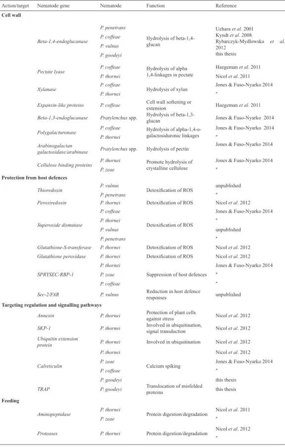
Table 1.1 Root-lesion nematodes genes and proteins related to infection and parasitism with known function.
Action/target Nematode gene Nematode Function Reference
Cell wall Beta-1,4-endoglucanase P. penetrans P. coffeae P. vulnus P. goodeyi
Hydrolysis of beta-1,4-glucan
Uehara et al. 2001 Kyndt et al. 2008
Rybarczyk-Mydlowska et al. 2012
this thesis
Pectate lyase P. coffeae
P. thornei
Hydrolysis of alpha 1,4-linkages in pectate
Haegeman et al. 2011 Nicol et al. 2011
Xylanase P. coffeae
P. thornei Hydrolysis of xylan
Jones & Fuso-Nyarko 2014
ʺ
Expansin-like proteins P. coffeae Cell wall softening or
extension Haegeman et al. 2011
Beta-1,3-endoglucanase Pratylenchus spp. Hydrolysis of
beta-1,3-glucan Jones & Fuso-Nyarko 2014
Polygalacturonase P. coffeae
P. thornei
Hydrolysis of alpha-1,4-o-galactosiduronic linkages
Jones & Fuso-Nyarko 2014
ʺ
Arabinogalactan
galactosidase/arabinase Pratylenchus spp. Hydrolysis of pectin
Jones & Fuso-Nyarko 2014
Cellulose binding proteins P. thornei
P. zeae
Promote hydrolysis of crystalline cellulose
Jones & Fuso-Nyarko 2014
ʺ
Protection from host defences
Thioredoxin P. vulnus
P. penetrans Detoxification of ROS
unpublished
ʺ
Peroxiredoxin P. thornei Detoxification of ROS Nicol et al. 2012
Superoxide dismutase
P. coffeae P. thornei P. vulnus P. penetrans
Detoxification of ROS
Jones & Fuso-Nyarko 2014
ʺ
unpublished
ʺ
Glutathione-S-transferase P. thornei Detoxification of ROS Nicol et al. 2012
Glutathione peroxidase P. thornei Detoxification of ROS Nicol et al. 2012
SPRYSEC-RBP-1
P. thornei P. zeae P. coffeae
Suppression of host defences
Jones & Fuso-Nyarko 2014
ʺ ʺ
Sec-2/FAR P. vulnus Reduction in host defence
responses unpublished
Targeting regulation and signalling pathways
Annexin P. thornei Protection of plant cells
against stress Nicol et al. 2012
SKP-1 P. thornei Involved in ubiquitination,
signal transduction Nicol et al. 2012
Ubiquitin extension
protein P. thornei Involved in ubiquitination Nicol et al. 2012
Calreticulin P. thornei P. zeae P. coffeae P. goodeyi Calcium spiking
Nicol et al. 2012 Jones & Fuso-Nyarko 2014
ʺ
this thesis
TRAP P. goodeyi Translocation of misfolded
proteins this thesis
Feeding
Aminopeptidase P. thornei
P. zeae Protein digestion/degradation
Nicol et al. 2011
ʺ
Proteases P. thornei Protein digestion/degradation Nicol et al. 2012
To understand the infection/parasitism mechanisms we select from the above list the effectors: calreticulin because it may be injected through the nematode stylet into plant tissues (Jaubert et al. 2005; Suchitra & Johi, 2005; Vieira et al. 2011)
and has been identified as playing an important role in infection and parasitism
Chapter 2
2. Material and Methods
2.1 Biological material
2.1.1 Pratylenchus goodeyi isolates
Samples of soil and banana roots were collected on the Southern coast of Madeira Island. The nematodes Pratylenchus goodeyi Sher & Allen 1953 were
extracted from soil by centrifugal flotation using the sucrose method(Jenkins 1964; Abrantes et al. 1976) and from roots by the maceration and sieving method (Abrantes et al. 1976; Hooper 1986). The roots were cut into pieces of approximately 1 cm and ground in a blender with 100 mL of water for 20 s. The suspension was transferred to a small sieve, with a mesh of approximately 45 µm, placed in a Petri dish. After 48 h the suspension was poured into a sieve of 38 µm and washed with sterile distilled water. The material retained on the 38 µm sieve was transferred to a beaker, ressuspended in sterile water, placed on a Doncaster plate and observed in a stereomicroscope (Nikon SMZ-U). The identification of P. goodeyi isolates based
on morphological characters was confirmed at the “Istituto per la Protezione delle
Piante Sezione di Bari” Italy.
2.1.2 Nematode multiplication
Two methods were used for in vitro nematode multiplication: Inoculation
on banana plants and carrots discs. On the first method nematode isolates were
transferred to 10 mL of sterile water and quantified. Populations of root-lesion
nematode P. goodeyi were then inoculated on banana plants (Musa acuminata
Colla) from in vitro culture, 20 cm height, in pots containing sterilized soil. Using a glass stick three holes were made around the plant, which were covered with soil after nematode inoculation. These potted plants remained in the laboratory, being watered whenever necessary. In addition, multiplication of P. goodeyi was also performed on carrot discs as described by Nico et al. (1999) and maintained in the laboratory at 24 ºC to be used in molecular analysis.
2.1.3 Plant material
15 kept in laboratory for the production of fruits and seeds.
Seeds of both plants were germinated in sterile peat and the plants were maintained in a greenhouse, under a 16 h photoperiod, day/night temperature of 35 ºC/18 ºC, respectively, and relative humidity of 70% until they reached 50-60 cm height. They were then collected and divided into two samples: one was weighed and frozen for further analysis (named fresh plant) and the other was placed in a ventilated drying chamber at 30 °C (named dry plant). Dried plants were ground in a cutting mill (Mod. 5KH35KG 254E, Arthur H. Thomas Co. Phila., PA., U.S.A.) and passed through sieves of 40 and 60 mesh (type AS200, Retsch, Germany). The 40-60 mesh size fraction (425-250 µm) was used to extract chemical compounds with water and organic solvents. Samples water content was determined on a moisture balance (Gibertini-Eurotherm).
2.2 Chemicals
All reagents used on plant extractions and chemical analysis were highly pure (p.a., 99% purity) and supplied by Sigma-Aldrich (Madrid, Spain): dichloromethane, trimethylchlorosilane, N,O-bis(trimethylsilyl)trifluoroacetamide, pyridine,
stigmasterol (95%), octadecanoic acid, nonadecan-1-ol, tetracosane, acetone (≥
99.5%) or Fluka Chemie (Madrid, Spain): ethanol absolute (≥ 99.8%).
2.3 Plant extractions
2.3.1 Solanum aqueous extractions
Two approaches were used to make the aqueous extractions: blender and
reflux. In the blender approach, S. nigrum and S. sisymbriifolium were extracted in water at a ratio of 1:4 or 1:20 (w/v, material/water) of fresh plant or dry plant, respectively. The plant material was gently shaken in water, ground for 10 min in the
blender and filtered using a G4 filter, pore size 10-16 µm (DURAN, Germany). This
procedure was performed using water either at room temperature (cold water) or
boiling (hot water). The same material/water ratio was used in the reflux approach. The plant material was refluxed for 1 h and the liquid fraction was obtained after filtration through a G4 filter.
All solid residues were lyophilized, quantified and stored at -20 ºC, in the
2.3.2 Solanum sequential extraction
Milled dried material placed in handmade cartridges (4cm diameter × 10 cm length) was Soxhlet extracted in a sequential extraction of at least 10 hours each with the following solvent sequence: dichloromethane, acetone, ethanol and water. Each extraction was followed by solvent evaporation in a rotative evaporator (R-200 Büchi, Sigma-Aldrich, Spain) combined with a vacuum pump (V-500 Büchi Vac, Sigma-Aldrich, Spain)and a bath (B-490 Büchi, Sigma-Aldrich, Spain) at a maximum temperature of 40 ºC. The extracts were collected and after drying under vacuum until constant weight, the percentage of extractives (compounds present at each extract fraction) was determined gravimetrically.
After ethanol extraction, the plant material that remained on the cartridges
was washed with ethanol and dried at 30 °C. This material was refluxed for 1 hour to obtain the extraction in water. These solutions were filtered under vacuum (G4 porosity), lyophilized and quantified gravimetrically.
All solid residues from each fraction were stored in cold and dark conditions until chemical analysis or mortality assessment on P. goodeyi.
2.4 Chemical analysis
2.4.1 Samples preparation and selection
Dichloromethane, acetone and water solid residues from S. nigrum and S.
sisymbriifolium were selected to carry out chemical analysis. The alkaline hydrolysis was performed as described by Oliveira et al. (2008).
2.4.2 Gas chromatography-mass spectrometry (GC-MS)
Dichloromethane solid residue (20 mg) from S. nigrum (SN) and S.
sisymbriifolium (SS) was dissolved in 250 µL of pyridine to convert compounds with hydroxyl and carboxyl groups into trimethylsilyl ethers and esters (TMS) following the protocol described by Oliveira et al. (2005). The derivatised extracts were analysed by GC-MS at chromatographic conditions used previously (Oliveira
et al. 2008; Vilela et al. 2014). Compounds were identified as TMS derivatives,
comparing mass spectra with GC-MS spectral library (Wiley-NIST Mass Spectral
Library 1999), retention times and fragmentation profiles with published data and
17 Duke’s Phytochemical and Ethobotanical database (http://www.ars-grin.gov/duke/) (Filimonov 1995) and the free Chemical structure database ChemSpider (http:// chemspider.com).
2.4.3 Ultra-high performance liquid chromatography (UHPLC)
The analyses of acetone and water solid residue from both plants were performed by an UHPLC system consisted of a variable loop Accela autosampler (200 vial capacity set at 15 °C), an Accela 600 LC pump and an Accela 80 Hz PDA
detector (Thermo Fisher Scientific, San Jose, CA, U.S.A). The separation of the compounds was carried out with a gradient elution program at a flow rate of 0.48
mL/min, at 45 °C, using a Kinetex C18 (50 mm × 2.1 mm × 1.7 μm) column supplied by Phenomenex. The injection volume in the UHPLC system was 10 μL and the mobile phase consisted in water:acetonitrile (99:1,v/v) (A) and acetonitrile (B), both with 0.1% of formic acid. The following linear gradient was applied: 0-4 min: 2%B, 4-7 min: 2-4.5%B, 7-20 min: 4.5-12%B, 20-22 min: 12-12.8%B, 22-25 min: 12.8-30%B, 25-30 min: 30-100%B, 30-34 min: 100-2%B followed by re-equilibration of the column for 4 min before the next run. Double online detection was carried out in the diode array detector, at 280 and 340 nm, and UV spectra in a range of 200-600 nm were also recorded. Before the injection, each solid residue was
dissolved in water or acetone HPLC grade, to obtain a final extract concentration between 10 and 30 mg/mL, and then filtered through a 0.2 μm PTFE syringe filter
(Santos et al. 2013).
2.4.4 Mass spectrometry
A LCQ Fleet ion trap mass spectrometer (ThermoFinnigan, San Jose, CA, U.S.A.) was used, equipped with an electrospray ionization source and operating in negative mode (Santos et al. 2013). The nitrogen sheath and auxiliary gas were 40 and 10 (arbitrary units), respectively. The spray voltage was 5 kV and the capillary temperature was 350 ºC. The capillary and tune lens voltages were set at -25 V and -125 V, respectively. CID-MSn experiments were performed on mass-selected
2.4.5 HPLC quantification
Calibration curves were obtained by UHPLC injection of gallic, caffeic and chlorogenic acids and catechin, quercetin, isorhamnetin and luteolin standard
solutions in methanol, with five different concentrations each, between 2 and 86.4 μg/mL. Besides the linearity, the limits of detection (LOD) and quantification
(LOQ) were also estimated using the S/N approach (n=5). The calibration curves
and additional relevant analytical data are shown in Table 2.1. The quantification of
individual compounds was accomplished with calibration data for the most similar standard, since for some of them no pure reference compounds were available (Santos et al. 2013).
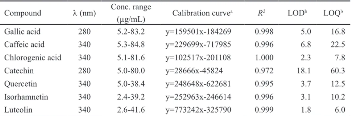
Table 2.1 Calibration data used for the UHPLC–UV quantification of phenolic compounds in
Solanumnigrum and S. sisymbriifolium acetone and water extracts.
Compound λ (nm) Conc. range
(µg/mL) Calibration curve
a R2 LODb LOQb
Gallic acid 280 5.2-83.2 y=159501x-184269 0.998 5.0 16.8 Caffeic acid 340 5.3-84.8 y=229699x-717985 0.996 6.8 22.5 Chlorogenic acid 340 5.1-81.6 y=102517x-201108 1.000 2.3 7.8 Catechin 280 5.0-80.0 y=28666x-45824 0.972 18.1 60.3 Quercetin 340 5.0-38.4 y=248648x-622681 0.995 3.7 12.5 Isorhamnetin 340 2.4-39.2 y=252963x-246614 0.996 3.1 10.2 Luteolin 340 2.6-41.6 y=773242x-325790 0.999 1.8 6.0
a y = peak area, x = concentration in µg/mL, LOD limit of detection, LOQ limit of quantification, b expressed
in µg/mL.
Bioactivities of phenolic compounds were predicted in silico through the online available tools: Dr. Duke’s Phytochemical and Ethobotanical database and free chemical structure database ChemSpider as described above (2.4.2).
2.5 Biological assays
2.5.1 Nematicidal activity
S. nigrum and S. sisymbriifolium solutions were prepared using the solid residue obtained from aqueous and sequential extractions at exact quantities of each fraction corresponding to the initial ratio of fresh and dry plant dissolved in water (basal concentration) and at the mean concentration of 10 mg/mL.
19 activity were tested in vitro against P. goodeyi. The biological assays were conducted using l mL of each extract in a Syracuse containing 10 adults P. goodeyi and left in the dark at room temperature (25 ± 1 ºC) for 10 days. Each assay was replicated
five times and sterile distilled water was used as control. Nematodes were observed
daily using a binocular microscope and numbers of inactive or dead nematodes
were recorded. Considering the efficiency of acetone extract nematode mortality
was monitored in this extract for a period of 24 h. Nematodes were considered dead when after being transferred to sterile water for 2 h and stimulated by prodding they remained inactive. Registered mortality was converted into cumulative mortality corrected by Abbott’s formula (Abbott 1925). These biological assays were performed twice.
2.5.2 Mobility and attraction
A solution of Pluronic gel (23% w/v; Pluronic F-127, Sigma) was prepared by stirring to dissolve the powder in sterile distilled water for 24 h at 4 ºC (Wang et al. 2009). The resulting gel was refrigerated at 4 ºC until used for nematode behaviour studies. A root tip (Rv), 1 cm long, from banana plants cultured in vitro was placed in the middle of a Syracuse (2 cm diameter) containing 1 mL of Pluronic gel. Ten
P. goodeyi, mainly adults, were hand-picked and put on the edge of each Syracuse and the Pluronic gel allowed to solidify at room temperature (Wang et al. 2009). A Syracuse containing Pluronic gel, but without a root, was used as control to assess nematode movement. Nematode behaviour as mobility and attractivity toward the root was recorded after 2, 4, and 6 h by observing the movement and the number of nematodes that reached the root.
2.5.3 Infection
Drops (150 µL) of S. nigrum acetone extract (10 mg/mL) were placed on microscopic glass chambered test slides to perceive the nematicide effects of the water soluble compounds on the P. goodeyi mobility and ability to infect the roots of banana plants. Ten nematodes, J2, females and males, were placed on the chambers of the microscopic glass and exposed to the extract for 0, 6, 12 and 18 h. After exposure, nematodes were transferred to Syracuses containing a root tip embedded in the Pluronic gel as mentioned above (2.5.2). The Syracuses were incubated at room temperature and mobility and attractivity were determined as before. The
percentage of infection was recorded and quantified (number of nematodes inside
the roots/total number of nematodes inoculated x 100) after 1, 2 and 3 h (Mukhtar
if they did not move when stimulated by prodding. The reduction in infection over control was calculated [(infection in control/infection in treated) - 1] (Mukhtar et al. 2013).
2.6 Molecular analysis
2.6.1 Biological samples
Roots from banana plants cultured in vitro (Rv), in sterilized soil (Rp) and infected with P. goodeyi (RPg) were washed with tap water, wiped and cut directly into a mortar containing liquid nitrogen, ground with a pestle and stored at -70 °C until the extraction of RNA.
Nematodes, mainly adults, obtained as previously described (2.1.1) from both infected roots and carrots discs were transferred manually to an Eppendorf tube containing sterilized distilled water in order to get P. goodeyi (Pg) samples of ca. 4000 individuals. These samples were quickly frozen in liquid nitrogen, ground with a micropestle and used immediately for the extraction of RNA.
2.6.2 RNA extraction and cDNA synthesis
Total RNA was extracted from 100 mg of crushed uninfected banana roots (Rv and Rp) using the RNeasy Plant Mini Kit (Qiagen, U.S.A.) upon a minor
modification. PVP40T at 1% and 0.4 volumes of 3 M potassium acetate (pH 6.5)
were added to the RLC extraction buffer. Samples were incubated on ice for 15 min and centrifuged at 14000 g at 4 °C for 15 min. Then 0.5 volumes of ethanol were added to the supernatant. The RNA was treated with DNase (Qiagen, U.S.A.) to remove possible DNA contamination and cleaned according to the manufacturer’s instructions.
The extraction of total RNA from nematodes (Pg) and from 5 mg of RPg samples were performed using the RNeasy Plus Micro Kit (Qiagen, U.S.A.) following the manufacturer’s instructions and treated with DNAse.
All RNA extractions were confirmed on a 1% agarose gel buffered with TAE
1x containing ethidium bromide (0.5 µg/mL) and photographed under UV light (DigiGenius, Syngene, U.K.).
21
2.6.3 cDNA amplification and cloning
Internal partial sequences of calreticulin (CRT) and beta-1,4-endoglucanase
(ENG) cDNAs from P.goodeyi were obtained by amplification through degenerate
oligonucleotides. CRTf1 and CRTr1 (Table 2.2) were designed from the conserved regions of MFGPIC and KWIHPEI, respectively, on amino acid sequences of calreticulin from different nematode species obtained through the National Center for Biotechnology Information (NCBI) database (Heligmosomoides polygyrus: CAL30086; Caenorhabditis elegans: CAA42159; Necator americanus: CAA07254;
Meloidogyne incognita: AAL40720; Onchocerca volvulus: AAA59056). ENG1 and ENG2 oligonucleotides designed for the gene encoding beta-1,4-endoglucanase
(Rosso et al. 1999) were used to amplify the correspondent internal partial sequence of the cDNA and a fragment coding for translocon-associated protein (TRAP) arose through serendipity.
Table 2.2 Primers used in this study for PCR amplification, RACE, RT-PCR, and expression
analysis.
Gene Primer name Sequences 5’ 3’ Cycles T. annealing (ºC)
Calreticulin CRTf1 ATGTTYGGICCIGAYATHTG 35 51
CRTr1 ATYTCIGGRTGDATCCAYTT 35 51
PgCRTf2 CGTTGGCTACAACGGGAAGAACC 35 65
PgCRTr2 TCAGGGATGTGCTCGGGCTTCT 35 65
Beta-1,4-endoglucanase ENG1 TAYGTIATHGTIGAYTGGCA 40 50
ENG2 GTICCRTAYTCIGTIACRAA 40 50
PgENGf3 CCACTGCCTACCCCGACAAAGC 35 67
PgENGr4 CACGAAGGGGCTGCTTGTGGGA 35 67
Translocon-associated protein PgTRAPf ATGCCGTCGTAAATGGCCAGAGC 35 64
PgTRAPr1 GTTGCTGACACCATTTTGGACGG 35 65
PgTRAPr2 GTTGTTGTTTGAGGTGATAGC 35 64
Ubiquitin UBI5 ATGCAGATYTTTGTGAAGAC 35 50
UBI3 ACCACCACGRAGACGGAG 35 50
Rubisco A5.2 GAGACCCTCTCCTACCTTCCT 35 50
B8.1 TTCCACATGGTCCAGTASCGC 35 50
The PCR amplification performed in a 50 µL reaction mixture contained 5 µL
oligonucleotide (Table 2.2). PCR products were gel purified through the QIAquick
gel extraction kit (Qiagen, U.S.A.) and cloned into pJET 1.2/blunt plasmid vector using the CloneJET PCR Cloning Kit (Fermentas, Lithuania) and as host Escherichia coli DH5α competent cells.
After sequencing specific oligonucleotides PgCRTf2, PgCRTr2, PgENGf3, PgENGr4, PgTRAPf, PgTRAPr1 and PgTRAPr2 were designed through the Primer 3 program (http://primer3.wi.mit.edu/). The oligonucleotides PgCRTf2 and PgCRTr2 were used to clone, respectively, the 3’ and the 5’ ends of CRT. The 3’ and 5’ ends of ENG were generated using PgENGf3 and PgENGr4 specific primers, respectively and PgTRAPf and PgTRAPr1 were used to clone, respectively, the 3’ and the 5’ remaining cDNA sequences. Rapid amplification of cDNA ends (RACE) were conducted using adaptors, oligonucleotides, enzymes and procedures from
the SMARTer™ RACE cDNA Amplification Kit (Clontech, U.S.A.). The amplified
5’ and 3’ cDNA fragments of the expected size were removed from the gel and
purified as before (QIAquick gel extraction kit) or using the High Pure PCR Product Purification Kit (Boehringer Mannhein, Germany) following the instructions of
their respective manufacturers. The 5’ ends were cloned into pJET 1.2/blunt cloning vector (CloneJET PCR Cloning Kit) and the 3’ cDNA ends into pGEM ®-T Easy vector (Promega, Spain) following the manufacturers’ guidelines.
2.6.4 Plasmid extraction, sequencing and sequence analysis
Plasmid DNA from transformed colonies was extracted using the GeneJET Plasmid Miniprep kit (Fermentas, Lithuania) and eluted in 50 µL of elution buffer (10 mM Tris-HCl, pH 8.5). Plasmids were sequenced on both directions using commercial AB 3739XL capillary sequence (Macrogen Europe, Amsterdam, The Netherlands). DNA sequence data were analyzed through the NCBI web site (www.ncbi.nlm.nih. gov/) (Benson et al. 2013). The BLAST program was used to search for sequence homology in nucleotide and amino acids database (Altschul et al. 1997). The Expasy server (http://web.expasy.org/) through the available translate tool was used to translate the cDNA sequences and to determine the physicochemical parameters (ProtParam tool) (Gasteiger et al. 2005) of the deduced proteins: calreticulin, beta-1,4-endoglucanase and translocon-associated protein. Multiple protein sequence alignments were generated by ClustalW2.1 at the European Bioinformatics Institute (EBI) website (http://www.ebi.ac.uk.) using standard parameters and an alignment plot was created by The Sequence Manipulation Suite (http://bioinformatics.org/ sms/). Predictions for the signal peptide were performed using SignalP-4.1 Server (http://www.cbs.dtu.dk/services/SignalP/) (Petersen et al. 2011). Proteins motifs
23 Domain Database from NCBI. EBI Phobius (Käll et al. 2007) and Interproscan (Quevillon et al. 2005) were used to scan proteins signatures. The tool pTarget (http://golgi.unmc.edu) (Guda & Subramaniam 2005; Guda 2006) and PSORT
(Prediction of Protein localization Sites) were used to confirm the possible location
of the proteins (Nakai & Kanehisa 1992). The PredictProtein Server (Rost & Liu 2003) and the TMHMM (http://www.cbs.dtu.dk) were employed to obtain the topology prediction. The program ProtFun 2.2 from the Center for Biological
Sequence Analysis (CBS) web site (http://www.cbs.dtu.dk) was used to find
possible functions of the proteins (Jensen et al. 2002, 2003).
2.6.5 RT-PCR analysis
RT-PCR analysis was performed with 5 µL of cDNA as template diluted 5x for nematode samples (Pg and RPg) and 10x for root samples (Rv and Rp)
in a 50 µL reaction mixture as described above (2.6.3). cDNA amplifications
were performed with the correspondent oligonucleotides (Table 2.2). PgCRTf2, PgCRTr2, PgENGf3, PgENGr4, PgTRAPf and PgTRAPr2 were used to amplify a fragment of CRT, ENG and TRAPδ, with 291, 315 and 320 bp respectively. UBI5 and UBI3 primers (Laplaze et al. 2000) were used to amplify a fragment of ubiquitin and as an internal control gene to normalize the amount of cDNA in semi-quantitative RT-PCR analysis. Also, A5.2 plus B8.1 primers designed from banana plant cDNA (Thomas-Hall et al. 2007) were used to amplify a fragment of rubisco. Amplicons were separated on a 1.5% agarose gel visualized under UV light as previously describe (2.6.2). The oligonucleotides used in this work were
synthesized by “STAB VIDA, Lda, FCT/UNL” (Caparica, Portugal) except the ubiquitin pair that was prepared by Invitrogen (Life Technologies, Spain).
2.6.6 Expression analysis
2.7 Statistical analysis
Unless otherwise stated at least three replicates were performed and data from
S. nigrum and S. sisymbriifolium sequential and aqueous extractions, P. goodeyi mortality, gene expression (calreticulin, beta-1,4-endoglucanase and translocon-associated protein delta subunit), infection and reduction of infection were subject to statistical analysis using the SPSS (Statistical Package for the Social Sciences) 15.0 software for Windows.
Data of the amount of extractives from each extract sequential fraction and from aqueous extractions were analysed to assess normal distribution by Kolmogorov-Smirnov and Shapiro-Wilk tests (P > 0.05) and one-way analysis
of variance (ANOVA) was used to find significant differences between extracts. Significant differences were further analyzed by Tukey’s multiple range test (P < 0.05). Registered mortality in S. nigrum and S. sisymbriifolium sequential extracts and aqueous extracts was converted into cumulative mortality and corrected by Abbott’s formula (Abbott 1925) prior to analysis. The previous statistical tests were
then carried out to analyzed normal distribution and to find significant differences
Chapter 3
Nematicidal activity of
Solanum
sisymbriifolium
and
S. nigrum
extracts against
the root-lesion nematode
Pratylenchus goodeyi
Margarida Pestana, Mónica Rodrigues, Lucília Teixeira, Isabel de O. Abrantes, Manuela Gouveia, Nereida Cordeiro
3.
Nematicidal activity of
Solanum sisymbriifolium
and
S.
nigrum
extracts against the root-lesion nematode
Pratylenchus
goodeyi
3.1 Abstract
The root-lesion nematode Pratylenchus goodeyi is a parasite of banana plants, frequently detected in Madeira Island (Portugal) affecting culture development and consequently the production, with economical damages. To identify the phytochemicals of Solanum sisymbriifolium and S. nigrum with nematicidal properties and determine the effect of those components on P. goodeyi, an extraction sequence of at least 10 hours each from dried plants was used. The chosen solvent sequence was: dichloromethane, acetone, ethanol and water. According to the results both plants have in their composition chemical components mainly found in water extracts, which affects the mobility and mortality of the root-lesion nematode. S sisymbriifolium and S. nigrum have potential to be used as a natural and environmentally friendly nematicide to control P. goodeyi.
3.2 Introduction
The root-lesion nematode Pratylenchus goodeyi Sher & Allen 1953 is very common in Madeira Island affecting banana culture. In order to control nematode populations farmers use phytopharmaceutical products, which also contribute to contaminate soil, groundwater and air. It is therefore of great importance to study alternative routes to those products by seeking less harmful chemicals to the environment and humans. Thus, some plants with nematicidal potential and its application have been analyzed (Musabyimana & Saxena 1999; Rahman & Somers 2005).
It is known that the incorporation of organic waste has a considerable impact on physical and biological properties of soil, promoting a favorable environment for the development of nematode antagonists (Badra et al. 1979; Bradow 1991; Bello
et al. 2000). In some cases, it can be also ascertain toxicity to some nematodes. Since plants are capable of producing a large variety of secondary metabolites with
multiple applications, much research has been conducted to find substances in plant
27
et al. 2003). Several benefits can result from the identification of phytochemicals
involved in these interactions, which may be used as nematicidal or can serve as a model for the development of synthetic products with positive activity on nematodes or on the environment around them (Chitwood 2002).
Several chemical compounds present in Solanum species as steroidal glycosides and alkaloids among others have a broad spectrum of activity (Perez
et al. 1998; Raju et al. 2003; Heo et al. 2004; Zhou et al. 2006; Jeong et al. 2007; Lin et al. 2007, 2008; Ji et al. 2008) and is therefore of great interest to develop studies for the application of this plant genus in different areas. Among this species
Solanum sisymbriifolium Lam., which does not exist in Madeira Island, has been successfully used to control populations of potato-cyst nematodes, Globodera spp. (Scholte 2000a) whereas S. nigrum L. very common in Madeira is believed to have therapeutic properties against some types of tumors since some compounds showed cytotoxic effects in tumor cells (Zhou et al. 2006). Recent studies revealed that S.
sisymbriifolium and S. nigrum are not good or non-hosts of P. goodeyi (Pestana et al. 2009). In addition, the incorporation of these plants into soil improved banana plant growth, directly through the release of exudates with nematicidal effect and indirectly by promoting the development of antagonists and making the rhizosphere unfavorable to the nematode.
In order to search for nematicidal substances plant extracts from S.
sisymbriifolium and S. nigrum were evaluated against P. goodeyi.
3.3 Results
3.3.1 Fractions of S. sisymbriifolium and S. nigrum dry material
Figure 3.1 shows the amount of extractives (compounds present in each solid residue) from sequential Soxhlet extraction of S. sisymbriifolium and S. nigrum plants that was determined after a complete extraction in each solvent. The results clearly indicate a predominant amount of polar fractions. Water solid residue revealed the highest amount of extractives from either S. sisymbriifolium or S. nigrum followed by ethanol solid residue. The compounds extracted in dichloromethane and acetone were 6 to 8 fold lower, respectively, than the compounds extracted in ethanol and water.
Normal distribution of the amount of extractives from each sequential
extract was confirmed (P > 0.05) through Kolmogorov-Smirnov and Shapiro-Wilk
normality tests. There were significant differences by ANOVA for S. sisymbriifolium
Figure 3.1 Percentage ofextractives from dry S. sisymbriifolium and S. nigrum plants obtained from solvents sequence: dichloromethane (DMC), acetone (Acet), ethanol (EtOH) and water. Results are the mean of 5 replicates ± SD.
3.3.2 P. goodeyi mortality and mobility in S. sisymbriifolium and S. nigrum
extracts
Bioassays of the S. sisymbriifolium and S. nigrum extracts solutions against the nematode P. goodeyi were madewith the different extractives fractions in concentration corresponding to 25 g of fresh plant per 100 mL of water (basal concentration). The extracts revealed differences at the toxicity level. The mobility of P. goodeyi was not or little affected in dichloromethane and acetone extracts from both plants, slightly affected in ethanol extracts but very affected in water extracts (Fig. 3.2). Nematodes placed in water extracts revealed lack of mobility on the second day and the recuperation of their mobility tested on sterile water diminished onwards. Further studies would be necessary to determine this effect through time (hours) in order to evaluate the nematicidal potential of both plants.
29 The values obtained for P. goodeyi mortality, subjected to different extracts showed a normal distribution by Kolmogorov-Smirnov and Shapiro-Wilk (P > 0.05)
normality tests. Tukey test showed significant differences in P. goodeyi mortality (P < 0.05) within the extracts, being water the most effective, reaching values of 99% for both plants. Dichloromethane and acetone extracts were statistically
insignificant on the nematodes death (P > 0.05)(Fig. 3.3).
Figure 3.3 P. goodeyi cumulative mortality for 10 days in S. sisymbriifolium (A) and S. nigrum (B) solvents sequence extracts.
3.4 Discussion