INTRODUCTION
Studies of cell proliferation are important for under-standing the biological processes involved in the forma-tion and development of multicellular organisms. Tradiforma-tion- Tradition-ally, the techniques used to measure DNA synthesis or cell proliferation were based on 3H-thymidine incorporation into cells, tissues and organs (Taylor et al., 1957), with the radioactivity incorporated being detected by autoradiogra-phy, fluorography or liquid scintillation. However, the long time required by these procedures and the need to use po-tentially dangerous and expensive materials have led to the development of non-radioactive methods.
One such non-radioactive approach is based on the use of 5-bromo-2’-deoxyuridine (BrdU). This thymidine analog is incorporated into DNA during the S phase of the cell cycle and is detected in histological sections using antibodies conjugated to enzymes or fluorescent dyes (Gratzner, 1982; Schutte et al., 1987; Mozdziak et al., 1994). Monoclonal antibodies against BrdU react with low affin-ity (Ka about 10-6 M) and only to single strands of DNA (Kitchin and Brown, 1995). Therefore, BrdU detection depends on avoiding conditions that may interfere with the antigen-antibody interaction.
Most procedures for immunohistochemical analy-sis are based on paraffin infiltration into biological ma-terial or cryostatic sectioning. Immunohistochemical lo-calization of cellular components depends on adequate tissue preservation with no destruction of antigenic sites,
and on the acquisition of sufficiently thin sections that provide a good resolution of subcellular structures (Kim-berly and Slpecky, 1995). Materials embedded in paraf-fin are generally well preserved, but the need for rela-tively thick sections makes high resolution analysis dif-ficult. Furthermore, excessive exposure to organic sol-vents and high temperatures during infiltration can com-promise the intactness of antigenic sites. In contrast, cryostatic sectioning of biological material is done at low temperatures with no need of exposure to organic sol-vents. Unfortunately, the sections are also thick, and cells can be damaged during freezing, resulting in injury to sub-cellular components and lower resolution of the final sig-nal. These negative effects are particularly important in delicate embryonic tissues.
An alternative to the above procedures, which pro-vides excellent preservation of the cellular morphology and antigens in delicate tissues, is the use of methacrylate-based plastic resins (Cole and Sykes, 1974). The use of these resins in immunohistochemistry depends on exposure of the antigenic site and on the antibody’s capacity to pen-etrate through the pores formed during polymerization of the resin. Plastic resins have been used successfully in his-tological studies in adult animals, but few investigations have examined their usefulness in embryonic tissue.
This paper describes the immunohistochemical de-tection of proliferating cells in high quality histological sections obtained from the neural tube and somites of chicken embryos embedded in plastic resin.
METHODOLOGY
Identification of proliferating cells in chicken embryos using
5-bromo-2’-deoxyuridine immunohistochemical detection
Mário Lúcio Lopes1, Gilberto Silber Schmidt2 and Luiz Lehmann Coutinho1
Abstract
Chicken embryos were incubated with BrdU, embedded in plastic resin, sectioned and screened immunohistochemically to identify prolifer-ating cells in the neural tube and somites. Fixation in 4% paraformaldehyde for 1 h was essential for detecting specific colorimetric signals of BrdU incorporation into cells during the S phase of the cell cycle. Transverse sections of the neural tube showed that the nuclei of proliferating cells (BrdU positive) had a uniform and centralized distribution, whereas unstained nuclei were found only along the extremities of the neural tube. Transverse sections of differentiated somites showed proliferating cells in the scleratome and dermatome. However, no incorporation of BrdU was observed in myotomic cells, which give rise to axial skeletal muscle. In spite of their proximity, the dermatome and myotome showed marked differences in cell proliferation. The excellent preservation of morphological characteristics in the embryonic tissues facili-tated identification of variations in BrdU incorporation.
MATERIAL AND METHODS
Incorporation of BrdU into chicken embryo DNA
Chicken embryos were collected after incubation for 56 h at 37°C and were maintained at this temperature in PBS solution (0.14 M NaCl, 2.7 mM KCl, 10 mM Na2 HPO4 and 1.8 mM KH2PO4, pH 7.4) containing 100 µmol of BrdU (Boehringer Mannheim). The embryos were subsequently washed twice in PBS (15 min each) to remove excess BrdU and then fixed in PBS with 4% paraformaldehyde for 1 h at 4°C, dehydrated in alcohol (25, 50, 70, 90 and 100% etha-nol, 5 min each) and embedded in JB-4 plastic resin (Polysciences).
Preparation of embryos for sectioning
Embryos were transferred to a microcentrifuge tube containing 750 µl of 100% ethanol and 750 µl of JB-4 resin A with 0.5% catalyzer. After mixing for 60 min at 4°C, the solution was removed and 1,500 µl of resin then added. Incubation was continued for 48 h at 4°C with eight changes every 2 h, and two more changes after 16 h each. Blocks were prepared in a plastic mold with polymerizing agent (Polysciences) at a ratio of 25:1 (JB-4 Sol A: polymeriz-ing agent) followed by 60 min for polymerizpolymeriz-ing at room temperature. Five- to six-µm sections were cut with a steel blade. BrdU was detected after transferring the sections to glass slides, followed by drying at 37°C.
BrdU detection
The sections were hydrated with PBT-01 (PBS plus 0.1% Triton X-100) for 5 min at room temperature. Excess pro-tein was removed by treatment with pronase E (100 µg/ml of PBT) for 60 min at 37°C, after which the slides were incu-bated at 65°C for 10 min in a water bath, followed by wash-ing in 30 ml of 0.5 M glycine (pH 2.0) and 20 ml PBT plus 0.1 M glycine (5 min each) at room temperature. After blot-ting with filter paper, the sections were incubated in 200 µl of incubation buffer (Boehringer Mannheim) containing monoclonal mouse antibody against BrdU (diluted 1:10), for 30 min at 37°C. The slides were washed twice with 20 ml PBT-01 (10 min each) to remove excess antibodies, incubated for 30 min at 37°C in 200 µl of PBT-01 containing polyclonal antibody conjugated with alkaline phosphate (1 unit/ ml) and directed against mouse monoclonal antibody. Following this incubation, the slides were washed twice in 20 ml PBT-01 (10 min each) and once in TSM (0.1 M Tris-HCl, 0.1 M NaCl, 0.05 M MgCl
2, pH 9.5) for 5 min. Sections were blotted with filter paper before colorimetric detection of BrdU. The substrates used were 5-bromo-4-chloro-3-indoly-1 phosphate plus nitroblue-tetrazolium (BCIP/NBT) at con-centrations of 0.17 mg/ml (BCIP) and 0.33 mg/ml (NBT) in 200 µl of TSM. The sections were developed in a dark room with rapid exposures to light for photomicrography.
Total nucleus detection
Specificity of the signals obtained in the immunohis-tochemical analyses was confirmed by total nucleus local-ization, through modification of the procedure described by Goodpasture and Bloom (1975). A 50% silver nitrate solution was applied to the slides, covered with another slide and kept at room temperature for 20 min. The cover slide was then removed and the sections washed in 25 ml deion-ized water. The histological sections were stained with 0.2 M NaOH and monitored by microscopy. When the nucleus turned brown, the reaction was interrupted by two washings in 25 ml of deionized water.
RESULTS AND DISCUSSION
Immunohistochemical analyses
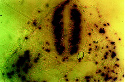
Proliferating cells that incorporated BrdU were clearly identified by the formation of dark signals in the nucleus (Figure 1). The detection process was quick, simple and did not require special equipment. However, to improve the signal specificity and intensity, it was necessary to re-duce the fixation time of the embryos in 4% paraformalde-hyde to 1 h and to remove excess proteins with pronase E in order to expose the antigenic sites over the DNA molecules. Washing with Triton X-100 (0.1%) reduced the background reactions caused by nonspecific absorption of the antibod-ies without decreasing the sensitivity for BrdU.
According to Schutte et al. (1987), the conditions of fixation can affect the immunoreactivity of the monoclonal antibody towards BrdU-modified DNA, whereas the type of resin can interfere with the antibody’s access to anti-genic sites. Methacrylate-based plastic resins allow the analysis of delicate embryonic tissues by preserving cyto-logical structures that are important for detecting BrdU in-corporation into proliferating cells. The infiltration process, done at 4°C, was quick and simple, and did not require sol-vents such as xylene, toluene or benzene. Moreover, 1- and 5-µm thick sections were easily obtained with a conven-tional microtome with a steel blade. The sections had ex-cellent transparency, and the nuclei that incorporated BrdU were easily detected. Staining with silver nitrate identified all the nuclei present in the section.
Identification of proliferating cells in the neural tube
nucleus is near the outer edge of the neural tube. During the G2 phase, the nuclei migrate towards the neural canal, where cell division occurs, and then return to the outer edge dur-ing the G1 phase. The short time allowed for BrdU incor-poration (60 min) meant that the labeled nuclei were con-centrated between the two borders of the neural tube, and rarely reached the extremities of the neural canal to initiate mitosis or the G1 phase.
The differences observed in signal intensity were prob-ably related to the period of DNA synthesis during which BrdU was available. Low intensity signals near the neural canal represented nuclei that partially incorporated BrdU in the final S phase of the cell cycle and migrated a greater distance. On the other hand, nuclei that completed the G1
phase and initiated DNA replication only at the end of the BrdU incubation period developed low intensity signals and remained closer to the outer edge of the neural tube. Pro-liferating cells that did not reach the S phase, including young glia cells and neurons, did not incorporate BrdU and showed no colorimetric reaction.
Neural tube formation is a very important process, because this structure will subsequently develop into the main regions of the adult animal’s brain and spinal medulla (Teillet and LeDouarin, 1983; Rong et al., 1992). The ori-gin of these structures is directly related to cell prolifera-tion and differentiaprolifera-tion. According to Fujita (1964), during the initial stages of neural tube formation cells incorporate 3H-thymidine into their nuclei, whereas during embryonic
Figure 1 - Transverse section of a chicken embryo (56-h incubation) showing proliferating cells in the neural tube (NT), notochord (N) and somite (SM).
development some cells stop incorporating precursors for DNA synthesis because they are no longer involved in ac-tive mitosis. Such cells include neurons and glial cells that are differentiated at the extremities of the neural tube.
Identification of proliferating cells in somites
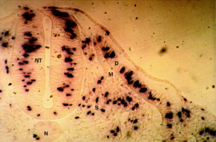
Somite analysis revealed a high density of proliferat-ing cells in the dermatome, whereas cells in the myotome showed no incorporation of BrdU. Signals were more uni-formly distributed in the scleratome, but at a lower density (Figure 3). These results agree with those of Langman and Nelson (1968), based on 3H-thymidine incorporation. Us-ing microautoradiography, these authors demonstrated that myotomic cells in chicken embryos did not divide, and that cellular multiplication occurred in the dermatome. Accord-ing to Philipson and Sorrentino (1992), cells from differ-ent tissues, species and embryonic developmdiffer-ental stage have cell cycles that vary considerably in duration. Such varia-tions were evident in transverse secvaria-tions of somites from embryos exposed to BrdU. Proliferating cells were easily identified in the dermatome and scleratome, but correspond-ing signals were not observed in the myotome which gives rise to axial skeletal muscle. In spite of their close proxim-ity, the dermatome and myotome exhibited marked differ-ences in cellular proliferation and were easily identified because of the good quality histological sections with well-preserved morphological characteristics.
In an immunohistochemical analysis done using an antibody against desmin and acetylcholinesterase activity, Kaehn et al. (1988) demonstrated myotome cells in chicken embryos originated from the dermatome, with no contribu-tion from the scleratome. This biological process explains the origin of skeletal muscle in chicken embryos. Our
re-sults suggest that dermatome cells proliferate intensively before migrating to the myotome where the cell cycle is interrupted and skeletal muscle gene expression is initiated. The identification of proliferating cells in the somite re-gion thus provides a means for studying the mechanisms involved in muscle fiber formation during the embryonic period.
ACKNOWLEDGMENTS
This research was supported by grants from the Secretaria de Ciência e Tecnologia do Estado de São Paulo, FAPESP and EMBRAPA. M.L.L. received a DTI scholarship from RHAE-MCT and G.S.S. and L.L.C. are recipients of Research Scholarships from CNPq. Publication supported by FAPESP.
RESUMO
Embriões de frango foram incubados na presença de BrdU e montados em resina plástica. A detecção de células em proliferação nos somitos e tubo neural foi feita através de anticorpos contra BrdU. Um ponto essencial para a otimização do método foi a fixação dos embriões por apenas uma hora em paraformaldeído a 4%. Análise de cortes transversais revelou que no tubo neural os núcleos marcados se posicionavam na região central. Cortes transversais em somitos diferenciados revelaram a presença de células em proliferação no dermátomo e esclerótomo, no entanto não foi observado nenhum sinal no miótomo. A metodologia aqui apresentada permitiu identificar com clareza e boa resolução as células em proliferação presentes em tecidos embrionários.
REFERENCES
Cole, M.B. and Sykes, S.M. (1974). Glycol methacrylate in light microscopy: a routine method for embedding and sectioning animal tissues. Stain Technol. 49: 387-400.
Fujita, S. (1964). Analysis of neuron differentiation in the central nervous system by tritiated thymidine autoradiography. J. Comp. Neurol.122: 311-328.
Gilbert, S.F. (1995). Início do desenvolvimento em vertebrado: neurulação e ectoderme. In: Biologia do Desenvolvimento. Sociedade Brasileira de Genética, Ribeirão Preto, pp. 267-345.
Goodpasture, C. and Bloom, S.E. (1975). Visualization of nucleolar organizer regions in mammalian chromosomes using silver staining. Chromosoma 53: 37-50.
Gratzner, H.G. (1982). Monoclonal antibody to 5-bromo and 5-iodo-deoxyuridine: a new reagent for detection of DNA replication. Science 218: 474-475.
Kaehn, K., Jacob, H.J., Christ, B., Hinrichsen, K. and Poelmann, R.E. (1988). The onset of myotome formation in the chick. Anat. Embryol. 177: 191-201.
Kimberly, A.H. and Slpecky, N.B. (1995). A simplified method for obtaining 0.5 mm sections of small tissue specimens embedded in PEG. Histochem. Cytochem.43: 637-643.
Kitchin, K.T. and Brown, J.L. (1995). Incorporation of 5-iodo-2’-deoxyuridine and 5-bromo-2’-deoxyuridine into rodent DNA as determined by neu-tron activation analysis. Anal. Biochem.229: 180-187.
Langman, J. and Nelson, G.A. (1968). A radioautographic study of the
de-velopment of the somite in the chick embryo. Embryol. Exp. Morphol. 19: 217-226.
Mozdziak, P.E., Schultz, E. and Cassens, R.G. (1994). Satellite cell mitosis in posthatch turkey skeletal muscle growth. Poult. Sci.73: 547-555.
Philipson, L. and Sorrentino, V. (1992). Growth control in animal cells. In:
Development: The Molecular Genetic Approach (Russo, V.E.A., Brody, S., Cove, D. and Ottolenghi, S., eds.). Springer-Verlag, Berlin, pp. 537-553.
Rong, P.M., Teillet, M.A., Ziller, C. and LeDouarin, N.M. (1992). The tube neural/notochord complex is necessary for vertebral but not limb and body wall striated muscle differentiation. Development115: 657-672.
Schutte, B., Reynders, M.M.J., Bosman, F.T. and Blijham, G.H. (1987). Ef-fect of tissue fixation on anti-bromodeoxyuridine immunohistochemis-try. Histochem. Cytochem.35: 1343-1345.
Taylor, J.H., Woods, P.S. and Hughes, W.L. (1957). The organization and duplication of chromosomes as revealed by autoradiographic studies using tritium-labeled thymidine. Proc. Natl. Acad. Sci. USA43: 122-127.
Teillet, M.A. and LeDouarin, N.M. (1983). Consequences of neural tube and notochord excision on the development of the peripheral nervous sys-tem in the chick embryo. Dev. Biol.120: 329-347.