535 535 535 535 535 Mem Inst Oswaldo Cruz, Rio de Janeiro, Vol. 100(5): 535-539, August 2005
Bacterial and fungal colonization of burn wounds
Jefferson Lessa Soares de M acedo/+, João Barberino Santos*
Hospital Regional da Asa Norte, Secretaria de Saúde do Distrito Federal, SMHN, AE 1, Bloco A, 70000-000, Brasília, DF, Brasil *Núcleo de Medicina Tropical, Universidade de Brasília, Brasília, DF, Brasil
A prospective study of fungal and bacterial flora of burn wounds was carried out from February 2004 to February 2005 at the Burns Unit of Hospital Regional da Asa Norte, Brasília, Brazil. During the period of the study, 203 patients were treated at the Burns Unit. Wound swab cultures were assessed at weekly intervals for four weeks. Three hundred and fifty four sampling procedures (surface swabs) were performed from the burn wounds. The study revealed that bacterial colonization reached 86.6% within the first week. Although the gram-negative organisms, as a group, were more predominant, Staphylococcus aureus (28.4%) was the most prevalent organism in the first week. It was however surpassed by Pseudomonas aeruginosa form third week onwards. For S. aureus and P. aeruginosa
vancomycin and polymyxin were found to be the most effective drugs. Most of the isolates showed high level resis-tance to antimicrobial agents. Fungi were found to colonize the burn wound late during the second week postburn, with a peak incidence during the third and fourth weeks. Species identification of fungi revealed that Candida tropicalis was the most predominant, followed by Candida parapsilosis. It is crucial for every burn institution to determine the specific pattern of burn wound microbial colonization, the time-related changes in the dominant flora, and the antimicrobial sensitivity profiles. This would enable early treatment of imminent septic episodes with proper empirical systemic antibiotics, without waiting for culture results, thus improving the overall infection-related morbidity and mortality.
Key words: burn wound - colonization - infection
Despite advances in the use of topical and parenteral antimicrobial therapy, and the practice of early tangencial excision, bacterial infection remains a major problem in the management of burn victims today. Few patients are as susceptible to the development of infections as burn patients. Severe dysfunction of the immune system, a large cutaneous colonization, the possibility of gastrointesti-nal translocation, a prolonged hospitalization and inva-sive diagnostic and therapeutic procedures, all contrib-ute to infections (Barreto et al. 1998, Macedo et al. 2000, Macedo 2003).
The experience accumulated over the past three de-cades, in the early interventional treatment of burns pa-tients has dramatically changed the cause of death; it is now estimated that about 75% of the mortality following burn injuries is related to infections, rather than osmotic shock and hypovolaemia. Therefore, knowledge of the responsible bacterial flora of burn wounds, its prevalence and bacterial resistance, is of crucial importance for fast and reliable therapeutic decisions.
Following colonization, the organisms on the surface start to penetrate the burn eschar to a variable extent, depending on their invasive capacity, local wound fac-tors, and the degree of patient’s immunosuppression. If viable subeschar tissue becomes invaded, disseminated infection is likely to occur.
+Corresponding author. E-mail: jlsmacedo@yahoo.com.br
Received 31 March 2005 Accepted 13 July 2005
Streptococcus pyogenes was the most frequently rec-ognized cause of burn wound sepsis in the early part of the last century. Over the years, however, Staphylococ-cus aureus and Pseudomonas aeruginosa have become the most frequently isolated organisms in most burn units (Lawrence 1992, Nasser et al. 2003, Agnihotri et al. 2004). It is generally known that the spectrum of infective agents varies from time to time and from place to place. It is, therefore, desirable to carry out periodic reviews of the bacterial flora of burn wounds so that preventive strate-gies could be modified as necessary. The aim of this pro-spective study is therefore to assess the current fungal and bacterial profile of burn wounds in Brasília, Brazil.
PATIENTS AND METHODS
Our analysis is based on a prospective study of bac-terial and fungal isolates from wound swabs taken from patients admitted to the Burn Unit of the Hospital Re-gional da Asa Norte, Brasília, Brazil, from February 2004 to February 2005.
Since 1980, patients having partial skin thickness burns covering less than 25% of the body surface area, were not generally admitted to the Burns Unit if they were adults, or less than 10% if they were children. Patients with full skin thickness burns of small extent (≤ 5% of the body surface area) were also treated as outpatients until the wound was ready for excision and grafting by members of plastic surgery team. Commonly admission to the Burns Unit only occurred with severely burned patients (> 25-30% of the body surface area).
536 536 536 536
536 Colonization of burn w ounds • JLS de M acedo, JB Santos
Silver sulphadiazine 1% was the topical antimicrobial of choice. The wound was inspected during change of dressing and swabs for culture from the burn wounds were taken weekly for four weeks or until all burn wounds were healed. Early excision of the burn eschar and graft-ing were practiced as soon as the patients were stabilized following the initial resuscitation.
At direct patient contact a protective gown and dis-posable gloves were used. Hands were washed with con-ventional soap when necessary, and disinfected with 70% ethanol/glycerol before and after patient contact.
The bacteriological isolation was carried out in the microbiology laboratory of the Hospital Regional da Asa Norte, Brasília. The swabs were dipped in Stuart´s trans-port medium then plated on blood agar, chocolate agar, MacConkey, and Sabouraud’s dextrose agar media (Difco). After incubation for 18-48 h at 37°C, the isolates were identified using convencional protocol.Afterwards, the sensitivity to the antibiotics was accomplished by auto-mated method Vitek-bioMérieux. The confirmation of pre-cision and accuracy of the procedures to evaluate the antimicrobial susceptibility was made using ATCC (Ameri-can Type Culture Collection) standard strains. When iso-lated S. aureus oxacillin resistant, Acinetobacter sp. and
P. aeruginosa multirresistants were confirmed by disc, by agar diffusion method according to the rules established by the National Committee for Clinical Laboratory Stan-dards (NCCLS 1999). Fungal cultures were obtained on Sabouraud dextrose agar (Difco) and on “mycogel” agar (Oxoid) at 37°C and observed daily for 20 days. The char-acterization of fungi was done by the germ tube test, morphological examination and automated method Vitek YBC yeast identification system (bioMérieux Vitek, Inc., MI, US) (Freydiere et al. 2001). However, antibiotic sensi-tivity of fungi can not be done due to technical problems.
RESULTS
Two hundred and three cases were treated as in-pa-tients at the Burns Unit of Hospital Regional da Asa Norte during the period of study. One hundred and twenty (59.1%) were males, 83 (40.9%) females, with a mean age of 24 years (range 1 to 82 years) and mean total body surface area burn of 15% (range 1 to 88%).
The flame burn was the predominant cause of burn among patients; 117 had flame burns, 58 a scald, and 19 had a electrical burn.
A total of 354 samples were processed from as many patients admitted to the burn unit. Single isolates were found in 316 (89.3%) samples, while 36 (10.2%) and 2 (0.5%) samples yielded double and triple isolates, respectively.
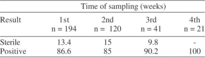
Tables I and II show the positive and sterile results of the wound swabs culture at weekly intervals postburn.
Table III shows the bacteriological isolates from the burn wounds at weekly intervals postburn. The predomi-nant organism was S. aureus which formed 28.4% of all isolates at the end of the first week after admisson. How-ever, by the end of the third week, P. aeruginosa had become more predominant (26.9%), while S. aureus formed only 12.2% of all isolates.
Burn wound sampling performed revealed further prevalence of negative bacilli (51.2%) over
gram-TABLE I
Percentage of bacteriological cultures from wound of patients treated at the Burns Unit of Hospital Regional da Asa Norte,
Brasília, from February 2004 to February 2005
Time of sampling (weeks)
Result 1st 2nd 3rd 4th
n = 194 n = 120 n = 41 n = 21
Sterile 13.4 15 9.8
-Positive 86.6 85 90.2 100
TABLE II
Percentage of fungal cultures from wound of patients treated at the Burns Unit of Hospital Regional da Asa Norte, Brasília,
from February 2004 to February 2005
Time of sampling (postburn weeks)
Result 1st 2nd 3rd 4th
n = 147 n = 97 n = 27 n = 11
Sterile 87.6 77.4 62.9 45.5
Positive 12.4 22.6 37.1 54.5
positive cocci (36%). Most of the gram negative isolates obtained were found to be multi drug resistant. All strains of staphylococci were susceptible to vancomycin.
However, the most common isolate overall was S. aureus (20.5%) followed by coagulase-nagative staphy-lococci (15.2%), P. aeruginosa (11.4%), Klebsiella sp. (11.2%), and Enterobacter sp. (10.4%).
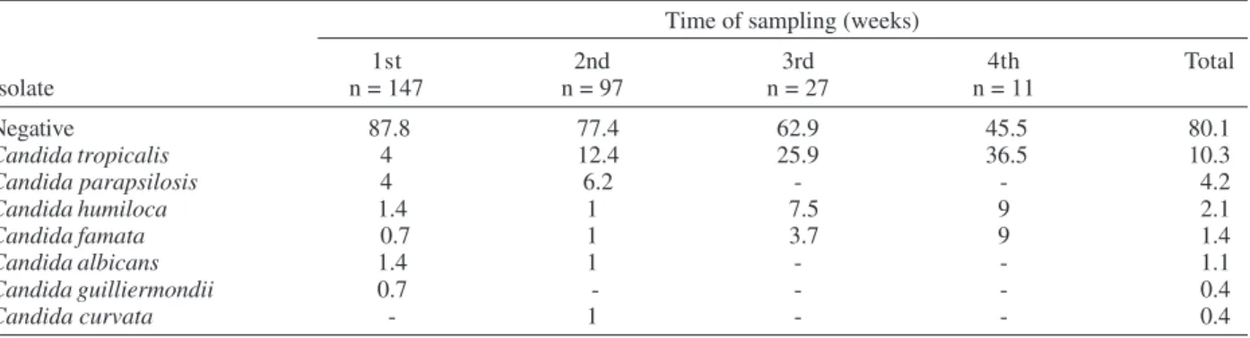
The only kind of fungi recovered on culture from swabs of burn wounds were Candida species. The fre-quency of fungal isolates increased steadily during the stay in hospital, peaking at third and fourth weeks (Table IV).
Table V presents the antimicrobial susceptibility of the gram-positive bacteria isolated from wound culture of burned patients. The incidence of oxacillin resistance among coagulase-negative staphylococci were high (44.6%) and among S. aureus were low (4.7%). However all staphylococci were susceptible to vancomycin. S. aureus showed high susceptibility to a wide range of antibiotics. Coagulase-negative Staphylococcus showed low to moderate susceptibility to amoxicillin/clavulanic acid, cephalothin, oxacillin, gentamicin, clindamycin, ciprofloxacin, ampicillin/sulbactan, and co-trimazole.
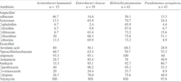
Table VI presents the antimicrobial susceptibility of the gram-negative bacteria isolated from wound culture of burned patients. More than 80% of A. baumannii iso-lated were susceptible only to ticarcillin/clavulanic acid and imipenem. More than 90% of E. cloacae and K. pneumoniae were sensitive to imipenem, amikacin, and ciprofloxacin. All strains of P. aeruginosa showed low susceptibility to a wide range of antibiotics, except only to polymyxin and amikacin.
DISCUSSION
537 537 537 537 537 Mem Inst Oswaldo Cruz, Rio de Janeiro, Vol. 100(5), August 2005
TABLE III
Percentage of bacteriological organisms isolated from wound cultures of patients treated at the Burns Unit of Hospital Regional da Asa Norte, Brasília, from February 2004 to February 2005
Time of sampling (weeks)
Isolate 1st 2nd 3rd 4th Total
n = 194 n = 120 n = 41 n = 21
Staphylococcus aureus 28.4 13.3 12.2 4.7 20.5
Coagulase-negative
staphylococci 15.5 15 14.6 14.4 15.2
Pseudomonas aeruginosa 6.7 10.8 26.9 28.7 11.4
Klebsiella sp. 9.8 14.3 4.9 19.1 11.2
Enterobacter sp. 9.8 10 14.6 9.5 10.4
Acinetobacter baumannii 2.1 4.2 9.8 9.5 3.9
Serratia sp. 2.1 5 - - 2.7
Aeromonas hydrophila 4.1 1.7 - - 2.7
Escherichia coli 1.5 3.3 2.4 4.7 2.3
Enterococcus faecalis 3.6 0.8 - - 2.1
Proteus mirabilis 0.5 2.5 2.4 - 1.3
Streptococcus pyogenes 0.5 - - - 0.3
Others 2.0 4.2 2.4 9.5 3.2
Total number of isolates 376
Total number of sampling procedures 354
Total number of patients studied 203
TABLE IV
Percentage of fungal organisms isolated from wound cultures of patients treated at the Burns Unit of Hospital Regional da Asa Norte, Brasília, from February 2004 to February 2005
Time of sampling (weeks)
1st 2nd 3rd 4th Total
Isolate n = 147 n = 97 n = 27 n = 11
Negative 87.8 77.4 62.9 45.5 80.1
Candida tropicalis 4 12.4 25.9 36.5 10.3
Candida parapsilosis 4 6.2 - - 4.2
Candida humiloca 1.4 1 7.5 9 2.1
Candida famata 0.7 1 3.7 9 1.4
Candida albicans 1.4 1 - - 1.1
Candida guilliermondii 0.7 - - - 0.4
Candida curvata - 1 - - 0.4
than an exception in major burns. In spite of the fact that all burned patients were routinely cleaned with an anti-septic solution (chlorhexidine) and had 1% silver sulphadiazine cream applied to their wounds, 86.6% of the patients studied had microorganisms isolated from their burn wounds at the end of the first week after admis-sion.
The susceptibility of burn wound to such opportunis-tic colonization by bacteria and fungi results from several factors including the presence of coagulated proteins, the absence of blood-borne immune factors, and the avascu-larity of the burn wound.
In this study, we evaluated the pattern of burn wound microbial colonization, as well as the time-related changes in the predominant flora throughout the patients’ hospi-tal stay.
There is no doubt that efforts at combating infection in burns must remain a continuing preoccupation, S. aureus was the most prevalent single organism (28.4%) colonizing the burn wounds in the first week following burn injuries. P. aeruginosa which came fourth in the sec-ond week surpassed staphylococci in all subsequent weeks.
The study has also shown that the flora of individual burn wounds changes over time. Gram-positive organ-isms are initially prevalent, then gradually become super-ceded by the gram-negative organisms that appear to have a greater propensity to invade.
538 538 538 538
538 Colonization of burn w ounds • JLS de M acedo, JB Santos
Our finding that S. aureus was the most common iso-late coincides with previous reports (Taylor et al. 1992, Vindenes & Bjerknes 1995, Lesseva & Hadjiiski 1996, Komolafe et al. 2003) but is in contrast to other studies which report P. aeruginosa as predominant organism (Atoyebi et al. 1992, Revathi et al. 1998, Singh et al. 2003, Nasser et al. 2003, Agnihotri et al. 2004).
Compared to several earlier reports on burn wound colonization and invasive infection, one of the most strik-ing differences is the high frequency of coagulase-nega-tive staphylococci throughout the hospital stay in this study. Even though the pathogenicity of these microor-ganisms in burn patients has been questioned, it should
TABLE V
Percentage of antimicrobial susceptibility of gram-positive bacteria isoladed from wound culture of patients treated at the
Burns Unit of Hospital Regional da Asa Norte, Brasília, from February 2004 to February 2005
Staphylococcus Coagulase-negative
aureus staphylococci
Antibiotic n = 77 n = 57
Amoxicillin/
Clavulanic acid 92.9 50.8
Cephalothin 90.6 50.8
Oxacillin 95.3 55.4
Gentamicin 94.1 80
Amikacin 100 92.3
Ciprofloxacin 96.5 80
Clindamycin 89.4 72.3
Co-trimoxazole 91.8 52.3
Vancomycin 100 100
Ampicillin/
Sulbactam 90.6 50.8
be noted that these patients are immunocompromised. Several studies have consistently suggested that coagu-lase-negative staphylococci should be considered a sig-nificant pathogen in both burn patients and critically ill surgical patients (Vindenes & Bjerknes 1995). As coagu-lase-negative staphylococci are also a bacteria frequently isolated from blood cultures in our ward (Macedo et al. 2003), it is of considerable concern that 44.6% of these isolates were resistant to oxacillin.
Contrary to findings in the beginning of last century before, the isolation of β-haemolytic streptococci from burn wounds has now become rare. It was also confirmed in this study that which the isolation rate of this bacteria was 0.3%.
History indicates that the relative importance and the cyclic pathogenicity of various microorganisms have changed and may be expected to continue changing as systemic and topical antibacterial treatment develops. The pattern of bacterial resistance is important for epide-miological and clinical purposes.
Even though some reports indicate a decrease in P. aeruginosa colonization of burn wounds, this microor-ganism has, since the mid-twentieth century, been held responsible for the majority of invasive burn wound in-fections in many burn centers (Vindenes & Bjerknes 1995). In our series, P. aeruginosa accounted for 11.4% of all burn-wound isolates, and only 60% were sensitive to imipenem.
In addtion, the nosocomial pathogen A. baumannii
was demonstrated in 3.9% of all isolates in this study.
Acinetobacter can cause infections in patients with burns, and these bacteria have been of much concern because of a rapid increase of resistance to a variety of antibacte-rial drugs. In our series 53.3% of these bacteria were resis-tant to ampicillin/sulbactam.
TABLE VI
Percentage of antimicrobial susceptibility of gram-negative bacteria isoladed from wound culture of patients treated at the Burns Unit of Hospital Regional da Asa Norte, Brasília, from February 2004 to February 2005
Acinetobacter baumannii Enterobacter cloacae Klebsiella pneumoniae Pseudomonas aeruginosa
Antibiotic n = 15 n = 39 n = 42 n = 43
Ampicillin/
sulbactam 46.7 14.6 56.1 13.3
Aztreonan 13.3 65.9 70.7 24.4
Cephalothin 6.7 2.4 65.9 4.4
Cefoxithin 6.7 9.8 87.8 6.7
Ceftriaxone 6.7 63.4 73.2 15.6
Ceftazidime 20 68.3 75.6 51.1
Ceftaxima 13.3 65.9 73.2 8.9
Ticarcillin/
clavulanic acid 80 56.1 68.3 28.9
Piperacillin/tazobactam 66.7 63.4 70.7 53.3
Imipenem 93.3 95.1 100 60
Gentamicin 26.7 85.4 78 48.9
Amikacin 33.3 95.1 92.7 86.7
Ciprofloxacin 40 90.2 95.1 53.3
Co-trimoxazole 20 24.4 53.7 8.9
Cefepime 26.7 78.0 75.6 48.9
Polymyxin ND ND ND 97.8
539 539 539 539 539 Mem Inst Oswaldo Cruz, Rio de Janeiro, Vol. 100(5), August 2005
Colonization of the burn wound with fungi is not a surprising phenomenon in view of the changes in micro-bial flora induced by systemic and topical chemotherapy. The origin of the fungi in these patients does not appear to be the gastrointestinal tract as suggested in other stud-ies of diseases complicated by fungemia (Colombo & Guimarães 2003). An epidemiological study demonstrated recovery of Candida from the wounds of 8 to 10% of severely burned patients studied and the absence of fungi in the stool or nasofharynx of these patients (Bruck et al. 1972). Colonization of fungi was found more commonly after third and fourth week postburn.
Species identification revealed that postburn patients harbored various species of Candida. These fungi spe-cies are the most common fungal organisms in burn wounds (Bruck et al. 1972, Vindenes & Bjerknes 1995)and in this study no other fungi were isolated. The most pre-dominant species obtained was C. tropicalis (10.3%), fol-lowed by C. parapsilosis (4.2%). This high incidence of
C. tropicalis observed in our study is specially alarming. As it is now well known that unlike C. albicans, which can be found as a commensal, C. tropicalis when present, is not a commensal and is almost always associated with the development of deep fungal infections.
C. albicans has always been considered as the most frequent pathogenic species causing nosocomial fungal infections in burn patients, with mortalility rates due to deep-seated infections raging from 38 to 50% (Macedo et al. 2003). However, recently other species of Candida, as
C. tropicalis, has emerged to be equally important in immunocompromised patients (Mathews et al. 2001, Leung et al. 2002, Gupta et al. 2004).
The colonization of the wounds with Candida spe-cies does not validate the start of antifungal therapy in burned patients. However, if the appearance of the wound is suggestive of invasive fungal infection, or if the patient has received intravenous antibiotics for bacterial infec-tions (specially older patients with burn larger than 40% total body surface area), or the patient is in a critical phase suggestive of a generalized breakdown on his/her host defense mechanisms, systemic antifungal therapy should be considered.
The high percentage of multi drug resistant isolates is probably due to empirical use of broad-spectrum antibi-otics. However, in the instances of imminent clinical burn wound sepsis, the success of treatment greatly depends on prompt administration of empirical i/v antimicrobial therapy.
Burns provide a suitable site for bacterial multiplica-tion and infecmultiplica-tion, mainly because of the larger area in-volved and longer duration of patient stay in the hospital. To ensure early and appropriate therapy in burn patients, a frequent evaluation of the wound is necessary. There-fore, a continuous surveillance of microorganisms and a regular update of their antibiotic resistance pattern is essencial to maintain good infection control programmes in the burn unit, thus improving the overall infection-re-lated morbidity and mortality.
REFERENCES
Agnihotri N, Gupta V, Joshi RM 2004. Aerobic bacterial
iso-lates from burn wound infections and their antibiograms: a five-year study. Burns30: 241-243.
Atoyebi OA, Sowemimo GOA, Odugbemi T 1992. Bacterial flora of burn wounds in Lagos, Nigeria: a prospective study.
Burns18: 448-451.
Barreto MX, Leonardi DF, Silva MA 1998. Infecção em queimaduras: estudo da flora predominante na UTI queimados do Hospital de Pronto-socorro de Porto Alegre. Rev Bras Ter Intens10: 177-180.
Bruck HM, Nash G, Stein JM, Lindberg RB 1972. Studies on the occurrence and significance of yeasts and fungi in the burn wound. Ann Surg 176:108-110.
Colombo AL, Guimarães T 2003. Epidemiologia das infecções hematogênicas por Candida spp. Rev Soc Bras Med Trop 36: 599-607.
Freydiere AM, Guinet R, Boiron P 2001. Yeast identification in the clinical microbiology laboratory: phenotypical meth-ods. Med Mycol 39: 9-33.
Gupta N, Haque A, Lattif AA, Narayan RP, Mukhopadhyay G, Prasad R 2004. Epidemiology and molecular typing of
Candida isolates from burn patients. Mycopathologia 158:
397-405.
Komolafe OO, James J, Kalongolera L, Makoka M 2003. Bac-teriology of burns at the Queen Elizabeth Central Hospital, Blantyre, Malawi. Burns29: 235-238.
Lawrence JC 1992. Burn bacteriology during the last 50 years.
Burns18 (Suppl. 2): S23-29.
Lesseva M, Hadjiiski OG 1996. Staphylococcal infections in the Sofia Burn Centre, Bulgaria. Burns22: 279-282.
Leung AY, Chim CS, Ho PL, Cheng VC, Yuen KY, Lie AK, Au WY, Liang R, Kwong YL 2002. Candida tropicalis fungaemia in adult patients with haematological malignancies: clinical features and risk factors. J Hosp Infect50: 316-319.
Macedo JLS 2003. Imunodepressão do queimado: patogênese e fator de risco para sepse. Rev Bras Queimadura3: 26-35.
Macedo JLS, Rosa SC, Castro C 2003. Sepsis in burned pa-tients. Rev Bras Med Trop36: 647-652.
Mathews MS, Samuel PR, Suresh M 2001. Emergence of Can-dida tropicalis as the major cause of fungaemia in India.
Mycoses 44: 278-280.
Nasser S, Mabrouk A, Maher A 2003. Colonization of burn wounds in Ain Shams University burn unit. Burns29: 229-233.
NCCLS-National Committee for Clinical Labaratory Standards 1999. Performance Standards for Antimicrobial Disk Sus-ceptibility Tests, Wayne (Approved Standard, M100-S92).
Revathi G, Puri J, Jain BK 1998. Bacteriology of burns. Burns 24: 347-349.
Singh NP, Goyal R, Manchanda V, Das S, Kaur I, Talwar V 2003. Changing trends in bacteriology of burns in the burns unit, Delhi, India. Burns29: 129-132.
Taylor GD, Kibsey P, Kirkland T, Burroghs E, Tredget E 1992. Predominance of Staphylococcus organisms in infections occurring in a burns intensive care unit. Burns18: 332-335.