589
TO SEE THE VIDEO GO TO THE RBCCV WEBSITE AT : http://www.rbccv.org.br/video2/fechamento.html
CONFLICT OF INTEREST STATEMENT: The authors declare that they have conflict of interests; Braile Biomédica® provided all the material used and supplied the video images of the operation, presenting its commercial products.
1. São José do Rio Preto Pediatric Cardiovascular Surgery Service – Hospital de Base – São José do Rio Preto Medical School, SP, Brazil.
2. University of Marília (UNIMAR), UNIMAR State Faculty of
Ulisses Alexandre CROTI1, Domingo Marcolino BRAILE1, Marcos Aurélio Barboza de OLIVEIRA1, Fábio Villaça
GUIMARÃES FILHO2
Rev Bras Cir Cardiovasc 2008; 23(4): 589-590
MULTIMEDIA
RBCCV 44205-1041
Fechamento de comunicação interventricular muscular de via de entrada do ventrículo direito
Closure of the interventricular communication of
right ventricular inflow tract
Medicine (FAMEMA) and Heart Institute of Marília, Marília, SP, Brazil.
Correspondence address: Ulisses Alexandre Croti
Hospital de Base – Faculdade de Medicina de São José do Rio Preto (FAMERP) – Avenida Brigadeiro Faria Lima, 5544 – São José do Rio Preto – SP – Brasil - CEP 15090-000
E-mail: [email protected]
Article receveid on October 15th, 2008 Article accepted on November 9st, 2008 PATIENT CHARACTERISTICS
The patient was a 9-month-old female child, born in Marília, São Paulo State, who had acyanotic congenital heart defect with both pulmonary hyperflow and hypertension diagnosed. She coursed with low weight and stature gain, reduced heart murmurs, and hyperphonesis of the second heart sound at a pulmonary focus. She was referred to surgical treatment.
The echocardiogram at our Medical service has confirmed the diagnosis of muscular interventricular communication (IVC) of right ventricle outflow tract (RVOT) [1], patent ductus arteriosus, and important pulmonary hypertension level with mean pressure of 45 mmHg.
Despite being young, an association with genetic syndrome was suspected. Thus, it was chosen to perform an investigation through a hemodynamic study and assessment of pulmonary reactivity. Pulmonary hypertension with adequate response to the vasodilators were found, that is, the mean pressure of 44 mmHg at the pulmonary artery, in ambient air, decreased to 32 mmHg with 100% oxygen, and to 34 mmHg with nitric oxide 10 ppm [2].
She underwent surgery to close the interventricular communication with removal and partial reinsertion of the septal cusp of tricuspid valve, and ligature of the ductus arteriosus. Cardiopulmonary bypass (CPB) time, at 32ºC, was 108 minutes; myocardial ischemia time was 76 minutes.
DESCRIPTION OF THE TECHNIQUE EMPLOYED An incision was made through the skin; the subcutaneous tissue was opened using an electric scalpel in order to perform a median transsternal thoracotomy. The pericardial sac was opened; the external cardiac structures were analyzed; the increased diameter of the pulmonary artery drew attention when compared to that of the aorta.
Bursae were surgically created in both the aorta and superior vena cava (SVC). Heparin was directly administered into the SVC. Pericardial sac was aspirated to remove possible preexisting clots. A tourniquet was made at the SVC; a purse was surgically made at and a thread for tourniquet was also passed around the inferior vena cava (IVC).
A demonstration of a 12F-arterial cannula and its features were made, such as its introduction into and attachment to the aorta. Air was removed from the interior of the cannula and a connection to the arterial line of the CBP circuit was performed. Then, a venous cannula was introduced into and attached to the SVC. Air was removed from the tubing and a demonstration of the Y venous drainage was performed. Similarly, another cannula was introduced into and attached to the IVC. Thus, the preparation to start the correction of the interventricular communication was finally concluded.
590
CROTI, UA ET AL - Closure of the interventricular communication of right ventricular inflow tract
Rev Bras Cir Cardiovasc 2008; 23(4): 589-590
performed: pull of the pulmonary trunk, dissection of the ductus arteriosus after the left pulmonary artery was identified, and ligature with a simple 4-0 polypropylene thread.
The operatory field was adjusted to start the closure of the interventricular communication, by positioning the cannula into the ICV, the aspirators at the pericardial sac were positioned; aorta cross-clamping was made; opening of the right atrium (RA) was performed, opening of a small orifice at the interatrial septum to introduce the aspirator, and left atrium (LA) decompression was performed.
A cardioplegic blood solution was administered into the aorta; aspiration of the coronary sinus was performed, and the adequate pressure at aorta was checked; a good flow was observed through the coronary arteries.
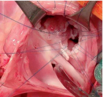
The right atrium was displayed with four 5-0 polypropylene repair threads; intracardiac structures with the border of oval fossa, crista terminalis, trabeculated portion of the RA were all shown; in addition, the components of the triangle of atrioventricular node (Koch’s triangle) such as the tendon of valve of IVC (Todaro’s tendon), the septal ring of the tricuspid valve, and the opening of coronary sinus were all seen. The site of the membranous septum and the region of atrioventricular block risk are indicated.
The septal cusp is pulled and a clamp was introduced through the right ventricle until the left ventricle, thus, identifying the site of interventricular communication and the presence of a papillary muscle centrally to the defect. Once the interventricular communication was characterized, the partial detachment of the septal cusp of tricuspid valve to proper presentation of the defect was performed [3]. U-suture (interrupted horizontal mattress U-suture) using 6-0 polypropylene stitches, at first along with the tricuspid annulus following around the entire orifice (Figure 1). Because it is a muscular defect, the posteroinferior border of the defect did not present risk of blockade, unlike the perimemabranaceous efet.
The preparation of the bovine pericardial patch was performed followed by the administration of cardioplegia, which was repeated in 20-minute intervals.
Stitches were sutured around the bovine pericardial patch and uniformly distributed. The positioning of the patch to occlude the orifice and the ligature of the stitches independently was performed.
The septal cusp was pulled along with the tricuspid annulus and reinserted with double continuous suture using 6-0 polypropylene thread. A saline solution was injected into the right ventricle (RV) to test the tricuspid valve competence.
The orifice made at the interatrial septum was closed and a saline solution was injected again to fill the left cavities resulting in air removal.
REFERENCES
1. Anderson RH, Wilcox BR. The surgical anatomy of ventricular septal defect. J Card Surg. 1992;7(1):17-35.
2. Working Group on Management of Congenital Heart Diseases in India. Consensus on timing of intervention for common congenital heart disease. Indian Pediatr. 2008;45(2):117-26.
3. Moreira Neto FF, Sgarbieri RN. Avaliação pós-operatória imediata da influência da desinserção da valva tricúspide no tratamento da comunicação interventricular. Rev Bras Cir Cardiovasc. 1998;13(4):330-4.
Fig. 1 – Opened right atrium, where it can be observed interventricular communication with polypropylene stitches around the orifice prior to the closure with the bovine pericardial patch
An orifice was made at the aorta and a 6-0 polypropylene thread was passed through. The introduction of a clamp and manual ventilation were limited to removal the remaining ar. Once the myocardial perfusion is started, the heart begins to beat, while the RA is closed using a 6-0 polypropylene thread. A strengthen suture was made around the aorta orifice.
The correction was completed; the heart reassumed its beats, the CPB support was discontinued, the arterial and venous cannulae were removed, strengthen stitches were made at the sutures.