RBCCV 44205-1551 DOI: 10.5935/1678-9741.20140065
The use of virtual resources in preoperative
preparation of infrarenal aneurysms: exploring the
OsiriX’s potential
O uso de recursos virtuais na preparação pré-operatória de aneurismas infrarrenais: explorando o
potencial do OsiriX
Giovani Jose Dal Poggetto Molinari
1, MD; Andréia Marques de Oliveira Dalbem
1, MD; Ana
Terezinha Guillaumon
1, PhD
1Universidade Estadual de Campinas (Unicamp), Campinas, SP, Brazil.
This study was carried out at Hospital de Clínicas da Universidade Estadual de Campinas (HC –Unicamp), Campinas, SP, Brazil.
No inancial support.
Correspondence address:
Giovani Jose Dal Poggetto Molinari
Hospital de Clínicas da Universidade Estadual de Campinas (HC- Unicamp) Rua Vital Brasil, 251- Cidade Universitária Zeferino Vaz - Barão Geraldo - Campinas, SP, Brasil – Zip code: 13083-888
E-mail: [email protected]
Article received on December 23rd, 2013 Article accepted on March 16th, 2014
Abstract
Introduction: In the past few years, the increase of endovas-cular surgeons' interest on tomography image edition through softwares is marked specially when it concerns to its use on pre-operatory study for endovascular aneurysm repair. It is presumed that the bigger the number of informations extracted from the tomography exam and its three-dimensional reconstruction, the smaller is the need of patient's exposure to contrast, as well as the its exposure and the surgical team to radiation. Concepts of image manipulation on the OsiriX software with volume reconstruction of tridimensional tomographic scans of virtual
luoroscopy were used.
Methods: Through manipulation of multi-slice tomography images under three-dimensional reconstruction on software, it was able to modify values of the exam's dose-irradiated distribu-tion. These volume reconstruction presets were saved as Virtual Fluoroscopy, reproducible upon any OsiriX platform. It was able to construct a biplanar image appearing to the patient's operatory
luoroscopy. When compared to the intraoperatory angiography,
the images were alike.
Discussion: Dose-irradiated distribution data manipulation allowed to visualize as opaque bone surfaces and transparent low-dose radiation's areas (viscerae). Thus, under previously marked renal arteries, it was possible to predict it's anatomical positioning
related to visualization under real luoroscopy. Foretelling the
better positioning of the C-arm through this technique enables to
obtain images with the minimum inluence of parallax effect. It is
believed that it supports to assess the renal arteries topographic positioning on a bi-dimensional intraoperatory image. The need of frequent angiographies to localize the renal arteries is reduced, decreasing the exposure to contrast on vulnerable patients.
Abbreviations, acronyms & symbols
DICOM Digital Imaging and Communications in Medicine AAA Abdominal Aortic Aneurysm
MPR Multiplanar reconstruction MIP Maximum Intensity Projection 3D Tridimensional
TC Computed Tomography
INTRODUCTION
Since last few years, the interest of vascular surgeons with endovascular surgery practice has grown in the use of image manipulation software DICOM (Digital Imaging and Communications in Medicine) on computed tomography (CT), especially when referring to its use in endovascular preoperative preparation of infrarenal abdominal aortic an-eurysm (AAA).
With the adoption of high-resolution multislice CT scans and the increased availability of applications reconstructions equipment protocols, postprocessing of images has become a great tool that assists the interpretation and documentation of changes, improving productivity and accuracy of infor-mation.
In multichannel detectors equipment, eficient transmis
-sion systems, processing and storage of data and the reined
engineering enable the reduction in acquisition time and im-proved spatial resolution in the longitudinal axis of the imag-es. The latter is dependent on the voxel size (the smallest unit
volume point in a digital image) that is in turn deined by the
slice thickness[1].
Currently, CT is one of the most important methods for diagnosis and monitoring of vascular diseases; and its perfor-mance is due to the spatial and temporal resolutions, associ-ated with inherent attenuation of the vascular lumen obtained by the administration of intravenous contrast. There is no type of reconstruction more effective than the other, but all have their features and directions, and often the use of more than one type suitable for the demonstration of a pathology is necessary[1]. Thinner CT sections allow the three-dimension-al reconstruction reaches a level of excellence in detail and quality, using increasingly smaller volumes of iodinated con-trast due to the increased speed of image capture obtained[2].
The exposure of the patient to a minimum volume of io-dinated contrast is one of the concerns more present during the planning and implementation of endovascular aneurysm repair, since the incidence of contrast-induced nephropathy in vulnerable patients - patients with renal dysfunction, di-abetic nephropathy, dehydration, hypotension, heart failure, octogenarians, among others - can vary from 12 50%. It is
deined as a 25% increase in baseline serum creatinine and is
usually transient. However, it can lead to undesirable clin-ical outcomes such as prolonged hospitalization, clinclin-ical
Resumo
Introdução: Desde os últimos anos, tem crescido o interesse dos cirurgiões vasculares com prática em cirurgia endovas-cular na utilização de softwares de manipulação de imagens
tomográicas, principalmente quando se refere à sua utilização
no reparo endovascular dos aneurismas de aorta abdominal infrarrenais. Assim, o pós-processamento das imagens tornou-se uma grande ferramenta na interpretação e documentação das alterações, melhorando a produtividade e a precisão das informações, utilizando volumes cada vez menores de contraste iodado no planejamento e execução do tratamento endovascular.
Da mesma forma, menor é a exposição à radiação ionizante no
intraoperatório. Divulgam-se os resultados iniciais da análise
da viabilidade da manipulação de imagens tomográicas no
software OsiriX por meio da luoroscopia virtual.
Métodos: Através da manipulação de imagens de cortes
to-mográicos inos sob-reconstrução tridimensional por volume, foi
possível manipular valores de projeção da distribuição de dose
irradiada. A esta coniguração, foi atribuído o nome de Virtual Fluoroscopy, formato reprodutível em qualquer plataforma Osi-riX. Com isto, obteve-se uma imagem biplanar aparentemente a
uma luoroscopia operatória do doente. Quando comparadas à angiograia e luoroscopia intraoperatória, estas imagens reve -laram-se equivalentes.
Discussão: A manipulação de dados de distribuição da dose ir-radiada em uma superfície permite que se visualizem como opacas áreas de alto contraste (como superfícies ósseas) e como transpa-rentes valores de baixa atenuação (partes moles). Orientados por marcações nas artérias renais, pode-se prever minuciosamente o
seu posicionamento anatômico em relação à sua visualização sob luoroscopia. Outrossim, a antecipação do correto posicionamen -to do aparelho de radioscopia com o uso desta técnica permite a obtenção da imagem com o mínimo de interferência do efeito parallax. Com isso, acreditamos ser possível estimar o
posiciona-mento topográico das artérias renais em imagem bidimensional intraoperatória. Consegue-se reduzir o número de angiograias na
tentativa de se obter a melhor imagem que forneça a localização das artérias renais e do colo do aneurisma, reduzindo sobrecarga renal em pacientes vulneráveis.
Descritores: Procedimentos Endovasculares. Aneurisma da
Aorta Abdominal. Tomograia Computadorizada Multidetectores.
complications during hospitalization and increased hospital mortality. While the risk of nephropathy is dose-dependent,
it is recommended in patients with glomerular iltration rate
less than 60% that the volume of administered contrast is less than 100 ml[3].
Thus, we can assume that the greater the amount of in-formation is extracted from the CT scan and its three-dimen-sional reconstruction, the less the need for contrast exposure during the intraoperative period, and the lower the exposure of the patient and the surgical team to radiation ionizing.
We believe that in addition to precise measurement - such as diameters, lengths and angles[1] - and the analysis of the characteristics of the aneurysm, it is possible to get better
use of information such as topographic positioning of vis-ceral arteries and their respective references of radioscopic viewing since the reconstruction of tomographic sections of smaller thicknesses allows the scanned virtual reproduction of the patient and his disease[4].
METHODS
We disclosed herein the initial results of the analysis of the feasibility of tomographic image manipulation software
(OsiriX MD) with the use of virtual luoroscopy. This tech
-nique has practical signiicance with easy incorporation into
routine endovascular planning. We used it as an aid in
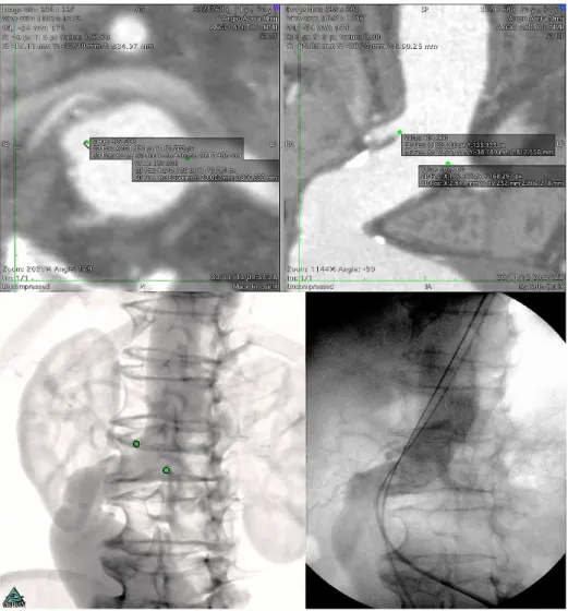
predic-Fig. 1 - Up - ostial renal artery markings in axial projection, with auxiliary view on longitudinal section (at right).
Below – At left, preoperative virtual luoroscopy, with representation of the markings of the renal
tion of the intra luminal placement of angiographic catheters
and luoroscopy unit during endovascular aortic aneurysms
repair. A number of cases from 14 studies were collected,
with promising results. The steps for preparing the conigu
-ration of virtual luoroscopy and illust-rations of the technique
used in two of our cases.
Multichannel CT scans of patients undergoing endovas-cular infrarenal AAA at the Center for Highly Complex En-dovascular Surgery, State University of Campinas, August to December 2013 were analyzed.
We used three-dimensional multiplanar reconstruction through DICOM images manipulation software (OsiriX MD)
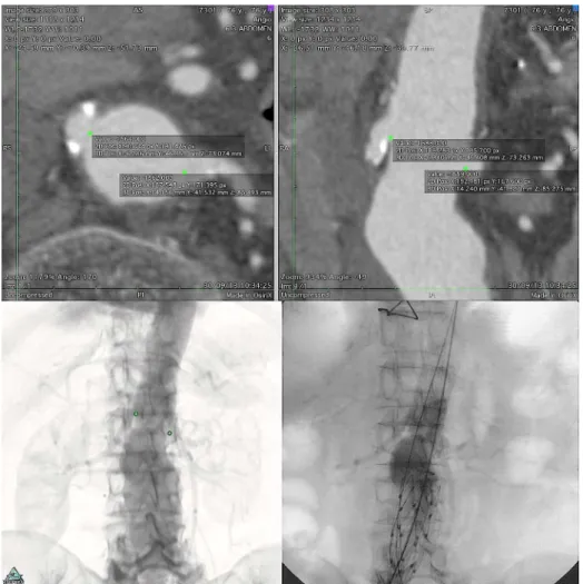
Fig. 2 - Up - ostial renal artery markings in axial projection, with auxiliary view on longitudinal section (at right).
Below – At left, preoperative virtual luoroscopy, with representation of the markings of the renal
arteries, previously performed on axial slices. At right, intraoperative angiography: Also note the
little inluence of the main body of the endoprosthesis on the angular position in the visualization
of renal arteries studied (in this case, Endologix AFX endoprosthesis)
for analysis of aneurysms in series of images with thin 1-3 mm CT slices, intravenous iodinated contrast in arterial phase. Initially, markings on the renal arteries on axial projections were made, through the point command. This feature also signals the voxel in the image examined, so that is subsequently represent-ed in any view, either axial or through both volume or multipla-nar three-dimensional reconstruction (Figures 1 and 2).
In a 3D reconstruction by volume rendering, using the
pre-deined Bone CT reconstruction and Pencil, we can
modify the tomographic values of windowing, CLUT (color
lookup table) and shading which in turn deine brightness,
At this coniguration, the name of Virtual Fluoroscopy
was assigned and reprinted in other OsiriX platforms, al-ways yielding the same two-dimensional image in a single tomographic volume, thus becoming a reproducible format. Once the series of images is subjected to a reconstruction using the software - with advance signaling of voxel through the point - this is shown in three-dimensional and multiplanar images, allowing a more detailed study of the reference point at odd angles of the human anatomy and its topography.
The images were reproduced and placements achieved intra-operatively, revealing themselves equivalent (Figures 1 and 2).
DISCUSSION
The reconstruction volume approaches the attenuation
coeficient of the voxel at a scale of color and degree of opac
-ity (transparency) along the axes. It preserves the information of depth, and shows a better spatial distribution of structures. In a traditional reconstruction, this three-dimensional effect is enhanced by light (shading)[1].
The manipulation of these data (the dose distribution ra-diating to a surface) allows visualization of the maximum
Fig. 3 - Manual coniguration of the preset 3D at OsiriX MD, named
as Virtual Fluoroscopy. Window width (WW - the window width) sets
the number of gray scales shown and window level (WL - the window level) sets the value of the average gray scale of this width. CLUT (color look up table - lookup table of colors) deines a mechanism
of the software used to transform a range of input color to another
color range[5]. When represented as No-CLUT in 8-bit, it displays images in grayscale. Turning off the shading feature the effect of light
is reduced (to enhance the three dimensional appearance) favoring
the perception of the inal image as biplanar, as it is conventional in a luoroscopy. This coniguration may be republished in any OsiriX
platform
intensity projection (MIP), which demonstrates the densest
voxel (higher attenuation coeficient) - which are displayed
as opaque areas of high contrast (as bone surfaces) and as transparent values of low attenuation (soft tissue). Even if in the plan above the overlay to structures that compete with the density of the aorta exists, this is a desirable effect when the aim is a three-dimensional reconstruction that simulates
a simple biplane luoroscopy in grayscale of the studied area. The ideal positioning of the luoroscopy unit during the
surgical procedure may be different than expected during the preoperative study, in that the aneurysm possibly short-en or lshort-engthshort-en higher than expected. However, although it is described that the angulation of the neck of the aneurysm can be changed, the angular position does not change even
under the inluence of the inserted guide wire or endopros
-thesis itself[6].
Thus, guided by the initial markings of the renal arteries, we can minutely predict its anatomical position in relation to
its visualization under luoroscopy, as well as estimate the lo
-cation of the aneurysm neck, considered the starting point for determining the intraoperative positioning of surgical arch and angiographic intraluminal catheter.
Additionally, one can associate corrections of projections of anteroposterior and rotational angle of the neck, since the tortuosity of the aneurysm causes typical changes in the anatomy of the patient and are particularly challenging in the endovascular treatment. Furthermore, the anticipation of the
correct positioning and centering of the luoroscopy device
using this technique allows obtaining image with minimal interference from the parallax effect (where there is a differ-ence in the apparent position of an object when viewed under overlay planes).
Thus we believe it is possible, through the virtual luo
-roscopy under manipulation of DICOM images in software, estimating the topographic position of the renal arteries in
intraoperative two-dimensional image (angiography and lu
-oroscopy). Similarly, when displaying a virtual luoroscopy
tomographically, one can reduce the number of intraopera-tive angiography in an attempt to obtain the best dimensional angiographic image that provides the location of the renal arteries and the aneurysm neck.
The closer this angiographic reproduction to virtual view
of luoroscopy the more careful is the surgeon search for po
-sitioning the renal arteries, and the better will be the use of the aneurysm neck for fastening and sealing of the endopros-thesis, being more accurate its release while the total volume of contrast used is smaller and reducing renal overload in vulnerable patients.
New ways to adapt this software has increasing by ex-panding its use to new tasks. Our proposal is to create
famil-iarity of professionals and encourage demystiied practice of
GJDPM Statistical analysis; final approval of the manuscript; conception and design of the study; implementation of operations and/or experiments; writing of the manuscript or revising it critically for its content
AMOD Realization of operations and/or experiments; writing of the manuscript or revising it critically for its content
ATG Final approval of the manuscript; writing of the manuscript or revising it critically for its content
Authors’ roles & responsibilities
REFERENCES
1. Kuroki IR, Magalhães FV, Rizzi P, Coreixas IMH. Angiotomograia. In: Brito CJ, ed. Cirurgia Vascular: cirurgia endovascular, angiologia. 3a ed. Rio de Janeiro: Revinter; 2013. p.438-96.
2. Ferreira MMV, Freitas AJ, Coelho LH, Zaniolo MRM, Sá J. Aneurismas da Aorta Torácica e Toracoabdominal - Tratamento
Endovascular. In: Brito CJ, ed. Cirurgia Vascular. 3rd ed. Rio de
Janeiro: Revinter; 2013. p.689-736.
3. Ribeiro PCA, Ribeiro MJS. Meios de contraste. In: Lobato AC, org. Cirurgia Endovascular. 2nd ed. São Paulo: Instituto de Cirurgia
Vascular e Endovascular de São Paulo; 2010. p.39-58.
4. Pitoulias GA, Donas KP, Schulte S, Aslanidou EA, Papadimitriou DK. Two-dimensional versus three-dimensional CT angiography in analysis of anatomical suitability for stentgraft repair of abdominal aortic aneurysms. Acta Radiol. 2011;52(3):317-23.
5. Blankensteijn JD, Kool LJS. Computed Tomography. In: Cronenwett JL, Johnston KW et. al. Rutherford’s Vascular Surgery. 7th ed. Philadelphia: Elsevier Inc.; 2010. p.329-43.