Electrognathographic evaluations of
rehabilitated edentulous patients
Avaliações eletrognatográficas em pacientes
edêntulos reabilitados
Abstract: This study investigated, through computerized electrognathographic evalua-tions (K6-I Diagnostic System, Myotronics-Noromed Inc., Tukwila, USA), the mandibu-lar movement pattern of 16 patients rehabilitated with complete dentures presenting no symptoms of stomatognathic functional alterations. The patients were instructed to wear an intra-oral appliance for occlusal plane coverage over their usual superior denture and were then rehabilitated with new dentures preserving a free-way space of 3 mm. After six-ty days, the occlusal vertical dimension was increased and the modiied inferior dentures were used for another 60 days. The obtained results were submitted to the Tukey Test and to the Friedman test, depending on which variable was under consideration, both at a sig-niicance level of 5%. The data revealed a signiicant decrease in free-way space when the irst and the last evaluations were compared. No signiicant differences were found during opening and closing. It was concluded that the presence of a free-way space at the end of the treatment conirms the importance of its existence for maintaining the balance of the masticatory system, assuming the occurrence of a postural repositioning.
Descriptors: Denture, complete; Masticatory muscles; Vertical dimension.
Resumo: O objetivo deste estudo foi avaliar por meio de eletrognatograia computado-rizada (K6-I Diagnostic System, Myotronics-Noromed Inc., Tukwila, EUA) o padrão dos movimentos mandibulares de 16 pacientes reabilitados com dentaduras completas, livres de alterações funcionais do sistema estomatognático. Os pacientes foram instruídos a utilizar um aparelho intra-oral de cobertura oclusal plana adaptado na prótese usual superior e então reabilitados com novas próteses preservando a existência de um espaço funcional livre de 3 mm. Após 60 dias promoveu-se um aumento da dimensão vertical de oclusão e as próteses inferiores assim modiicadas foram utilizadas por mais 60 dias. Os resultados foram submetidos ao Teste de Tukey e ao Teste de Friedman, dependendo da variável em estudo, ambos com 5% de signiicância. Constatou-se decréscimo signiicante do espaço funcional livre quando compararam-se a primeira e a última avaliações. Não foram encontradas diferenças durante abertura e fechamento. Concluiu-se que a presença de um espaço funcional livre ao inal do tratamento vem comprovar a importância de sua existência na manutenção do equilíbrio do sistema mastigatório, admitindo-se que ocorre o reposicionamento postural.
Descritores: Prótese total; Músculos mastigatórios; Dimensão vertical. Henrique Casselli (a)
Alexandre Brait Landulpho(b) Wilkens Aurélio Buarque e Silva(c) Frederico Andrade e Silva(c)
(a) PhD Student; (b)Assistant Professor; (c)Professors – Department of Prosthodontics and Periodontics, School of Dentistry of Piracicaba, State University of Campinas.
Corresponding author: Henrique Casselli Av. 41, 290 B Cidade Jardim Rio Claro - SP - Brazil CEP: 13501-190
E-mail: [email protected]
Introduction
There is a consensus that the determining factors in the functional unbalance of the stomatognathic system are those that alter the relationship between teeth, masticatory muscles and the temporomandib-ular articulations. With this in mind, determination of a therapeutic position before oral rehabilitation is of fundamental importance.3,11
Technological development has led to the opti-mization and application of computerized diagnos-tic systems such as electrognathography. It is used to corroborate the neuropsychological analysis of the factors linked to prosthetic rehabilitation pro-cedures.9 Among these factors, it is important to
point-out the maxillomandibular relationship and the occlusal vertical dimension, considering both vertical and horizontal mandibular movements.1,5,8
We have therefore considered pertinent to perform a study, with patients rehabilitated with total superior and inferior dentures, directed to monitor the pat-tern of vertical, sagittal and horizontal movements of the mandible during four pre-established periods through computerized electrognathography.
Materials and Methods
Sixteen completely edentulous patients were se-lected for this study, including 6 males and 10 fe-males, with an average age of 53 years, all wearers of total dentures for more then 10 years with no signs of functional stomatognathic disorders.13 The patients
presented themselves for rehabilitation treatment at the School of Dentistry of Piracicaba, State Univer-sity of Campinas (UNICAMP), and were submitted to the clinical and physical evaluations required for maintaining clinical records at the Functional Sto-matognathic Disorders Study Center (Cetase).6
This study was revised and approved by the Re-search Ethics Committee, School of Dentistry of Pi-racicaba, State University of Campinas (protocol nº. 76/2005).
The sample consisted of patients that had clini-cal indications of alterations of the occlusal verticlini-cal dimension, such as a pronounced impression of the nasolabial fold, wear of the occlusal surfaces of the current total denture teeth and deicient lip contour.
After the irst evaluations, the patients were
in-structed to wear an intra-oral appliance for occlusal plane coverage, waxed over the superior cast of the current dentures previously mounted on an articu-lator (Gnatus modelo 9600, Gnatus equipamentos Médico-Odontológicos Ltda., Serrana, SP, Brazil) in maximum intercuspation. The appliance was con-structed in thermopolymerized acrylic resin (Clássico Ind. e Com. Ltda., São Paulo, SP, Brazil) respecting the coniguration of the occlusal plane in the antero-posterior and latero-lateral direction. Patients were instructed to use the appliance during a period of thirty days during which weekly adjustments were to be made. They were also instructed to remove the appliance only during meals.6
Together with the use of the appliance, new su-perior and inferior total dentures were constructed through the conventional technique. A superior wax plane was adjusted until it became parallel to Camp-er’s plane in a lateral view and parallel to the bipupil line in a frontal view. The superior lip tubercle was used as a reference for the maximum height of the an-terior occlusal plane. The free-way space was kept at 3 mm to determine the occlusal vertical dimension.17
An intra-oral recording apparatus was made within the inter-arch distance of the inal casts mounted on the articulator.11 To trace Gysi’s gothic arc record4,13
the set was taken to the mouth for the patient to per-form the bordering and intra-bordering movements of protrusion, retrusion, and right and left laterality. To establish the point of centric occlusion, the me-tallic platform was marked with a multi-laminated, 2-mm-in-diameter spherical drill, anteriorly to the vertex of the trace and inside the area corresponding to the bordering movements in order to be tangent to the limits of the trace.11 The superior and inferior
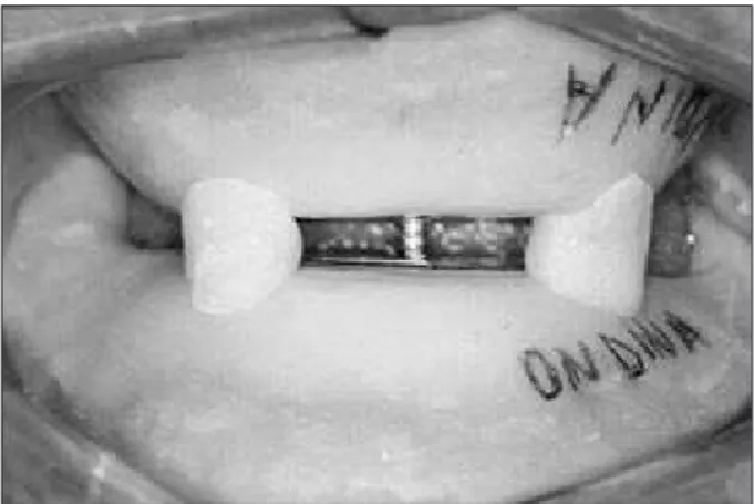
baseplates were ixed to each other intra-orally with a small amount of chemically-activated acrylic resin (Duralay, Reziance Dental MFG Company, Worth, IL, USA) and with the gimlet over the marking, therefore remounting the inferior cast (Figure 1). The selected artiicial teeth (Trubyte Biotone, Dent-sply Ind. e Com. Ltda., Petrópolis, RJ, Brazil) were mounted observing the establishment of a balanced bilateral occlusion.15 The dentures were installed and
were then worn for a period of 60 days.
occlusal vertical dimension, performed by molding the superior denture with hydrocolloid (Jeltrate, Dentsply Ind. e Com. Ltda., Petrópolis, RJ, Brazil) and obtaining a cast in hard plaster (Herostone, Dentsply Ind. e Com. Ltda., Petrópolis, RJ, Bra-zil). With the aid of a low-speed steel disc, a block with the teeth of the new inferior denture was re-moved preserving the integrity of the denture base. The remaining inferior base was repositioned in the patient’s mouth and was molded with hydrocolloid, so that an inferior cast of this base was obtained in hard plaster. A pink wax rim (Clássico Ind. e Com. Ltda., São Paulo, SP, Brazil) was adapted to this base, adjusting it with a delicate contact against the occlusal plane of the superior denture in an occlu-sal vertical dimension equal to that present before the removal of the block of teeth. The superior den-ture cusp impressions on the wax rim were rebased
with small amounts of zinc oxide-eugenolate paste (Lysanda Produtos Odontológicos Ltda., São Paulo, SP, Brazil) to conirm the relation with greater pre-cision. The facial arc was adjusted and the relation between teeth and wax rim were transferred to the articulator (Gnatus modelo 9600, Gnatus equipa-mentos Médico-Odontológicos Ltda., São Paulo, SP, Brazil) with the incisal guide pin graduated at “0”. Separation of the superior and inferior base-plates from the metallic gimlet/platform set of the intra-oral record apparatus was performed using a carborundum disk, preserving the maxillomandibu-lar relation. The gravitational center of the superior cast was determined and the intra-oral record plates were ixed with chemically-activated acrylic resin (Clássico Ind. e Com. Ltda., São Paulo, SP, Brazil) in the superior cast and in the baseplate of the inferior denture, respecting the following criteria:
the gimlet was positioned upon the gravitational center of the superior model;
the superior plate was positioned parallel to the superior ramus of the articulator, and
the trace vertex was directed to the middle point between the superior central incisors.
The record plates were separated and after verify-ing that the increase of the vertical dimension was equivalent to the free-way space suppression and that the esthetic requests were satisied by the adjustments of the gimlet height, the set was transferred from the articulator to the mouth (Figure 2). The patient was requested to repeat the bordering movements obtain-ing a new centric occlusion point13 (Figure 3). 1.
2.
3.
Figure 1 - Baseplates fixed to each other with the set in the
centric occlusion point.
Figure 2 - Record plates repositioned in the mouth with
free-way space suppression.
1ª OC
2ª OC
The set of intra-oral record plates was ixed with chemically-activated acrylic resin (“Duralay”) and was transferred to the articulator with the incisal guide pin graduated at “0”. The inferior cast was remounted. The block of inferior teeth was ixed to the base of the inferior denture, interposing bite wax (Clássico Ind. e Com. Ltda., São Paulo, SP, Brazil) between them, and respecting a condition of maximum intercuspation with the antagonist cast. Installation of the denture was accomplished after clinical intra-oral try-in, inclusion, polymerization, inishing and polishing. During a period of approxi-mately 2 days, when the procedures involving occlu-sal vertical dimension increase were performed, the patients were instructed to use the old dentures with interposition of the occlusal appliance.
Each patient was submitted to a total of four computerized electrognatographic evaluations dur-ing the followdur-ing moments: 1st) initial (T0), with the
usual dentures in position; 2nd) 30 days (T1) after
the installation of the occlusal plane appliance; 3rd)
60 days (T2) after the installation of the new den-tures preserving the free-way space of 3 mm; 4th) 60
days (T3) after the increase of the occlusal vertical dimension. During evaluations, the patients were placed in a calm environment and seated on a pad-ded chair with both feet totally touching the ground. Arms were put to rest on lateral supports and they were positioned to keep their backs erect and the Frankfort horizontal plane parallel to the loor. A computerized diagnostic system (K6-I, Myotronics, Noromed Inc., Tukwila, WA, USA), which possess-es an electrognathograph with eight electromagnetic sensors (K6-I Computerized Mandibular Scanning - CMS), connected to a conventional computer was used to perform the evaluations. While performing the electrognathographic evaluations, the electro-magnetic sensors, disposed in a similar structure as that of the facial arc placed on the nasal support, create an electromagnetic ield which tracks the movements made by a magnet, glued with its own adhesive (Stomahesive, Convatec-Squibb Co., Princ-eton, NJ, USA) at the cervical region of the inferior incisors. The protocol to calibrate the eletrogna-thograph suggested by Jankelson in 198016 was
fol-lowed during all the evaluations. The magnet was
to be placed with the patient in maximum habitual intercuspation or, during the second evaluation, in closure upon the occlusal plane appliance, so that its long axis remained parallel to the incisal borders of the superior incisors and so that the magnetic north would be directed to the right side of the patient. Once ixed on the head, the sensors were lined bilat-erally parallel to Camper’s plane.
Cycle of maximum mouth opening and closing
Starting at the position of maximum habitual in-tercuspation, patients were requested to perform a movement of maximum opening followed by clos-ing, and to end again in maximum habitual inter-cuspation.
Final mandibular closing
The patients were instructed to remain at rest for a period of approximately seven seconds. After this period the operator requested the patient to perform three subsequent movements of mandibular closing, from rest to maximum intercuspation, covering all the dimension of the free-way space, and inally re-turning to rest.
For each evaluation, three electromyographic records were made and only the arithmetic average between the values obtained were considered for tabulation of the results. A period of thirty seconds was observed between each record.
Results
The electrognathographic data for Scan #1 was submitted to variance analysis, Tukey and Friedman tests with a 5% signiicance level; for scan #3, the Friedman test with a 5% signiicance level was ap-plied.
Final mandibular closing
Cycle of maximum mouth opening and closure
No signiicant statistical differences were found between the different periods for the four variables studied (Table 2).
Discussion
When the cycle of maximum opening and closing is observed, the maintaining of a constant vertical dimension opening, considering an occlusal vertical dimension increase of around 8-10 mm (clinically
measured), reveals the great capacity that muscles have to alter their longitudinal height. Hence, they have the capacity to adapt to a condition of vertical dimension regardless of it being clinically favorable or not. Mastication muscles are able to suffer length and volume alterations when submitted to a stimu-lus, which also relect great density alterations.10
Even when considering the existing physiological adaptations, it is possible to conclude that the ver-tical amplitude of mouth opening does not restrict itself to a high or low occlusal vertical dimension.7
This dimension seems to be used as a reference to establish the minimum suficient dimension for food to be captured. However, a decrease of 2.06 mm in amplitude occurred and this may be related to a mechanism of self-preservation of the muscles. This was observed when the opening dimension present during the last period was close to the limit for neu-romuscular spindle activation.
A greater tendency that the mandible had to turn towards the left when performing maximum open-ing duropen-ing all periods was observed. The appliances were constructed with the smallest thickness pos-sible to prevent interference with the values of ver-tical dimension. This unfortunately limited directly its action towards articulations and muscles.12 The
description made by the studied patients in practic-ing left unilateral mastication before the beginnpractic-ing of the rehabilitation process and its continuity af-ter the new dentures were installed revealed a high-er demand of the lathigh-eral pthigh-erygoid at the opposite
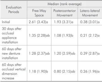
Table 1 - Median electrognathographic values, in
millime-ters, rank averages and statistical analysis of the three vari-ables studied for the different evaluation periods during final mandibular closing.
Evaluation Periods
Median (rank average)
Free-Way Space
Posteroanterior Movement
Latero-lateral Movement
Initial 2.61 (3.43)a 1.93 (3.31)a 0.38 (3.01)a
30 days after occlusal appliance installation
1.35 (2.28)ab 1.08 (1.93)b 0.21 (2.12)a
60 days after new denture installation
1.28 (2.37)ab 1.20 (2.59)ab 0.39 (2.87)a
60 days after occlusal vertical dimension increase
1.18 (1.90)b 0.80 (2.15)ab 0.26 (1.96)a
Note: The median values followed by different letters (lowercase for col-umns) are different by the Friedman test at the 5% significance level.
Table 2 - Average and median electrognathographic values, in millimeters, standard deviations, rank averages and statistical
analysis of the variables studied for the different evaluation periods, during the cycle of maximum mouth opening and closing.
Evaluation Periods
Average (standard deviation) Median (rank average)
Maximum vertical opening
Anteroposterior Movement
Maximum deviation to the left
Maximum deviation to the right
Initial 33.47 (5.04)a 18.91 (7.63)a 3.40 (1.86)a 0.40 (2.31)a
30 days after occlusal
appliance installation 33.52 (4.86)a 16.71 (4.23)a 3.63 (1.55)a 0.31 (2.87)a
60 days after new denture
installation 32.09 (4.53)a 18.28 (5.80)a 2.76 (1.69)a 0.30 (2.46)a
60 days after occlusal
vertical dimension increase 31.41 (6.18)a 15.57 (5.06)a 3.54 (1.45)a 0.30 (2.34)a
side. This implies an anticipated trigger in relation to the ipsilateral pterygoid when simultaneously de-manded.16 Another factor that can be related with
this tendency is that the appliance did not provide intra-articular space gain, did not favor a better synchronization of the articular structures and, spe-cially, did not favor an adequate repositioning of the articular bands with the disc correctly placed. Although the same intra-oral record was used for maxillomandibular relation measurement for the irst and second occlusal vertical dimensions, the system was not appropriately prepared for the great intra-articular space gain that occurred in a sudden manner.12 Such evidence suggests that the beneits
that the therapy with this appliance provides may also be related to its employment considering the deinitive occlusal vertical dimension in which the patient shall be rehabilitated, since the articular me-chanic optimization can also offer a better muscular response by the liberation of the disc.2
Analysis of the values obtained for the free-way space in the situation of terminal mandibular clos-ing shows a signiicant decrease of this space durclos-ing the last evaluation (1.18 mm) in relation to the irst evaluation (2.61 mm). As previously described, a free-way space of 3 mm was established for the irst occlusal vertical dimension; however, at the end of 60 days using the new dentures, this space present-ed much smaller values (around 1.28 mm). This
di-mensional decrease may be related to the detected optimization of the masseter muscles during this phase which, after the improvement of clinical con-ditions offered by the new dentures, directed the mastication stimulus as an auto-preservation vec-tor, stiffening itself. Because of the 1.26 mm free-way space difference between the second and irst periods and the gain of 1.18 mm during the fourth period when this space was totally suppressed, it is possible to conclude that the average electromyo-graphic values in the situation of rest did not suffer inluence of the thickness with which the occlusal appliance was constructed, nor had any relation with the vertical dimension in which it was adapt-ed, as the same did not interfere in the longitudinal length of the muscles.
Conclusions
Nature seems to always seek a situation in which the free-way space is preserved and, although not presenting a constant mathematical dimension pat-tern throughout the interventions performed, the free-way space always tried to establish itself within the most economic and healthy dimension for every established occlusal vertical dimension. This proves once again that the free-way space is not a safe ref-erence to determine occlusal vertical dimension and its maintenance at the end of the treatment high-lights the occurrence of a postural repositioning.11,14
References
1. Carr AB, Donegan SJ, Christensen LV, Ziebert GJ. An elec-trognathographic study of aspects of “deprogramming” of human jaw muscles. J Oral Rehabil. 1991;18(2):143-8. 2. Dawson PE. New definition for relating occlusion to varying
conditions of the temporomandibular joint. J Prosthet Dent. 1995;74(6):619-27.
3. Gelb H. Evaluation of static centric relation in the temporo-mandibular joint dysfunction syndrome. Dent Clin North Am. 1975;19(3):519-30.
4. Gysi A. The problem of the articulation. Part I. Dent Cosmos. 1910;52:1-19.
5. Johnson A, Wildgoose DG, Wood DJ. The determination of freeway space using two different methods. J Oral Rehabil. 2002;29(10):1010-3.
6. Landulpho AB, e Silva WA, e Silva FA, Vitti M. The effect of the occlusal splints on the treatment of
temporomandibu-lar disorders – a computerized electromyographic study of masseter and anterior temporalis muscles. Electromyogr Clin Neurophysiol. 2002;42(3):187-91.
7. McNamara Jr JA, Carlson DS. Functional adaptations in the temporomandibular joint. Dent Clin North Am. 1975;19(3):457-71.
8. Miralles R, Dodds C, Palazzi C, Jaramillo C, Quezada V, Ormeno G et al. Vertical dimension. Part 1: comparison of clinical freeway space. Cranio. 2001;19(4):230-6.
9. Okeson J. Fundamentos de Oclusão e Desordens Temporoman-dibulares. 2ª ed. São Paulo: Artes Médicas; 1992.
10. Raustia AM, Salonen MAM, Pyhtinen J. Evaluation of masti-catory muscles of edentulous patients by computed tomography and electromyography. J Oral Rehabil. 1996;23(1):11-6. 11. Silva FA, Silva WAB. Dimensão vertical de oclusão: um método
12. Silva FA, Silva WAB. Reposicionamento mandibular. Contribui-ção técnica por meio de férulas oclusais duplas com puas. Rev Assoc Paul Cir Dent. 1990;44(5):283-6.
13. Solberg WK. Background e problemas clínicos. In: Disfunção e desordens temporomandibulares. São Paulo: Santos, 1989. cap. 1, p. 8-13.
14. Tallgren A. The reduction in face height of edentulous and partially edentulous subjects during long term wear. Acta Odontol Scand. 1996;24(2):195-239.
15. Tamaki T. Dentaduras Completas. 3ª ed. São Paulo (SP): Sarvier; 1977.
16. Vitti M, Basmajian JV. Integrated actions of masticatory mus-cles: simultaneous EMG from eight intramuscular electrodes. Anat Rec. 1976;185:173-90.