1
Arquivos Brasileiros de Cardiologia - Volume 84, Nº 2, Fevereiro 2005
Case Report
Ablation of Idiopathic Ventricular Tachycardia
with Left Bundle-Branch Block Morphology
Located in the Pulmonary Trunk
Luiz Roberto Leite, José R. Barreto, Roberval Melo, Edmur C. Araujo, Luciano Nogueira,
Lucas A. Fonseca, Edson D’Ávila, César Gonzáles, Fernando Cruz, Angelo A. V. de Paola
Brasilia, DF / Rio de Janeiro, RJ / São Paulo, SP - Brazil
Hospital Santa Luzia, Brasília, and Escola Paulista de Medicina - UNIFESP Mailing address: Luiz Roberto Leite - SQSW 104 - Bloco F - 402 Cep 70670-406 - Brasília, DF, Brazil
E-mail: leite.luiz@brturbo.com.br Sent for publication: 12/09/2003 Accepted for publication: 06/04/2004 English version by Stela Maris Costalonga
We report the case of a 26-year-old female patient with palpitations and presyncopes due to nonsustained ventricular tachycardia, who had no structural heart disease. The patient underwent electrophysiological study in an attempt to ablate the arrhythmogenic focus, whose location was determined by using mapping criteria. Because mapping of the right ventricular outflow tract was not successful, the catheter was placed inside the pulmonary artery with satisfactory mapping of the arrhyth-mogenic focus, and tachycardia was eliminated as soon as ra-diofrequency was initiated. The patient has remained asympto-matic for 14 months, with no treatment with antiarrhythmic drugs, and no arrhythmias on serial 24-hour Holter.
More than 30 years ago, Zipes and Knope 1 reported the
presence of cardiac fibers with the capacity to generate an elec-trical impulse within thoracic veins. It has only been recently, however, that special attention has been given to the clinical meaning of this finding. The pulmonary veins, and, less often, the superior vena cava and the Marshall vein (embryonic remnant of the left superior vena cava) have been identified as arrhythmogenic foci that induce atrial fibrillation 2-6. In addition, ablation of those
foci has been shown to cure the arrhythmia 2-6.
Like the thoracic veins, the pulmonary artery and aorta also have a myocardial extension with electrical activity. Asirvatham et al 7, in an anatomic study with 230 human hearts, showed the
presence of cardiac fibers within the pulmonary artery, and reported that 13% of the hearts studied had cardiac muscle, on average, 3 mm above the pulmonary valve. In addition, application of ra-diofrequency in the coronary leaflet has been reported to be able to eliminate certain types of extrasystoles 8,9.However, the ablation
of arrhythmogenic foci in the pulmonary artery has rarely been explored, and, so far, only one group has reported the origin of that arrhythmia above the pulmonary valveplane 10,11. In our study,
we report the case of a patient with idiopathic ventricular tachy-cardia, whose arrhythmogenic focus was eliminated with appli-cation of radiofrequency in the pulmonary trunk.
Case Report
The patient is a 26-year-old female who has had palpitations and presyncopes for 4 years although using propranolol, propafe-none, and sotalol. The clinical examination showed irregular cardiac rhythm due to the presence of arrhythmia. The 12-lead electro-cardiogram showed sinus rhythm intercalated with nonsustained ventricular tachycardia with left bundle-branch block morphology, lower deviation of the axis in the frontal plane, and S-R transition in V4 (fig.1). The presence of structural heart disease was ruled out through a chest X-ray, echocardiography, and magnetic reso-nance imaging. The latter did not show replacement of muscle tissue by adipose tissue in the right ventricle. The signal-averaged electrocardiogram was negative for the presence of late potentials. The 24-hour Holter showed 52% ventricular beats, 1672 of which were in the form of nonsustained ventricular tachycardia of up to 15 beats. The diagnosis of idiopathic ventricular tachycardia ori-ginating in the right ventricular outflow tract was suspected, and the patient was referred for electrophysiological study for ablation of the arrhythmogenic focus.
2
Arquivos Brasileiros de Cardiologia - Volume 84, Nº 2, Fevereiro 2005
Ablation of Idiopathic Ventricular Tachycardia with Left Bundle-Branch Block Morphology Located in the Pulmonary Trunk
that site, the ventricular tachycardia and extrasystoles were eli-minated, without recurrence even after isoproterenol infusion. The patient has remained asymptomatic for 14 months, with no treat-ment with antiarrhythmic drugs, and no arrhythmias on serial 24-hour Holter monitoring.
Discussion
Although the presence of electrical activity in myocardial fibers within thoracic vessels may be an arrhythmogenic focus, it was only recently that catheter ablation of those triggering foci was shown to cure arrhythmia 2-6,8-10.In the case of arrhythmias with
wide QRS, specific extrasystoles with right bundle-branch block morphology and axis deviation to the inferior quarter of the frontal plane may be eliminated in the coronary leaflets, demonstrating that the origin of the arrhythmia is out of the cardiac chambers9.
Less frequently, the extrasystoles of the left ventricular outflow tract may have the left bundle-branch block morphology and be confounded with those originating from the right ventricular outflow tract. Differentiation by using electrocardiography has been per-formed with the R/S ratio in the V3 lead, correlating an early transition with the left ventricular outflow tract, and a late transition (R/S < 1.0 in V3) with the right ventricular outflow tract 8.
Based on that criterion, the morphological pattern of ventricular tachycardia here presented suggested a right ventricular outflow tract origin. However, the application of radiofrequency that cured the arrhythmia was performed in the pulmonary artery. Recently, Timmermans et al 10,11 have reported the cure of ventricular
ta-chycardia with morphology similar to that here presented by using ablation in the pulmonary trunk.
The pulmonary artery origin of the arrhythmia may still be corroborated by the fact that the arrhythmogenic mechanisms of idiopathic ventricular tachycardia of the outflow tract are the automatism or triggering activity, or both, characteristics that make it focal. Therefore, it is acceptable that the myocardial fibers located in the great vessels may be foci of arrhythmias, and, due to their focal nature, the stimulation of the site has the same morphology of the QRS of the spontaneous arrhythmia. Thus, it is also understandable that the punctual application of radiofrequency at the site of arrhythmia origin definitely eliminates it. Our patient, after 14 months of clinical follow-up with serial Holter, remains asymptomatic with no ventricular arrhythmia.
The difficulty in achieving success in ablating that type of arrhyth-mia has been attributed to supposed epicardial foci. Epicardial map-ping has proved to be an option for successful treatment. However, it is worth noting that, as in our case and in that of Timmermans et al 10,11, the arrhythmia focus may originate in the pulmonary trunk,
and it may be advantageous to map that site before attempting other forms of mapping or other access routes.
Therefore, ventricular tachycardia with the pattern of left bundle-branch block and axis deviation to the lower quarter in the frontal plane, a morphology that suggests the existence of a focus in the right ventricular outflow tract, may also have its origin in the pulmonary trunk. Radiofrequency application in that site may definitively cure the arrhythmia, and in cases in which ablation in the right ventricular outflow tract failed, mapping of the pulmonary trunk may be a useful alternative before other methods are used.
Fig. 1 - Twelve-lead electrocardiogram showing sinus rhythm and frequent episodes of nonsustained ventricular tachycardia with left bundle-branch block morphology in V1 and lower axis deviation in the frontal plane (positive D1, D2, D3, and AvF). Note that QRS complex is predominantly negative until the V3 lead, therefore showing late transition (R>S in V4).
I II
III
AVR AVL
AVF
V1
V2
V3
V4 V5
V6
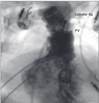
Fig. 2 - Peripheral electrocardiogram in the D1, D2, D3, AvF, V1, and V6 leads and intracavitary electrogram (RVd) recorded at 100-mm/s and 10 mm/mV velocities, respectively. Note that the intracavitary electrogram (distal RV) is the potential recorded by the distal poles of the ablation catheter positioned in the pulmonary trunk, showing 30-ms precocity in regard to the earliest QRS during ventricular tachycardia.
I
II
III
AVF
V1
V6
RV d
Catheter tip
PV
3
Arquivos Brasileiros de Cardiologia - Volume 84, Nº 2, Fevereiro 2005
Ablation of Idiopathic Ventricular Tachycardia with Left Bundle-Branch Block Morphology Located in the Pulmonary Trunk
1. Zipes DP, Knope RF. Electrical properties of the thoracic veins. Am J Cardiol 1972;29:372-6.
2. Hsu LH, Jaïs P, Keane D, et al. Atrial fibrillation originating from persistent left su-perior vena cava. Circulation 2004;109:828-32.
3. Haissaguerre M, Jais P, Shah DC, et al. Spontaneous initiation of atrial fibrilla-tion by ectopic beats originating in the pulmonary veins. N Engl J Med 1998; 339:659-66.
4. Oral H, Scharf C, Chugh A, et al. Pulmonary vein isolation for paroxysmal and per-sistent atrial fibrillation. Circulation 2002;105:1077-81.
5. Oral H, Knight BR, Ozaydin M, et al. Segmental ostial ablation to isolate the pul-monary veins during atrial fibrillation: feasibility and mechanistic insights. Circu-lation 2002;106:1256-62.
6. Tsai CF, Tai CT, Hsieh MH, et al. Initiation of atrial fibrillation by ectopic beats ori-ginating from the superior vena cava: electrophysiological characteristics and re-sults of radiofrequency ablation. Circulation 2000;102:67-74.
References
7. Asirvatham SJ, Friedman PA, Packer DL, Edwards WD. The presence of ventricular muscular extensions into the pulmonary artery and aorta beyond the semilunar valva. PACE 2001;24:734.
8. Kamakura MD, Shimisu W, MatsuoK, et al. Localization of Optimal Ablation Site of Idiopathic Ventricular Tachycardia from Right and Left Ventricular Outflow Tract by Body Surface ECG. Circulation 1998;98:1525-33.
9. Hachiya H, Aonuma K, Yamauchi Y, et al. Electrocardiographic characteristics of left ventricular outflow tract tachycardia. Pacing Clin Electrophysiol 2000;23(11 Pt 2):1930-4.
10. Timmermans C, Rodriguez LM, Crijns HJ, Moorman AF, Wellens HJ. Idiopathic left bundle-branch block-shaped ventricular tachycardia may originate above the pul-monary valve. Circulation 2003;108:1960-7.