progression of cervical lesions in a
group of Colombian patients
Associação da atividade da telomerase com a progressão
de lesões cervicais em um grupo de pacientes da Colômbia
BAyron mAnuel ruiz-hoyospAtriCiA lAndázuri2
Abstract
PURPOSE: To analyze the relation between the cytological indings and telomerase activity (TA). METHODS: Cervical samples were evaluated and classiied according to the Bethesda System. Telomerase activity was measured total product generated values (TPG) using the TRAP assay (telomeric repeat ampliication protocol); data were analyzed statistically using the χ2 test, with the level of signiicance set at p<0.05. RESULTS: The study was conducted on 102 patients.
Of these, 3.9% showed normal cytological indings, 8.8% showed cervicitis; 2% showed Atypical Squamous Cells of Undetermined Signiicance (ASCUS); 67.6% showed Low Grade Squamous Intraepithelial Lesion (LSIL); 11.8% showed High Grade Squamous Intraepithelial Lesion (H-SIL) and 5.9% showed Squamous Carcinoma. Among telomerase-positive samples, the TPG values were cervicitis<normal<ASCUS<L-SIL<H-SIL<Carcinoma. CONCLUSION: Results show increased telomerase activity with increasing severity of lesion, supporting the association between TA and type of lesion.
Resumo
OBJETIVO: Analisar a relação entre os achados citológicos e atividade da telomerase (AT). MÉTODOS: Amostras cervicais foram avaliadas e classiicadas pelo sistema Bethesda. A AT foi medida como valores de produto total gerado (PTG), utilizando o protocolo de ampliicação repetida da telomerase (TRAP); os dados foram analisados estatisticamente usando o teste do χ2,com nível de signiicância de p<0,05. RESULTADOS: Cento e dois pacientes
foram analisados: 3,9% com achados citológicos normais, 8,8% com cervicite, 2% com células escamosas atípicas de signiicado indeterminado (ASCUS), 67,6% com lesão escamosa intraepitelial baixo grau (LEI-BG), 11,8 % com lesão intraepitelial escamosa alto grau (LEI-AG) e 5,9% com carcinoma escamoso. Valores PTG para amostras positivas AT foram: cervicite<normal<ASCUS<LEI-BG<LEI-AG<Carcinoma. CONCLUSÃO: Os resultados mostram um aumento na AT com o aumento da lesão, sustentando a associação entre a AT e o tipo de lesão.
Universidad Del Quindío, Colombia.
1Biology Program; School of Basic Sciences and Technologies, Universidad Del Quindío – Colombia. 2Medicine Program; School of Health Sciences, Universidad Del Quindío – Colombia.
Conlict of interests: none.
Keywords
Telomere Telomerase Cervix uteri Preneoplastic conditions Squamous intraepithelial lesions
Palavras-chave
Telômeros Telomerase Colo do útero Condições pré-neoplásicas Lesões intraepiteliais escamosas
Correspondence Patricia Landázuri Carrera 15 # 12N Armenia – Quindío, Colombia
Received 07/27/2015
Accepted with modiications 09/14/2015
DOI: 10.1590/SO100-720320150005462
Introduction
Telomeres, which form a protective cap on chro-mosome ends, are usually composed of short G-rich Deoxyribonucleic acid (DNA) repeats (TTAGGG)
com-plexed with proteins1,2 with estimated lengths of 5-15 kb
in humans3. Their main function is to protect chromosomes
from incomplete replication, nuclease degradation, and
end-to-end fusion during replication3.
During DNA replication, telomeric DNA shortens progressively, mainly due to the end-replication problem, that is, the inability of the DNA replication machinery
to fully replicate DNA ends2,3. That is why eukaryotic
cells have evolved with a specialized reverse transcriptase enzyme, called telomerase, that is responsible for the new
telomere extension in most adult tissues2,3. Telomerase
is a ribonucleic acid (RNA)-dependent polymerase that synthesizes telomeric DNA sequences and provides the
molecular basis for unlimited proliferative potential2-4.
Telomerase contains a template region that is
comple-mentary to the telomeric DNA repeat (TTAGGG),to
counteract the continuous degradation of telomeres by adding the telomeric DNA repeats to the 3’-ends of
chromosomes2,3.
In certain multicellular organisms, including humans, telomerase is strongly repressed in normal somatic tissues, but expressed in highly proliferative cells, including ovaries, testes and hematopoietic
tis-sues2, Continued proliferation of human cells requires
maintenance of telomere length, usually accomplished
by telomerase1,2. Telomerase activity can also be detected
in most primary human tumor specimens and tumor-derived cell lines, and its activity gradually increases with
the progression of cancer4-6. In fact, telomerase activity
is detected in 85 to 95% of the most common cancers, such as prostate, breast, colon, lung, neuroblastoma,
pancreatic, uterine and liver cancer4-7. This association
between telomerase (speciically the telomerase reverse transcriptase subunit) and cancer has opened new paths
for cancer and anticancer therapy research7,8.
Cervical cancer develops from precursor lesions9,
which can be classified by the Bethesda System10,11.
This classification system is used for reporting cyto-logical findings during a cervical cytology procedure. Cytological findings are classified as normal/benign reactive changes (as cervicitis), squamous cell abnor-malities, and glandular cell abnormalities. Squamous cell abnormalities include: atypical squamous cells of undetermined significance (ASCUS); atypical squamous cells which cannot exclude high-grade intraepithelial lesions (ASC-H); low grade squamous intraepithelial lesions (L-SILs), encompassing evidence of human papilloma virus (HPV) infection and/or mild dysplasia;
and high grade intraepithelial lesions (H-SILs), in-cluding moderate dysplasia, Cervical Intraepithelial Neoplasia grade II (CINII) and Cervical Intraepithelial
Neoplasia grade III (CINIII)11. Thus, in the natural
process of cervical carcinogenesis, lesions can be classified into three grades: low-grade squamous intraepithelial lesion (L-SIL), high-grade squamous intraepithelial lesions (H-SIL), and cervical
carci-noma10. The Bethesda system for cervical cytology
reporting should be used universally as it provides a
standardized interpretation10,11.
Telomerase has also shown to be reactivated in
premalignant lesions such as SILs and other tumors6,12.
Some studies have shown that telomerase activity is higher in tumor tissue compared with normal tissue, but its role in precancerous lesions and its relation with
disease progression are yet to be deined13, although the
analysis of telomerase as a potential biomarker of cervical dysplasia has been the focus of intense study for several
years12-14. In this way, a rapidly expanding body of data
suggests that the detection of telomerase expression could play a useful role as a diagnostic adjunct in the practice of cervical cytopathology. There appears to be a corresponding increase rate of detection of telomerase activity with increasing severity of cytological
abnor-malities15. The induction of telomerase expression in
normal human epithelial cells and ibroblasts result-ing in ininite replication and telomerase activity is evident in approximately 90% of human solid tumors, suggesting that the expression of telomerase has a role
in malignant transformation4,6.
Telomerase activity can be measured in vitro by using
the telomeric repeat ampliication protocol16.This assay
has been used to test telomerase activity in numerous cancer specimens, and in uncultured and cultured samples
of normal tissue from many cell types16.
The aim of the present study was to analyze the rela-tion between cytological reports and telomerase activity using samples from women with a cytological abnormality in a Colombian population.
Methods
Patients and cytological samples
After written consent was obtained, the cervix of voluntary women was scraped with an endocervical brush. Cervical scrapings were evaluated by a pathologist through a pap smear staining procedure in order to classify them according to the Bethesda system. The scraped cells were then suspended in saline solution at room temperature, and inally frozen and stored at -20ºC until used for the telomerase activity assessment by means of the TRAP assay. Telomerase activity assays and cytological/histo-logical examinations were performed independently in a blinded manner.
For the TRAP assay, the biopsy samples were centrifuged, and thus concentrated. Pellets were ho-mogenized in 100 µL of ice-cold lysis buffer — 10 mM
Tris-HCl (pH 7.5), 1.5 mM MgCl2, 10 mM KCl,
1 mM EGTA, 0.1 mM phenylmethylsulfonyl luoride,
5 mM β-mercaptoethanol, 0.5% CHAPS, and 10%
glycerol (Chemicon International). After 30 min of incu-bation on ice, the lysate was centrifuged at 13,000 rpm. for 30 min at 4ºC, and the supernatant was frozen and stored at -80ºC. The protein concentration in the
extract was measured by Bicinchoninic Acid assay17.
For the TRAP assay, 0.5 µg of protein were used. Each extract was assayed in 25 µL of reaction mixture con-taining 5X TRAP reaction mix concon-taining TS primer (5’-AATCCGTCGAGCAGAGTT-3’), and a reverse primer (RP) with a modiied sequence (instead of a CX
primer), 2U Taq Polymerase, dH2O, and tissue extract
according to the manufacturer. Each reaction mixture contained an Internal Telomerase Assay Standard for the quantitative estimation of telomerase activity levels used for the identiication of false negative tumor samples
containing Taq inhibitors18. All samples had an
addi-tional negative control consisting of RNAse treatment for each one before the performance of the PCR step. Besides, two positive controls were included per each PCR setting, which were: synthetic oligonucleotide with 8 telomeric repeats (TSR8) serving as a template of the PCR reaction, and telomerase-positive cell extract provided in the kit. PCR cycles consisted of two steps: a 30 min incubation step at 30ºC for telomerase-mediated extension of the TS primer, and then 33 cycles at 94ºC for 30s, and at 55ºC for 30s. PCR products were elec-trophoresed on a 10% polyacrylamide gel and visualized
by silver staining19.
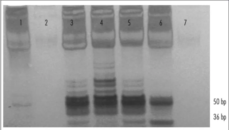
The presence of telomerase activity was established by inspecting the ilms for the presence of the character-istic 6-base pair increment ladder, as seen in Figure 1. The level of telomerase activity is expressed as an
ab-solute value20 and called TPG (total product generated)
value. One unit of TPG was deined as 0.001 mole, or 600 molecules, of TS primer extended for at least three
telomeric repeats by telomerase present in the extract20.
One TPG corresponds approximately to telomerase activity from one immortal cell and is determined by the formula TPG=(Telomerase sample – RNAse treated sample)/Internal control of sample (TSR8-negative
control)/(Internal control of TSR8) x 10021.
Statistical analysis was performed using the χ2 test
to evaluate signiicant differences. Findings with p<0.05 were considered statistically signiicant. All analyses were performed using the SPSS statistical analysis program, version 14.0.
Results
This study included 102 patients. The age range was between 17 and 71 years. Four of them had normal cytological indings (3.9% of all the cases), while 96.1% had some abnormal cytological indings described as follows: 9 cases of cervicitis (8.8%); 2 cases of Atypical Squamous Cells of undetermined signiicance (2%); 69 cases of low grade squamous intraepithelial lesion 69 (67.6%); 12 cases of high grade squamous intraepithe-lial lesion (11.8%); and 6 cases of squamous carcinoma (5.9%). Thus, L-SIL was the more frequent lesion in our sample, followed by H-SIL, cervicitis, carcinoma and ASC-US.
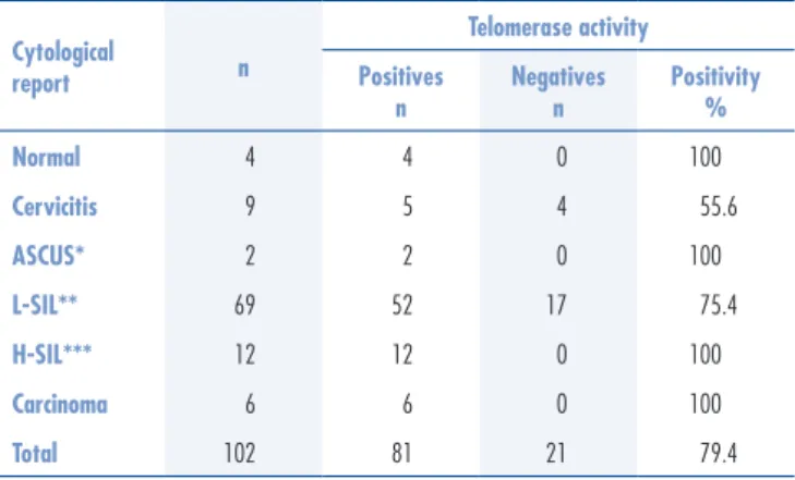
Table 1 shows the cytological results, inal histological diagnosis for the cervical scraping and telomerase activity. The higher frequencies (100%) of positivity for telom-erase activity were found in normal (4/4), ASCUS (2/2), H-SIL (12/12) and carcinoma (6/6) reports, followed by L-SIL with a 75.4% of positivity (52/69), and cervicitis reports with 55.6% (5/9).
Telomerase activity was determined as the total product generated value (TPG). Population TPG average was 31.6 with values ranging from zero, in those cases
Figure 1. Telomerase activity in human cervical cancer using the TRAP assay (Silver staining). (1) negative sample; (2) sample treated with Proteinase K; (3) sample with increased activity; (4) Telomerase Substrate template oligonucleotide (TSR8) Quantiication Control; (5) positive cells provided by the kit; (6) sample with decreased activity; (7) positive cells treated with proteinase K
1 2 3 4 5 6 7
with telomerase activity absence, to 133.3 in carcinoma. Telomerase activity measured in TPGs showed a dif-ferential tendency (p<0.05) according to cytological reports (Figure 2).
Among telomerase-positive cases, the lower TPG values were found in cervicitis with a minimum value of 11.1 and a maximum value of 50 TPGs. Then, following cervicitis cases, the increase of TPG values is followed by normal, ASC-US, L-SIL, H-SIL, and carcinoma ind-ings. Carcinoma showed the higher values of telomerase activity as shown in Figure 2.
There were signiicant differences in telomerase
activ-ity levels among the groups (p<0.05; L-SIL versus H-SIL,
L-SIL versus carcinoma, H-SIL versus carcinoma). However,
when comparing patients with normal reports with car-cinoma, the differences seem to be stronger (p<0.005).
Discussion
Telomerase activity has been demonstrated in a wide variety of human tissues, through malignant tumors or
precancerous cells, such as cervical lesion12-15. However,
we found negative telomerase activity in 20.6% of the samples of this study. Related literature describes nega-tive telomerase activity despite the presence of cervical
lesions14,22-24. Accordingly, a study on a Chinese
popula-tion, found a negativity percentage close to that obtained
for our population22. However, 52.9% of negativity was
found in a German population14;while in two Japanese
populations, the negativity percentage was 50 and 54.9%,
respectively18,23. On the other hand, negativity rates reached
75% for a Venezuelan population24. The development of
a malignant phenotype among certain types of cancers is independent of telomerase, and telomerase activation may not be a strict requirement of carcinogenesis for all
cervical cancers24, which seems to be the argument that
explains these negativity percentages.
Related literature illustrates that about 10 to 15% of cancers and cancer-derived cell lines are telomerase nega-tive but instead maintain telomere length by a homologous recombination-based pathway called alternative lengthening of
telomeres (ALT)25,26. Likewise, it is also possible that samples
might be contaminated with excessive blood or necrotic cells, possibly including telomerase inhibitors, leading to false nega-tive results. Studies suggest that the inhibitory effects observed
in the TRAP assay were due to the Taq inhibitors used27.
A correlation between telomerase positivity and cytological reports could not be found. However, when quantifying telomerase activity, it became signiicantly higher according to cervical lesion reported. These results support previous reports, in which telomerase activity levels seem to be related to the pathologic stages of dif-ferent types of cancers including hepatocellular carcinoma
(HCC)28, where human telomerase reverse transcriptase
(hTER) RNA expression showed a statistically signiicant relationship with tumor size in HCC patients regarding
a cervical lesion24,29. A correlation between telomerase
activity or hTER expression and the varying degrees of cervical lesions was observed regarding potentially ma-lignant oral lesions. A correlation in telomerase activity
and severity of the lesion30 was also found.
Furthermore, our study results are not only in ac-cordance with those of several other groups who have identiied telomerase activity in the majority of malignant tumors, but also in line with those who have identiied telomerase activity in proliferative diseases, predisposing
the development of malignant tumors28-32.
Studies reporting a positive correlation between
telomerase activity and pathologic degree of tumor29-30
suggest that the cells with higher telomerase activity Cytological
report n
Telomerase activity
Positives n
Negatives n
Positivity %
Normal 4 4 0 100
Cervicitis 9 5 4 55.6
ASCUS* 2 2 0 100
L-SIL** 69 52 17 75.4
H-SIL*** 12 12 0 100
Carcinoma 6 6 0 100
Total 102 81 21 79.4
Table 1. Cytology results of cervical scraping and telomerase activity
*ASCUS: Atypical Squamous Cells of Undetermined Signiicance;**L-SIL: Low grade Squamous Intraephitelial Lesion; ***H-SIL: High grade Squamous Intraephitelial Lesion.
Figure 2.Telomerase activity (in TPG unites) in different cervical lesions. Series 1;
Normal;
31.2 35
27.6
35.6
58.1 75.2
Total Product Generated (TPG)
Cytological Report
1. Schmidt JC, Cech TR. Human telomerase: biogenesis, traficking, recruitment, and activation. Genes Dev. 2015;29(11):1095-105. 2. Sandin S, Rhodes D. Telomerase structure. Curr Opin Struct Biol.
2014;25(100):104-10.
3. Kupiec M. Biology of telomeres: lessons from budding yeast. FEMS Microbiol Rev. 2014;38(2):144-71.
4. Jesus BB, Blasco MA. Telomerase at the intersection of cancer and aging. Trends Genet. 2013;29(9):513-20.
5. Akincilar SC, Low KC, Liu CY, Yan TD, Oji A, Ikawa M, et al. Quantitative assessment of telomerase components in cancer cell lines. FEBS Lett. 2015;589(9):974-84.
6. Li Y, Tergaonkar V. Noncanonical functions of telomerase: implications in telomerase-targeted cancer therapies. Cancer Res. 2014;74(6):1639-44.
7. Mocellin S, Pooley KA, Nitti D. Telomerase and the search for the end of cancer. Trends Mol Med. 2013;19(2):125-33.
8. Bollmann FM. Physiological and pathological signiicance of human telomerase reverse transcriptase splice variants. Biochimie. 2013;95(11):1965-70.
9. Buseman CM, Wright WE, Shay JW. Is telomerase a viable target in cancer? Mutat Res. 2012;730(1-2):90-7.
10. Verma I, Jain V, Kaur T. Application of Bethesda system for cervical cytology in unhealthy cervix. J Clin Diagn Res. 2014;8(9):OC26-30. 11. Nayar R, Wilbur DC. The Pap test and Bethesda 2014. “The
reports of my demise have been greatly exaggerated.” (after a quotation from Mark Twain). Acta Cytol. 2015;59(2):121-32. 12. Wang PH, Chen GD, Chang H, Yang SF, Han CP, Lin LY, et al.
High expression of human telomerase reverse transcriptase in
high-grade intraepithelial neoplasia and carcinoma of uterine cervix and its correlation with human papillomavirus infection. Reprod Sci. 2007;14(4):338-48.
13. Bravaccini S, Sanchini MA, Amadori A, Medri L, Saragoni L, Calistri D, et al. Potential of telomerase expression and activity in cervical specimens as a diagnostic tool. J Clin Pathol. 2005;58(9):911-4. 14. Saretzki G, Fischer H, Kaufmann IG, Schewe C, Nadjari B, Blohmer
J, et al. Telomerase activity in cervical smears. Anal Cell Pathol. 2001;23(1):39-43.
15. Tu Z, Zhang A, Wu R, Jiang J, Li Y, Wulan N, et al. Genomic ampliication of the human telomerase RNA gene for differential diagnosis of cervical disorders. Cancer Genet Cytogenet. 2009;191(1):10-6.
16. Liu Y, Wu BQ, Zhong HH, Xu ML, Fang WG. Detection of telomerase activity in cultured cells and tumor tissue of lung carcinoma by modiied telomeric repeat ampliication protocol. Pathol Int. 2010;60(5):386-94.
17. Walker JM. The Bicinchoninic Acid (BCA) assay for protein quantitation. In: Walker JM, editors. The protein protocols handbook. Totowa: Humana Press; 2002. p. 11-4.
18. Kyo S, Takakura M, Ishikawa H, Sasagawa T, Satake S, Tateno M, et al. Application of telomerase assay for the screening of cervical lesions. Cancer Res. 1997;57(10):1863-7.
19. Sanguinetti CJ, Dias Neto E, Simpson AJ. Rapid silver staining and recovery of PCR products separated on polyacrylamide gels. Biotechniques. 1994;17(5):914-21.
20. Kim NW, Wu F. Advances in quantiication and characterization of telomerase activity by telomeric repeat ampliication protocol (TRAP). Nucleic Acids Res. 1997;25(13):2595-7.
References
levels have a selective growth advantage due to improved stability of chromosomes with restored telomeres.
The fact that samples with normal indings showed telomerase activity was not expected, since normal samples have not shown positivity in other populations, such as
Japan23 Venezuela24, and India33 among others. Nevertheless,
all the normal samples in our study were telomerase-positive. By checking each normal sample individually, it was found that, in previous cytological reports from those women (dated 6 to 12 months before the study), cytological indings were abnormal. One of them showed L-SIL while the others exhibited ASC-US indings. It is possible that, for this study, normal reports rendered a false-negative result for cervical lesion in the screening report. This observation could be a irst explanation as to why telomerase activity was found in samples with normal reports. Nonetheless, in some studies, telomerase activity has also been detected on other populations with normal
reports14,22. This has been attributed to the sensibility of
the technique22, or the presence of activated lymphocytes
or granulocytes, which can also induce a positive result
since both have active telomerase14.
Given that predicting the evolution of ASCUS into another type of injury remains a controversial issue, it is not easy to explain the cause of telomerase positivity in samples with normal indings. However, according to some authors, the emergence of HSIL is common in cases where changes that deine the ASCUS on immature
squamous cells occurred10,11.
Through the analysis of both samples with ASCUS reports included in this study, it was evident that one of them presumed metaplasia. Thus, it can be said that detecting telomerase activity, in cases with ASCUS re-ports, is not an eventuality, since cells could be showing a metaplastic abnormality. However, there is not enough evidence for this afirmation. As far as we know, this is the irst study about telomerase activity and cervical cytology report in Colombia.
21. Gammaitoni L, Weisel KC, Gunetti M, Wu KD, Bruno S, Pinelli S, et al. Elevated telomerase activity and minimal telomere loss in cord blood long-term cultures with extensive stem cell replication. Blood. 2004;103(12):4440-8.
22. Wang SZ, Sun JH, Zhang W, Jin SQ, Wang HP, Jin YS, et al. Telomerase activity in cervical intraepithelial neoplasia. Chin Med J (Engl). 2004;117(2):202-6.
23. Kyo S, Takakura M, Tanaka M, Kanaya T, Inoue M. Telomerase activity in cervical cancer is quantitatively distinct from that in its precursor lesions. Int J Cancer. 1998;79(1):66-70.
24. Pinto-Tang J, Castro T, Premoli G. Detección de la actividad de telomerasa en lesiones cervicales, mediante el Protocolo de Ampliicación de Repeticiones Teloméricas (PART) no radioactivo. Invest Clin. 2005;46(3):255-63.
25. Neumann AA, Watson CM, Noble JR, Pickett HA, Tam PP, Reddel RR. Alternative lengthening of telomeres in normal mammalian somatic cells. Genes Dev. 2013;27(1):18-23.
26. Pal D, Sharma U, Khajuria R, Singh SK, Kakkar N, Prasad R. Augmented telomerase activity, reduced telomere length and the presence of alternative lengthening of telomere in renal cell carcinoma: plausible predictive and diagnostic markers. Gene. 2015;562(2):145-51. 27. Chen YC, Huang FC, Lin JJ. The addition of a spin column step
in the telomeric repeat application protocol removes telomerase inhibitors. Anal Biochem. 2015;478:49-51.
28. El-Mazny A, Sayed M, Sharaf S. Human telomerase reverse transcriptase messenger RNA (TERT mRNA) as a tumour marker for early detection of hepatocellular carcinoma. Arab J Gastroenterol. 2014;15(2):68-71.
29. Wang HY, Kim G, Cho H, Kim S, Lee D, Park S, et al. Diagnostic performance of HPV E6/E7, hTERT, and Ki67 mRNA RT-qPCR assays on formalin-ixed parafin-embedded cervical tissue specimens from women with cervical cancer. Exp Mol Pathol. 2015;98(3):510-6.
30. Dorji T, Monti V, Fellegara G, Gabba S, Grazioli V, Repetti E, et al. Gain of hTERC: a genetic marker of malignancy in oral potentially malignant lesions. Hum Pathol. 2015;46(9):1275-81. 31. Ruiz-Hoyos BM, Loango-Chamorro N, Landázuri P. Exactitud
de la actividad de la telomerasa para el diagnóstico del virus del papiloma humano en mujeres con patología cervical en Armenia, Colombia, 2007. Rev Colomb Obstet Ginecol. 2012;63(3):207-14.
32. El Belbesy SF, El Aggan HA, Sultan HK, El Naggar AA, Ahmed HKF. Telomere length and human telomerase reverse transcriptase (hTERT) level in patients with acute myeloid leukemia: impact on clinical outcome Acta Haematol Pol. 2015;46(4):304-11. 33. Reddy VG, Khanna N, Jain SK, Das BC, Singh N. Telomerase-A