abstract
Lack of correlation between tubular dentine
cement penetration, adhesiveness and leakage in
roots illed with gutta percha and an endodontic
cement based on epoxy amine resin
Ricardo MACHADO1, Ulisses Xavier da SILVA NETO1, Everdan CARNEIRO1, Luiz Fernando FARINIUK1, Vânia Portela
Ditzel WESTPHALEN1, Rodrigo Sanches CUNHA2
1- Department of Endodontics, Pontiical Catholic University of Paraná, Curitiba, PR, Brazil. 2- School of Dentistry, Restorative Dentistry, University of Manitoba, Winnipeg, Canada.
Corresponding address: Ricardo Machado - Rua Anibal Gaya, 898 - Casa 14 - Condomínio Nova Era - Centro -Navegantes - SC - Brazil - 88.375-000 -
Phone/Fax: (55 47) 3319-1625 - Cel: (55 47) 8409-1561 - e-mail: [email protected]
Submitted: March 15, 2013 - Modiication: October 10, 2013 - Accepted: October 28, 2013
O
bjective: To analyze possible correlations among tubular dentine cement penetration,adhesiveness and apical leakage in illings performed with gutta percha and an
endodontic cement based on epoxy amine resin. Material and Methods: Sixty similar,
extracted human mandibular central incisors were irrigated, instrumented and illed following the same protocol. First, apical leakage was quantiied by luid iltration tests. Then,
these same specimens were sectioned for analysis of tubular dentine cement penetration and the middle thirds were submitted to push-out tests to analyze the adhesiveness of the
illings. Results: In brief, the means and standard deviations with a conidence interval of
95% were as follows: tubular dentine cement penetration (8.875±4.540), adhesiveness (4.441±2.683) and apical leakage (0.318±0.215). The data were confronted using the Pearson’s test (P>0.05), and it was possible to prove that there was no correlation between
(1) tubular dentine cement penetration and apical leakage (r2: 0.08276), (2) tubular
dentine cement penetration and adhesiveness (r2: -0.2412) and (3) adhesiveness and
apical leakage (r2: 0.1340). Conclusion: After analysis of these data, it could be observed
that there exists no correlation among the variables analyzed in this study.
Keywords: Dentin. Adhesiveness. Leakage.
INtrODUctION
In recent years, the evolution of Endodontics has
broken several paradigms, driven by technological and techniques advances in all its phases of execution. However, despite all these technical
and scientiic developments, some concepts have
not changed. The main objectives of root canal treatment continue to be the elimination of or reduction in the number of microorganisms within the root canal space, and the prevention of possible infection or reinfection18,28.
With this in mind, there is a clear interest in
improving the effectiveness of the root canal illing
techniques. Proof of this can be found in the great number of different systems recently developed to
limit microbiological action capable of inducing or even causing resistance of a periapical lesion7,25.
The illings failures observed in several studies,
resulting from many different methodologies, have given rise to a global trend, evidenced by the literature, toward enhancing the ability of
endodontic illing materials to project themselves
into the dentinal tubules2,7,31. These materials
not only act as antibacterial agents, theoretically speaking, but can also optimize the quality of the seals provided by these penetrations. This hypothesis is based on the concept that tubular dentine cement penetration considerably increases
the contact surface of the illing material with the
root canal walls, thereby improving the seal6,19,24.
the illing materials also has the ultimate goal of
preventing leakage13.
However, these hypotheses have not yet been
completely clariied in the literature, and few studies
have been performed to investigate these possible correlations4,6,12,13,19,23,24.
Based on the above, this study aimed to analyze possible correlations between tubular dentine cement penetration, adhesiveness and leakage in
roots illed with gutta percha and an endodontic
cement based on epoxy amine resin.
MatErIaL aND MEtHODs
specimen selection
After approval of the Ethics Committee (process
5314), sixty mandibular central incisor teeth with single straight canals, complete rhizogenesis, with no resorption or previous endodontic treatment, and free foraminal access, were provided by the university tooth bank and selected for this study.
All this information was veriied by clinical analysis
and vestibulo-lingual and mesiodistal radiographs. The crowns were then removed using a low-speed
steel cutting disc (Isomet-Buehler, Lake Bluff, IL,
USA), standardizing all roots at 13 mm in length.
specimen preparation
The accesses were performed using a tapered-tip bur 3082 (KG Sorensen, Barueri, SP, Brazil). Working length was established by subtracting 1
mm from the point where the ile was visible at the
apical foramen. The coronal and middle thirds of each canal were prepared using Gates Glidden drills (Dentsply-Maillefer, Ballaigues, Switzerland) sizes 4, 3 and 2, by placing each instrument 2 mm deeper than the previous one. The apical foramina were standardized using real length instrumentation of
the teeth up to instrument 25 K-Flexoile
(Dentsply-Maillefer, Ballaigues, Switzerland), and the apical
thirds were prepared with the Proile 04 System
(Dentsply-Maillefer, Ballaigues, Switzerland) up to size 35 at working length. The canals were irrigated between each instrumentation with 2 mL of freshly
prepared 2.5% NaOCl plus a lush of 3 mL of 17% EDTA for 3 min. Five milliliters of sterile water was used as a inal rinse.
Canal illing
The prepared canals were illed using the lateral
compaction technique. After drying with paper
points, a size 20 ile was used to place 10 µL of an
endodontic cement based on epoxy amine resin – AH Plus (Dentsply-DeTrey, Konstanz, Germany) – into the canal, using a counterclockwise rotation.
The sealer was labeled with a 0.1% Rhodamine
B dye (Sigma-Aldrich, St. Louis, MO, USA) for the purpose of further analysis by optical light
microscopy.
A preitted size 35, 0.04-taper gutta-percha
cone (Dentsply-Maillefer, Ballaigues, Switzerland) was used as the master cone, and two accessory
cones were used in addition. The illed roots were
stored at 37°C and 100% humidity for 7 days, to allow the sealer to set.
Apical leakage analysis by luid iltration
tests
The luid iltration method was used to determine
apical leakage. The root apex was connected to a Luer-type metal needle by means of a plastic tube. The allowed leakage margin for the tested
groups was quantiied according to the movement of a small air bubble inside a 25 µL micropipette (Microcaps-Fisher Scientiic, Philadelphia, PA, USA).
The inside of the pipette and the entire system
was illed with distilled water and a pressure of
10 psi was applied. After making sure there was no leakage in the connections, the system was activated and balanced for 4 minutes. The volume
of luid was calculated by observing the air bubble displacements, expressed in µL/min-¹.10 psi.
Measurements were made at 2-minute intervals in a period of 8 minutes22,27.
Tubular dentine cement penetration analysis by optical microscopy
Each specimen was sectioned horizontally at 3,
6 and 8 mm from the apex, using a low-speed steel
cutting disc (Isomet-Buehler, Lake Bluff, IL, USA).
Three slices per root were created, resulting in a total of 180 slices. A standard polishing procedure using SiC paper (200, 300, 400, 600) followed by 3
µm diamond paste was employed on the coronally
facing surface of each slice to produce a
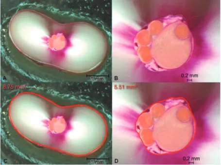
high-relection surface (Figures 1A and B), and each slice
was observed in a high-resolution stereomicroscope to acquire images at 1048x1048 pixels, covering the entire root surface. For each image, the outer perimeter of the root and the inner perimeter of the root canal walls were outlined and measured using the AxioVision Software 4.11 (Carl Zeiss, Jena, Germany) (Figures 1C and D). The total cross-sectional area of the canal wall was obtained for each section by subtracting the value of the outer perimeter from the inner perimeter. The absolute cement penetration values (Figure 2) for each section were then converted into a percentage of cement penetration into dentinal tubules, by calculating the total cross-sectional area of the canal wall previously obtained in the high-resolution stereomicroscope. The percentages were averaged for each specimen.
Adhesiveness analysis by mechanical push-out tests
Firstly, the thickness of each slice of the 60 sections corresponding to the middle third was
measured with a digital caliper (Mitutoyo
IP67-Mitutoyo, Neuss, Germany). Then, the specimens were submitted individually to push-out bond strength tests using a universal-testing machine
(EMIC DL200MF, São José dos Pinhais, PR, Brazil) at a speed of 0.5 mm/min up to bond failure using
a 0.50-mm diameter stainless steel cylindrical
plunger. The plunger tip was sized and positioned so
that it contacted only the illing material. Because
of the convergence of the root canal sections, the push-out force was applied from apical to coronal. The bond strength expressed in MPa at failure was calculated by dividing the load in newtons by the area of the bonded interface. The area of the bonded interface was calculated according to the following formula: area=2πr X h, where π is kept
constant at 3.14, and r and h are the radius and
height measured in millimeters of the illing material
Figure 1- Coronal surfaces of the same slice after metallographic treatment. Total cross-sectional area (A) and canal
cross-sectional area (B). The outer (C) and the inner (D) perimeter of the root canal walls outlined and measured using AxioVision Software 4.11 (Carl Zeiss, Jena, Germany)
Figure 2- Absolute cement penetration values outlined and measured using AxioVision Software 4.11 (Carl Zeiss, Jena,
that was pushed out1,23. statistical analysis
The results obtained were submitted to the Pearson test with a significance level of 95% (p<0.05) to perform the correlation analysis among the variables of the study using the statistical
software SPSS 11.5 (SPSS Inc., Chicago, IL, USA).
rEsULts
In isolated analyses of the three variables, the means and standard deviations, with a conidence
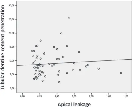
interval of 95%, were the following: tubular dentine cement penetration (8.875±4.540), adhesiveness (4.441±2.683) and apical leakage (0.318±0.215). The results obtained in the correlation analysis among these study variables were confronted and are shown in Figures 3, 4 and 5, respectively.
There was no statistically signiicant correlation
Figure 3- Representative image of the correlation analysis between tubular dentine cement penetration and leakage
Figure 4- Representative image of the correlation analysis between adhesiveness and tubular dentine cement penetration
between tubular dentine cement penetration and apical leakage (r2: 0.08276), adhesiveness and
tubular dentine cement penetration (r2: -0.2412)
and adhesiveness and apical leakage (r2: 0.1340).
DIscUssION
The hypothesis that tubular dentine cement
penetration could improve sealability was irstly
proposed in 198429. Later, other studies were
performed to address this premise3,9,16,20. These
other studies were based on the original hypothesis that a better cement penetration into the dentinal tubules would somehow improve the quality of
endodontic illings. Therefore, previous reports
that tubular dentine cement penetration was one of the most important factors to be considered in
choosing the illing material justify the importance
of conducting comparative studies on this matter10.
Most of the papers have analyzed the three variables studied in our paper – tubular dentine cement penetration2,21, adhesiveness5,8, and
leakage14,15,22, merely comparing different materials.
Onlya few articles have tried to analyze possible correlations among these variables4,6,12,13,19,23,24.
Comparing four endodontic cements (Diaket,
Endomethasone, CRCS and Ketac Endo) in relation
to their projections into the dentinal tubules and
their sealability using the same illing technique,
Sen, et al.19 (1996) showed that the best results
were obtained by different materials in isolated analyses. As in our results, it was not possible to observe any correlation between these two variables. The methods used (scanning electron microscopy and dye leakage test) were different
from those adopted in this study (optical microscopy
and luid iltration test), but the main methodological
point was to use the same specimens to analyze different points, an approach considered essential for analyzing possible correlations among two or more variables4.
Using a different protocol, another study investigated the dentine permeability obtained by
two substances used in the inal irrigation of root
canals, namely, sodium hypochlorite and ethanol 95%. This permeability was measured by analyzing the cement penetration using optical microscopy.
In addition, specimens from the different study groups were also submitted to the luid transport test to analyze the sealability of their illings.
Although a greater cement penetration and a lower level of leakage were observed in the group that
used ethyl alcohol, this correlation was not veriied
statistically24, thereby showing results similar to
those of our study (r2: 0.08276).
After analyzing our results and most of the studies performed to date, it appears that no correlation exists between tubular dentine cement penetration and sealability, or else the research methods used were unable to detect any correlation.
The majority of the leakage tests, including luid iltration, showed leakage only when there was
at least one void extending from the apical to the
coronal thirds. A root canal illing that looks poorly
condensed on the radiograph may contain many “cul-de-sac”-type voids and no leakage. On the other hand, very small “through-and-through”-type voids that are invisible on radiographs may
be detected by the luid iltration test and show
leakage rates would probably not be smaller if a major amount of sealer penetrated into the dentinal
tubules. It seems that a more plausible hypothesis
would be that this variable may be improved when there is a better adaptation of the cement to the canal walls.
Addressing adhesiveness and cement penetration, a classic study correlating these variables was performed to compare the adhesiveness of two
illing systems (Gutta Percha/Kerr Pulp Canal Sealer EWT and Resilon/Epiphany)and the authors found
more favorable results for the Resilon/Epiphany system. However, an analysis of images by SEM
demonstrated no large cement penetrations into the dentinal tubules23. In view of this inding, the
authors suggested that there was no effective correlation between cement penetration and adhesiveness, a conclusion corroborated by the results of this study. Our correlation results (r2:
-0.2412) and those mentioned above suggest that better adhesiveness is not related to a possible mechanical overlap provided by cement penetration
or adhesive tags into the dentinal tubules. It
seems legitimate to state that the establishment of a consistent hybrid layer in intratubular dentine plays a more important role in achieving better adhesiveness to the root dentine11,17.
Nagas, et al.12 (2007) analyzed the adhesiveness
and leakage in root illings comparing different
methods of photoactivation (quartz halogen light for 40 seconds, light-emitting diode for 20 seconds, and
plasma ARC for 6 seconds). It is worth highlighting
that there were some important methodological differences in relation to the current study. The authors used different specimens to perform the push-out and leakage tests, and also carried out different statistical tests to analyze each variable
individually. In the results related to adhesion, there were signiicant statistical differences among the
three groups compared. However, when the leakage was analyzed separately, the differences between the two groups with the best results were not
signiicant, suggesting a lack of correlation between
these variables. This conclusion was demonstrated by the correlation results of our study as well (r2:
0.1340).
To date, in the only study that has shown a positive correlation between adhesiveness and leakage13, there was an important methodological
difference from those used in this research. The authors did not use gutta-percha or any solid material associated to the cement, which was
used alone. Considering that the luid iltration
test shows leakage only when there is at least one void extending from the apical to the coronal thirds, it is possible that the sealer alone may be able to improve the sealability observed by this methodology.
The main point of our study was designed to produce a large experimental group. This is an important experimental design feature, because standard correlation analysis posits that any random factor affects only one subject, and not others. This requirement is violated when two or more different experimental groups are created, as
was the case in all the aforementioned studies. In
fact, there was no rationale to justify the creation of two or three experimental groups when the main goal was restricted to verifying a potential cause-and-effect correlation. Therefore, when attempting to verify a potential cause-and-effect correlation, a single sizeable group should be created. However, even when a single well-standardized group was used, assessed through updated and refined experimental models, a correlation could not be established among the variables4.
cONcLUsION
Based on the experimental conditions and the results observed in this in vitro study, it is possible to prove that there are no correlations among tubular dentine cement penetration, adhesiveness and leakage.
acKNOWLEDGEMENts
The authors deny any conlicts of interest related
to this study.
rEFErENcEs
1- Carneiro SM, Sousa-Neto MD, Rached FA Jr, Miranda CE, Silva SR, Silva-Sousa YT. Push-out strength of root illings with or without thermomechanical compaction. Int Endod J. 2012;45:821-8.
2- Chandra SS, Shankar P, Indira R. Depth of penetration of four resin sealers into radicular dentinal tubules: a confocal microscopic study. J Endod. 2012;38:1412-6.
3- De-Deus GA, Gurgel-Filho ED, Maniglia-Ferreira C, Coutinho-Filho T. The inluence of illing technique on depth of tubule penetration by root canal sealer: a study using light microscopy and digital image processing. Aust Endod J. 2004;30:23-8. 4- De-Deus G, Brandão MC, Leal F, Reis C, Souza EM, Luna AS, et al. Lack of correlation between sealer penetration into dentinal tubules and sealability in nonbonded root illings. Int Endod J. 2012;45:642-51.
5- Ebert J, Leyer A, Günther O, Lohbauer U, Petschelt A, Frankenberger R, et al. Bond strength of adhesive cements to root canal dentin tested with a novel pull-out approach. J Endod. 2011;37:1558-61.
6- Ghoddusi J, Dibaji F, Marandi S. Correlation between sealer penetration and microleakage following the use of MTAD as a inal irrigant. Aust Endod J. 2010;36:109-13.
7- Hammad M, Qualtrough A, Silikas N. Evaluation of root canal obturation: a three-dimensional in vitro study. J Endod.
2009;35:541-4.
8- Imai Y, Komabayashi T. Properties of a new injectable type of root canal illing resin with adhesiveness to dentin. J Endod. 2003;29:20-3.
9- Malyk Y, Kaaden C, Hickel R, Ilie N. Analysis of resin tags formation in root canal dentine: a cross sectional study. Int Endod J. 2010;43:47-56.
10- Mamootil K, Messer HH. Penetration of dentinal tubules by endodontic sealer cements in extracted teeth and in vivo. Int Endod J. 2007;40:873-81.
11- Mastoras K, Vasiliadis L, Koulaouzidou E, Gogos C. Evaluation of push-out bond strength of two endodontic post systems. J Endod. 2012;38:510-4.
12- Nagas E, Cehreli ZC, Durmaz V, Vallittu PK, Lassila LV. Regional push-out bond strength and coronal microleakage of Resilon after different light-curing methods. J Endod. 2007;33:1464-8. 13- Neelakantan P, Subbarao C, Subbarao CV, De-Deus G, Zehnder M. The impact of root dentine conditioning on sealing ability and push-out bond strength of an epoxy resin root canal sealer. Int Endod J. 2011;44:491-8.
14- Oddoni PG, Mello I, Coil JM, Antoniazzi JH. Coronal and apical leakage analysis of two different root canal obturation systems. Braz Oral Res. 2008;22:211-5.
15- Ozok AR, van der Sluis LW, Wu MK, Wesselink PR. Sealing ability of a new polydimethylsiloxane-based root canal illing material. J Endod. 2008;34:204-7.
16- Patel DV, Sherriff M, Ford TR, Watson TF, Mannocci F. The penetration of RealSeal primer and Tubliseal into root canal dentinal tubules: a confocal microscopic study. Int Endod J. 2007;40:67-71.
17- Prado M, Simão RA, Gomes BP. Effect of different irrigation protocols on resin sealer bond strength to dentin. J Endod. 2013;39:689-92.
18- Ricucci D, Loghin S, Siqueira JF Jr. Exuberant Bioilm infection in a lateral canal as the cause of short-term endodontic treatment failure: report of a case. J Endod. 2013;39:712-8.
19- Sen BH, Pişkin B, Baran N. The effect of tubular penetration of root canal sealers on dye microleakage. Int Endod J. 1996;29:23-8.
20- Sevimay S, Dalat D. Evaluation of penetration and adaptation of three different sealers: a SEM study. J Oral Rehabil. 2003;30:951-5.
21- Shokouhinejad N, Sabeti M, Gorjestani H, Saghiri MA, Loti M, Hoseini A. Penetration of Epiphany, Epiphany self-etch and AH Plus into dentinal tubules: a scanning electron microscopy study. J Endod. 2011;37:1316-9.
22- Silva Neto UX, Moraes IG, Westphalen VP, Menezes R, Carneiro E, Fariniuk LF. Leakage of 4 resin-based root-canal sealers used with a single-cone technique. Oral Surg Oral Med Oral Pathol Oral Radiol Endod. 2007;104:53-7.
23- Skidmore LJ, Berzins DW, Bahcall JK. An in vitro comparison of the intraradicular dentin bond strength of Resilon and gutta-percha. J Endod. 2006;32:963-6.
24- Stevens RW, Strother JM, McClanahan SB. Leakage and sealer penetration in smear-free dentin after a inal rinse with 95% ethanol. J Endod. 2006;32:785-8.
25- Tanomaru-Filho M, Sant'Anna A Jr, Berbert FL, Bosso R, Guerreiro-Tanomaru JM. Ability of gutta-percha and Resilon to ill simulated lateral canals by using the Obtura II system. J Endod. 2012;38:676-9.
26- Van der Sluis LW, Wu MK, Wesselink PR. An evaluation of the quality of root illings in mandibular incisors and maxillary and mandibular canines using different methodologies. J Dent. 2005;33:683-8.
27- Vasiliadis L, Kodonas K, Economides N, Gogos C, Stavrianos C. Short and long-term sealing ability of Gutta-low and AH-Plus using an ex vivo luid transport model. Int Endod J. 2010;43:377-81. 28- Vieira AR, Siqueira JF Jr, Ricucci D, Lopes WS. Dentinal tubule infection as the cause of recurrent disease and late endodontic treatment failure: a case report. J Endod. 2012;38:250-4. 29- White RR, Goldman M, Lin PS. The inluence of the smeared layer upon dentinal tubule penetration by plastic illing materials. J Endod. 1984;10:558-62.
30- Wu MK, Bud MG, Wesselink PR. The quality of single cone and laterally compacted gutta-percha illings in small and curved root canals as evidenced by bidirectional radiographs and luid transport measurements. Oral Surg Oral Med Oral Pathol Oral Radiol Endod. 2009;108:946-51.