PROPOSAL OF A METHODOLOGY FOR INDIVIDUALIZED
IODINE-131 THERAPY FOR GRAVES’ DISEASE IN PATIENTS
WITH HYPERTHYROIDISM*
Francisco de Araujo1
, Rossana Corbo de Melo2
, Ana Maria de Oliveira Rebelo3
, Bernardo
Maranhão Dantas4
, Ana Letícia A. Dantas4
, Eder Augusto de Lucena5
OBJECTIVE: Several methods are utilized for determining the radioiodine activity in the treatment of Graves’ disease (hyperthyroidism). Some of this methods do not take into consideration the thyroid absorbed dose or the necessary parameters for this estimation. The association between absorbed dose and administered activity depends on effective half-life, iodine uptake and thyroid mass of each patient. The present study was aimed at developing a methodology for individualized 131
I therapy for Graves’ disease in patients with hyperthyroidism of the Graves’ disease. MATERIALS AND METHODS: A neck-thyroid phantom developed at Instituto de Radioproteção e Dosimetria, containing a standard solution of 131
I, was utilized for calibrating the scintillation gamma camera and uptake probe installed in the Department of Nuclear Medicine of the Hospital Universitário Clementino Fraga Filho da Universidade Federal do Rio de Janeiro. RESULTS: The view angle of the collimator/detector assembly presented values compatible with the thyroid gland size for dis-tances of 25 cm (uptake probe) and 45.8 cm (scintillation gamma camera). Calibration factors were 39.3 ± 0.78 and 4.3 ± 0.17 cpm/kBq, respectively. The 14–30-hour interval in the retention curve allows the estimation of activity between two points for determining the effective iodine half-life in the thyroid. CON-CLUSION: The utilization of equipment usually available in nuclear medicine clinics is feasible, so this is a simple, effective and low cost methodology.
Keywords: Graves’ disease; Hyperthyroidism; Iodine therapy; Iodine-131; Scintillation gamma camera; Up-take probe.
Proposta de metodologia para tratamento individualizado com iodo-131 em pacientes portadores de hiper-tireoidismo da doença de Graves.
OBJETIVO: Diferentes métodos são usados para determinar atividade do radioiodo para tratamento de hiper-tireoidismo (doença de Graves). Alguns não consideram a dose absorvida pela tireóide ou os parâmetros necessários para este cálculo. A relação entre dose absorvida e atividade administrada depende da meia-vida efetiva, da captação do iodo e da massa da tireóide de cada paciente. O objetivo deste trabalho é pro-por uma metodologia para tratamento individualizado com 131
I em pacientes portadores de hipertireoidismo da doença de Graves. MATERIAIS E MÉTODOS: Usou-se um simulador de tireóide-pescoço desenvolvido no Instituto de Radioproteção e Dosimetria contendo solução de 131
I, para calibração da gama-câmara e sonda cintilométrica do Serviço de Medicina Nuclear do Hospital Universitário Clementino Fraga Filho da Universi-dade Federal do Rio de Janeiro. RESULTADOS: O campo de visão colimador-detector apresentou valores compatíveis com o tamanho da glândula para as distâncias de 25 cm (sonda de captação) e 45,8 cm (gama-câmara). Os fatores de calibração (cpm/kBq) foram 39,3 ± 0,78 e 4,3 ± 0,17, respectivamente. O intervalo entre 14 e 30 horas da curva de retenção permite o cálculo de atividade entre dois pontos, para determina-ção da meia-vida efetiva do iodo na tireóide. CONCLUSÃO: A utilizadetermina-ção de equipamentos usualmente dispo-níveis em serviços de medicina nuclear é viável, tornando esta metodologia simples, eficaz e de baixo custo. Unitermos: Doença de Graves; Gama-câmara; Hipertireoidismo; Iodoterapia; Iodo-131; Sonda cintilométrica. Abstract
Resumo
* Study developed at Instituto de Radioproteção e Dosimetria/ Conselho Nacional de Energia Nuclear (IRD/CNEN), Rio de Ja-neiro, RJ, Brazil. Sponsorship: Conselho Nacional de Desenvol-vimento Científico e Tecnológico – CNPq (PCI Scholarship) and Fundação Carlos Chagas Filho de Amparo à Pesquisa do Estado do Rio de Janeiro – Faperj (APQ1 Project).
1. Master in Radioprotection and Dosimetry, Instituto de Ra-dioproteção e Dosimetria/Conselho Nacional de Energia Nuclear (IRD/CNEN), Rio de Janeiro, RJ, Brazil, Researcher, Conselho Nacional de Desenvolvimento Científico e Tecnológico – CNPq (PCI Scholarship).
2. PhD of Medicine (Radiology), Universidade Federal do Rio de Janeiro (UFRJ), Rio de Janeiro, RJ, Brazil, MD, Endocrinolo-gist.
3. Master in Nuclear Energy – Coordinator for the Programs of Post-graduation in Engineering/Universidade Federal do Rio de
INTRODUCTION
The first studies about the thyroid func-tion have been developed with 131I, that up
to the present time has been utilized in nuclear medicine for therapy of hyperthy-roidism and, particularly in cases of thyroid ablation for treatment of cancer. Therapeu-tic 131I doses are orally administered in the
form of liquid or capsules(1). Radiodine
advantages include: easy administration,
Janeiro (COPPE/UFRJ), Physics, Division of Nuclear Medicine, Universidade Federal do Rio de Janeiro (UFRJ), Rio de Janeiro, RJ, Brazil.
4. PhD of Biology (Nuclear Biosciences), Universidade do Es-tado do Rio de Janeiro (UERJ), Researchers at Instituto de Radio-proteção e Dosimetria/Conselho Nacional de Energia Nuclear (IRD/CNEN), Rio de Janeiro, RJ, Brazil.
5. Master in Biology (Nuclear Biosciences), Radioprotection Technician, Universidade do Estado do Rio de Janeiro (UERJ), Rio de Janeiro, RJ.
Mailing address: Dr. Francisco de Araujo. Instituto de Radio-proteção e Dosimetria (IRD/CNEN). Avenida Salvador Allende, s/ nº. Rio de Janeiro, RJ, Brazil, 22780-160. E-mail: faraujo@ird. gov.br
thyroid gland after 131I administration
var-ies among patients, depending on several factors such as: degree of iodine uptake, mass of uptaking tissue, effective iodine half-life in the thyroid, distribution of ra-dioactivity throughout the tissue, and cells radiosensitivity.
However, scarce conclusive informa-tion is available in the literature regarding the patients absorbed dose. As a function of the magnitude of the administered activ-ity for treatment with radioiodine, the main risks for patients involve increase in the probability of developing cancer in differ-ent organs or tissues, and the effects on the descendants of women at childbearing age(4). The realistic evaluation of these risks
requires a biokinetic analysis of the 131I
behavior in the organism, followed by the calculation of the patients absorbed dose. A consensus is still to be achieved on the ideal protocol for treating hyperthyroid-ism. Frequent discussions have been held on which protocol would contribute with the best clinical results. Currently, differ-ent protocols are utilized to define the ac-tivity to be administered in the radioiodine therapy for hyperthyroidism, but not all of them take the thyroid absorbed dose into consideration. Some protocols utilize a fixed administered activity (standard), without considering biokinetic parameters such as the thyroid gland volume, the io-dine effective half-life in the thyroid, and the individual uptake(5). When a 10 mCi
standard activity is utilized in a patient af-fected by Graves’ disease, the thyroid ab-sorbed-dose may range from 60 Gy to 600 Gy(5). In patients with a short iodine
half-life, the absorbed dose per activity unit will be low and, possibly, the treatment will not
roidism are diffuse toxic goiter, also know as Graves’ disease, multinodular toxic goi-ter, and toxic adenoma. Graves’ disease is the most frequent one (80%)(6). The
effec-tive iodine half-life in the thyroid of pa-tients affected by Graves’ disease is low, while the uptake is high as compared with the multinodular and uninodular goiters(5).
The typical thyroid iodine retention curves show that the highest level of iodine uptake in patients affected by diffuse toxic goiter occurs, on average, 12 hours after the ra-dionuclide administration. This character-istic is extremely relevant in the calculation of the effective half-life in the interval be-tween 14 and 30 hours following the radio-iodine administration to the patients. The utilization of the available tools and routine procedures in the division of nuclear medi-cine has made this low cost methodology feasible, simple and effective.
MATERIALS AND METHODS
Production of the thyroid phantoms
The experimental part of this study was initially developed in the Laboratory of In Vivo Measurements of Instituto de Radio-proteção e Dosimetria (IRD), where the thyroid phantom was constructed with a 100 mm-diameter filter paper (Whatman) trimmed in a thyroid shape and size(7). This
phantom was impregnated with a mass consisting of 241.86 mg of 131I solution,
with specific activity of 3.075 MBq/g (83.1
µCi/g) and 1.1% total uncertainty. This solution was previously calibrated by the National Laboratory of Ionizing Radiation Metrology (Laboratório Nacional de Me-trologia das Radiações Ionizantes) of Ins-tituto de Radioproteção e Dosimetria da
This phase was developed in the Divi-sion of Nuclear Medicine of Hospital Universitário Clementino Fraga Filho da Universidade Federal do Rio de Janeiro (HUCFF-UFRJ), and consisted in the cali-bration of the Siemens Diacam Gamma camera equipped with a NaI(Tl) crystal, with 2" × 2" and 59 photomultiplier thick-nesses, pinhole-type lead collimator (Fig-ure 2), and a SCT-13004 scintillation probe (Figure 3). This priority of this methodol-ogy is the optimization of the in vivo de-tection system, for calculating the effi-ciency of 131I detection in the thyroid.
Collimator-detector assembly field of view
For establishing the best working dis-tance between the source and the detector, curves typical of the collimator-detector assembly field of view regarding the equip-ment involved in this experiequip-ment were traced. The method utilizes isocounting or
isoresponse curves for each of the
sys-tems(9). These curves were obtained by the
positioning of the detector at different dis-tances from a punctiform 131I source, with
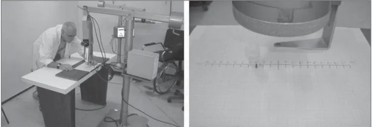
collimator-de-Figure 1. Sequence of the construction of the physical-anthropomorphic thy-roid-neck phantom of IRD and positioning for calibration of the detector sys-tem.
Figure 2. Diacam gamma-camera, with a pinhole-type collimator of the HUCFF-UFRJ Division of Nuclear Medicine.
Figure 4. Collection of isocounting curves of the collimator-detector system field of view, for selection of the best distance between the detector and the 131I
source.
Figure 3. SCT-13004 scintillation probe of the HUCFF-UFRJ Division of Nuclear Medicine.
tector assembly, without the reducing ring, five different distances (42.8 cm, 43.8 cm, 44.8 cm, 45.8 cm and 46.8cm) were mea-sured with the aid of a gauging spacer de-veloped at IRD (Figure 5). For the SCT-13004 detector-collimator assembly three different distances (20 cm, 25 cm and 30 cm) were measured with the ruler coupled with the assembly (Figure 4). The function of the ruler and “spacer” is to keep the ac-curacy and reproducibility of the measure-ments.
Calibration factor for gamma camera and scintillation probe
Initially, the thyroid-neck phantom was positioned for counting the background radiation with the thyroid phantom free
from contamination, and later with the phantom impregnated with the 131I solution,
as previously described. In both cases, the detector was positioned at 42.8 cm, 43.8 cm, 44.8 cm, 45.8 cm and 46.8 cm from the phantom, the countings being performed in five minutes for each distance. Each mea-surement was repeated for three times for a better statistical result. The same proce-dure was adopted for obtention of the cali-bration factor of the scintillation probe, only with a variation of the distances be-tween the detector and the phantom (20 cm, 25 cm and 30 cm).
Determination of biokinetic data
reten-tion curve based on measurements in the thyroid of patients with Graves´ disease has allowed the evaluation of convenient time intervals for calculating of activities values for determination of the radioiodine effec-tive half-life and the initial uptake in the thyroid gland. The relation between the thyroid absorbed dose and administered activity required for the therapy was calcu-lated by means of an equation developed by Marinelli-Quimby(11).
D/A = 0.043 UoTef/V
These curves allow the determination of the collimator-detector system field of view for the equipment involved in the present experiments, indicating the best source-detector distance. The most informative method for demonstrating these character-istics for each device is based on the isocounting or isoresponse curves. In this region named “field of view”, the values from any region of the thyroid gland are accounted with an uniform sensitivity, be-sides reducing not only the environment background radiation, but also the back-ground radiation coming from other re-gions of the patient’s body.
Table 1 demonstrates mean values of three countings for each source position in relation to the collimator central axis, and percent values of counting rates regarding
sition of maximum counting with the re-spective associated uncertainties.
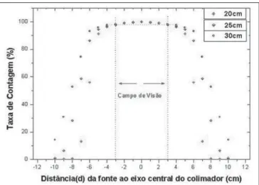
Figure 7 presents the isocounting curve of the SCT-13004 system detector-collima-tor for distances of 20 cm, 25 cm and 30 cm, where the percent counting rate is shown as a function of the distance be-tween the 131I punctiform source and the
central axis of the collimator.
Optimization and determination of the calibration factor
For the Diacam gamma camera system with a pinhole lead collimator, without re-ducing ring – Table 3 shows the calibration factors for 131I and uncertainties associated
with the respective distance between the detector and the phantom. For the Siemens Diacam gamma camera system, the
experi-Figure 5. Gauging spacer for measurements de-veloped in the IRD.
Table 1 Field of view data of the gamma camera system with pinhole collimator without reducing ring for five source-detector distances, utilizing a 131I
punc-tiform source in three 30-second countings.
Mean counting ± σm
Distance d (cm) 0 ± 1 ± 2 ± 3 ± 4 ± 5 ± 6 ± 7 ± 8 ± 9 ± 10
28950 ± 98
28847 ± 69
28448 ± 69
28381 ± 68
27753 ± 68
26943 ± 67
26068 ± 66
25378 ± 65
24327 ± 63
22871 ± 61
21837 ± 60
Tc (%)* 100.00 99.65 98.26 98.03 97.78 94.93 91.85 89.41 85.71 79.00 75.43
27240 ± 95
27213 ± 67
26883 ± 67
26081 ± 66
25591 ± 65
24735 ± 64
24254 ± 63
23536 ± 63
22425 ± 61
21301 ± 59
21334 ± 59
Tc (%) 100.00 99.90 98.69 98.23 95.62 93.74 91.94 88.80 84.03 80.40 78.32
24624 ± 91
24611 ± 64
24601 ± 64
24240 ± 63
23675 ± 63
23181 ± 62
22395 ± 61
21907 ± 60
20676 ± 59
19812 ± 57
19203 ± 56
Tc (%) 100.00 99.94 99.90 98.44 96.10 94.14 90.94 88.96 83.96 80.46 78.01
9987 ± 58
9980 ± 41
9972 ± 41
9968 ± 41
9835 ± 40
9615 ± 40
9413 ± 39
9327 ± 39
9255 ± 38
8206 ± 37
7907 ± 36
Tc (%) 100.00 99.92 99.84 99.80 98.47 95.27 92.25 90.36 85.04 82.17 79.18
9386 ± 56
9380 ± 39
9372 ± 39
9363 ± 39
9275 ± 39
9054 ± 38
8729 ± 38
8585 ± 37
8331 ± 37
7626 ± 36
7333 ± 35
Tc (%) 100.00 99.94 99.86 99.82 98.83 96.47 92.91 91.47 89.40 81.25 78.13 43.8 cm
42.8 cm 44.8 cm 45.8 cm 46.8 cm
* Tc (%), percent counting rate as a function of the distance between the 131I punctiform source and the collimator central axis.
Figure 6. Isocounting curves of the field of view of the Diacam gamma-cam-era system with pinhole-type collimator, without reducing ring, for five distances between the source and detector window, with a 131I punctiform source.
Figure 7. Curves representing the field of view of the SCT-13004 system detector-collimator assembly for 20 cm-, 25 cm-, and 30 cm-distances be-tween the 131I punctiform source and the detector window.
Table 3 Variation of the calibration factor for the Diacam gamma camera with pinhole-type lead collimator, without the reducing ring, of the HUCFF-UFRJ Division of Nuclear Medicine, with the gaug-ing spacer for 131I in relation to five distances
be-tween the detector and the phantom.
Distance 42.8 cm 43.8 cm 44.8 cm 45.8 cm 46.8 cm
CF (cpm/kBq) ± σ
5.3 ± 0.19
4.9 ± 0.18
4.6 ± 0.17
4.3 ± 0.17
4.1 ± 0.16
The associated relative uncertainties ranged between 3.52% and 3.95%. CF, calibration factor.
Table 4 Variation of the calibration factor of the SCT-13004 system for 20 cm-, 25 cm-, and 30 cm-distances between the 131I punctiform source
and the detector window.
Distance
20 cm
25 cm
30 cm
CF (cpm/kBq) ± σ
55.0 ± 0.98 39.3 ± 0.78
28.9 ± 0.63
The associated relative uncertainties ranged between 1.78% and 2.25%. CF, calibration factor.
ments were developed with a pinhole-type lead collimator without the reducing ring and with a gauging spacer.
For the scintigraphic probe SCT-13004
– Table 4 presents a variation of the cali-bration factor of the SCT-13004 system for 20 cm-, 25 cm-, and 30 cm-distances be-tween the 131I punctiform source and the
detector window.
Calculation of biokinetic data
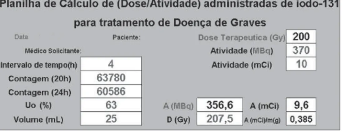
Data on the worksheet (Figure 8) were worked out with resources from
mathemat-ics and physmathemat-ics, for obtention of biokinetic parameters.
A = cpm /CF (1)
λef = ln (A0/A)/t (2)
Tef = ln 2/lef (3)
In the measurements with gamma-cam-era or scintillation probe for 131I uptake
(test activity) by the thyroid of patients af-fected by Graves’ disease, the counting rates (cpm) are collected through two suc-cessive tests in a time interval (t), and the initial activity (A0) and final activity (A) are
calculated for the considered time interval, utilizing the values for calibration factor (CF) obtained in the present study (equa-tion 1). These values will be applied to the equation (2), for obtention of λef (decay constant), and successively to the equation (3), for calculating the effective half-life in the organ (Tef). The initial uptake (U0) will
be calculated by extrapolation with the values of the previously mentioned uptake tests. The thyroid gland volume can be
es-Mean counting ± σm
Distance
Table 2 Field of view data of the SCT 13004 scintigraphic probe system for three source-detector dis-tances, utilizing a 131I punctiform source in three 30-second countings.
20 cm 25 cm 30 cm
d (cm) 0 ± 1 ± 2 ± 3 ± 4 ± 5 ± 6 ± 7 ± 8 ± 9 ± 10
72616 ± 110
72090 ± 110
71921 ± 109
70858 ± 109
69789 ± 108
66228 ± 105
40647 ± 82
9618 ± 40
620 ± 10
345 ± 8
261 ± 7
Tc (%)* 100.0 99.3 99.0 97.6 96.1 91.2 55.9 13.2 0.8 0.5 0.4
51052 ± 92
50897 ± 92
50621 ± 91
50102 ± 91
49403 ± 91
48283 ± 90
44020 ± 86
29905 ± 70
14486 ± 49
3375 ± 24
446 ± 9
Tc (%) 100.0 99.7 99.1 98.1 96.7 94.6 86.2 58.6 28.3 6.6 0.9
37536 ± 79
37331 ± 79
37065 ± 78
36871 ± 78
36577 ± 78
35970 ± 77
35016 ± 76
28007 ± 68
19770 ± 57
11670 ± 44
5409 ± 30
Tc (%) 100.0 99.4 98.7 98.2 97.4 95.8 93.2 74.6 52.6 31.0 14.4
* Tc (%), percent counting rate as a function of the distance between the 131I punctiform source and the
collima-tor central axis.
timated by palpation or ultrasonography. These data and the corresponding therapeu-tic activity will be conveniently recorded in a Microsoft Excel worksheet for calcula-tion of the desired absorbed-dose or vice-versa, as shown on the worksheet in Fig-ure 8.
DISCUSSION
Figure 6 shows the curves with a small plane segment, where the system will present as a result the same number of countings. This plane segment increases in diameter as the distance between the source and the detector window increases. The field of view is represented by the segment between the vertical dotted lines, and its size is indicated on the abscissas axis. For the evaluation of the thyroid radioiodine uptake with the Diacam gamma camera system, the source-detector distances of 45.8 cm and 46.8 cm where those that in-dicated a better radiation counting re-sponse, presenting a field of view compat-ible with the thyroid gland size of approxi-mately six centimeters (abscissas –3 and +3) for patients affected by Graves’ dis-ease. The distances of 42.8 cm, 43.8 cm and 44.8 cm, although with countings number higher than the distances of 45.8 cm and 46.8 cm, but with a field of view < 6 cm, the system will not record all of the radia-tions from the thyroid gland, as it can be observed on the isocounting curves. For distances above 46,8 cm, the detector sys-tem will record, besides the radiation from the thyroid gland, a significant quantity of the environment background radiation, and those from other regions of the patient’s body. Between distances of 45.8 cm and
46.8 cm, the first one is preferred for pre-senting a higher number of countings con-sidering the proximity of the source and, as a result, a lower associated uncertainty. The calibration factor of 4.3 ± 0.17 (cpm/kBq) utilized in the present study is associated with this distance. Notwithstanding, the calibration factor value corresponding to the 46.8 cm distance also can be utilized in the calculation of the absorbed-dose (Gy)/ administered activity (MBq) ratio, for the patients submitted to radioiodine therapy for hyperthyroidism. These calibration fac-tor will be a function of the thyroid gland enlargement (with a field of view > 6 cm), depending on the disease severity.
On the isocounting curves in Figure 7, one can observe that the best responses for the SCT-13004 system are those in the 25 cm and 30 cm. In this case, the 25 cm dis-tance is preferred for, besides providing a higher number of countings and, conse-quently a low associated uncertainty, is the most utilized in the measurements of thy-roid radioiodine uptake, in routine proce-dures of nuclear medicine. In this distance, it can be observed that the procedure is more comfortable for the patients, the sys-tem sensitivity is appropriate for the mea-surements in relation to the 30 cm distance, besides presenting a field of view compat-ible with the thyroid size in patients with Graves’ disease. The calibration factor ob-served for this distance was 39.3 ± 0,78 (cpm/kBq).
The thyroid-neck phantom as well as the calibration protocol developed have shown to be appropriate for the purposes of the present study. Both the gamma camera and the uptake probe may be utilized for deter-mining the 131I activity in the patients´
thy-roid. This procedure will be later applied for optimization of the individualized ac-tivity to be administered to each patient.
This methodology is feasible and low-cost, considering that the patients will visit the hospital for only two times, the first visit for receiving the test-activity, and the second one for being submitted to the up-take processes (%) between the14-hour and the 30-hour time intervals, where a data collection will be performed including counting rates required for calculating biokinetic data, (effective iodine half-life in the thyroid and initial uptake), and im-mediately after, the calculated activity ad-ministration. The methodology is effective and reliable because utilizes all of the biokinetic parameters required for calcula-tion of the thyroid absorbed-dose (thera-peutic dose).
Acknowledgements
The authors thank Instituto de Radio-proteção e Dosimetria da Comissão Nacio-nal de Energia Nuclear (IRD/CNEN) and Hospital Universitário Clementino Fraga Filho da Universidade Federal do Rio de Janeiro (HUCFF-UFRJ), for the technical support.
This research was evaluated and ap-proved by the HUCFF-UFRJ Committee for Ethics in Research, on May 15, 2006.
REFERENCES
1. Thompson MA. Radiation safety precautions in the management of the hospitalized 131I therapy
patient. J Nucl Med Technol 2001;29:61–66. 2. United Nations Scientific Committee on the
Ef-fects of Atomic Radiation. UNSCEAR 2000 Re-port to the General Assembly, with scientific an-nexes. Volume I: Sources. Vienna, Austria: UNSCEAR, 2000.
Pro-tection. Release of patients after therapy with un-sealed radionuclides. ICRP Publication 94. Ox-ford: Pergamon Press, 2004.
4. Brandão CDG, Antonucci J, Correa ND, Corbo R, Vaisman M. Efeitos da radioterapia nas gerações futuras de mulheres com carcinoma diferencia-do de tireóide. Radiol Bras 2004;37:51–55. 5. Jönsson H. Radioiodine therapy of
hyperthyroid-ism. Simplified patient-specific absorbed dose planning. Malmö: Department of Radiation Phys-ics, Lund University, 2003.
6. Nyström E, Berg G, Jansson S, et al.
Thyreoto-xikos hos vuxna. Klippan, Sweden: Ljungbergs Tryckeri AB, 1999.
7. International Commission on Radiological Pro-tection. General principles for the radiation pro-tection of workers. ICRP Publication 75. Oxford: Pergamon Press, 1997.
8. Dantas BM. Bases para a calibração de corpo inteiro utilizando simuladores físicos antropo-mórficos. (Tese de Doutorado). Rio de Janeiro, RJ: Universidade do Estado do Rio de Janeiro, 1998.
9. Silva CB. Medida da captação de iodo pela
tireói-de: análise comparativa entre sistema gama-câ-mara com colimador tipo “pinhole” e sistema 13S002. (Dissertação de Mestrado). Rio de Ja-neiro, RJ: Instituto Militar de Engenharia, 2001. 10. Williams RH. Textbook of endocrinology. 4th ed. Philadelphia-London-Toronto: WB Saunders, 1968.