Alpha in Urothelial Cancer
Rebecca L. Ross
1, Julie E. Burns
1, Claire F. Taylor
2, Paul Mellor
3, Deborah H. Anderson
3, Margaret A.
Knowles
1*1 Section of Experimental Oncology, Leeds Institute of Cancer and Pathology, St James’s University Hospital, Leeds, United Kingdom, 2 Leeds Cancer Research United Kingdom Centre Genomics Facility, Leeds Institute of Cancer and Pathology, St James’s University Hospital, Leeds, United Kingdom,
3 Cancer Research Unit, Saskatchewan Cancer Agency and the University of Saskatchewan, Saskatoon, Canada
Abstract
Bladder cancers commonly show genetic aberrations in the phosphatidylinositol γ-kinase signaling pathway. Here we have screened for mutations in PIK3R1, which encodes p85α, one of the regulatory subunits of PIγK. Two hundred and sixty-four bladder tumours and 41 bladder tumour cell lines were screened and 18 mutations were detected. Thirteen mutations were in C-terminal domains and are predicted to interfere with the interaction between p85α and p110α. Five mutations were in the BH domain of PIKγR1. This region has been implicated in p110α-independent roles of p85α, such as binding to and altering the activities of PTEN, Rab4 and Rab5. Expression of these mutant BH-p85α forms in mouse embryonic fibroblasts with p85α knockout indicated that all forms, except the truncation mutants, could bind and stabilize p110α but did not increase AKT phosphorylation, suggesting that BH mutations function independently of p110α. In a panel of 44 bladder tumour cell lines, 80% had reduced PIKγR1 mRNA expression relative to normal urothelial cells. This, along with mutation of PIK3R1, may alter BH domain functioning. Our findings suggest that mutant forms of p85α may play an oncogenic role in bladder cancer, not only via loss of ability to regulate p110α but also via altered function of the BH domain.
Citation: Ross RL, Burns JE, Taylor CF, Mellor P, Anderson DH, et al. (β01γ) Identification of Mutations in Distinct Regions of p85 Alpha in Urothelial Cancer. PLoS ONE 8(1β): e84411. doi:10.1γ71/journal.pone.0084411
Editor: Karl X Chai, University of Central Florida, United States of America
Received September γ0, β01γ; Accepted November 18, β01γ; Published December 18, β01γ
Copyright: © β01γ Ross et al. This is an open-access article distributed under the terms of the Creative Commons Attribution License, which permits unrestricted use, distribution, and reproduction in any medium, provided the original author and source are credited.
Funding: Financial support provided by Yorkshire Cancer Research (http://yorkshirecancerresearch.org.uk/) (Lγ6β) and University of Leeds MRC studentship (MK, RR), Canadian Institutes of Health Research (http://www.cihr-irsc.gc.ca/e/19γ.html) (MOP84β77)(DA), and Saskatchewan Health Research Foundation Fellowship (http://shrf.ca/)(PM). The funders had no role in study design, data collection and analysis, decision to publish, or preparation of the manuscript.
Competing interests: The authors have declared that no competing interests exist. * E-mail: [email protected]
Introduction
The phosphatidylinositol γ-kinase (PIγK) signaling pathway plays a critical role in the regulation of cell growth, proliferation and survival [1] and mutations that lead to aberrant activation of the pathway are found in virtually all types of cancer. In urothelial carcinoma (UC) of the bladder, several genomic alterations that lead to deregulation of the pathway have been identified. These include inactivating mutations in PTEN and
TSC1 and activating mutations in PIK3CA and AKT1
[β,γ,4,5,6,7,8]. Previously we showed that several of these events are non-redundant, suggesting that mutation of more than one pathway member may have additive or synergistic effects [4]. These alterations are found both in low-grade, non-invasive and muscle-non-invasive UC, indicating that this pathway plays a critical role in UC development and suggesting that it represents an important therapeutic target in these cancers.
PIγ kinases phosphorylate the γ` position on the inositol ring of phosphinositol-4,5-bisphosphate (PIPβ) to generate the lipid second messenger phosphinositol-γ,4,5-triphosphate (PIPγ) that activates AKT downstream signaling. The class IA PIγKαis an obligate heterodimer consisting of the p110αcatalytic subunit (p110α), encoded by the PIK3CA gene, and a regulatory subunit, encoded by one of γ genes, PIK3R1, PIK3R2 and PIK3R3. The regulatory subunits are essential for stability of p110α and in the resting state suppress its catalytic activity [9]. Each has two SHβ domains that can bind activated membrane-bound growth factor receptors or adaptor molecules, altering its conformation, relieving inhibition of p110α and allowing p110α to phosphorylate PIPβ [10].
lack the critical inter-SHβ (iSHβ) region required for inhibition of p110α activity [1β]. A similar truncated form was reported in a human Hodgkin’s lymphoma cell line [1γ] and 4 smaller deletions, within exons 14 or 15 that encode the iSHβ region, in ovarian and colon cancers [14]. Two splicing mutations were also identified, both of which led to skipping of exon 15. Expression of one of these mutant forms with a deletion of exon 1γ resulted in constitutive activation of the PIγK pathway in cells, providing evidence that mutant p85 can act as an oncogene in human cancer. A 9 bp deletion encompassing the exon-intron junction of exon 1β [15] and 9 other mutations, of which 8 were in the iSHβ region were identified in glioblastomas [16] and 15 mutations were found in colon cancer, the majority of which were shown to reduce its p110α-inhibitory activity whilst retaining ability to stabilise the complex [17]. A single PIK3R1 mutation was recently reported in a study of UC that screened exons 1β, 14 and 15, (<1% frequency)[18]. In contrast to these low mutation frequencies, it has recently been reported that β0 - 40% of endometrioid endometrial cancers contain PIK3R1 mutations, the majority in the nSHβ and iSHβ domains [19,β0].
Our previous finding of mutations in several components of the PIγK pathway in UC, and the finding of PIK3R1 mutations in other cancer types, prompted us to search for mutations in
PIK3R1. As evidence has emerged that that N-terminal domains of p85α have oncogenic functions, we chose to screen the entire coding sequence of PIK3R1, rather than focus on exons 1β, 14 and 15. Here we report a series of mutations in UC-derived cell lines and primary UC tumours, including deletions in the iSHβ region and a series of missense mutations, several in the breakpoint cluster region homology (BH) domain, which has GTPase activity towards Rab5 [β1] and can bind PTEN [ββ,βγ]. Our findings suggest that mutant forms of p85α may play an oncogenic role in bladder cancer, not only via loss of ability to regulate p110α but also via altered function of the BH domain.
Materials and Methods
Ethics statement
The study was approved by the Leeds East Research Ethics Committee (99/156) and written informed consent was obtained from all patients.
Patient Samples and DNA Isolation
Cold cup biopsies of β64 urothelial carcinomas (UC) were collected, snap-frozen and stored in liquid nitrogen. The remainder of the tissue was embedded in paraffin for diagnostic assessment. The tumour panel consisted of 10 pTaG1, 4β pTaGβ, β4 pTaGγ, 7 pT1G1, β7 pT1Gβ, 57 pT1Gγ, β pTβG1, 1γ pTβGβ, 54 >pTβGγ, 5 G1, 8 Gβ and 4 Gγ tumours with no underlying stroma (pTx) and 11 tumours with no information [β4,β5]. All were transitional cell carcinoma. DNA was extracted from frozen sections and venous blood samples as described previously [4].
Urothelial cell lines
Forty-four UC cell lines (β5γJ, 56γ7, 6γ9V, 647V, 9β-1, 94-10, 96-1, 97-1, 97-18, 97-β4, 97-7, BC-γC, BFTC905, BFTC909, CALβ9, DSH1, HT1197, HT1γ76, J8β, JMSU1, JO’N, KU-19-19, LUCC1, LUCCβ, LUCCγ, LUCC4, LUCC5, LUCC6, LUCC7, LUCC8, MGH-Uγ, RT11β, RT4, SCaBER, SD, SW780, SW1710, Tβ4, TCCSUP, U-BLC1, UM-UCγ, VM-CUB-1, VM-CUB-β and VM-CUB-γ) were investigated (Table S1). LUCC1-LUCC8 lines were established in the authors’ laboratory from bladder tumour tissues obtained with written informed consent. Cell line identity was verified by short tandem repeat DNA typing using Powerplex 16 kit (Promega). Profiles were compared to publically available data (ATCC, DSMZ) or where no reference profile was available, were confirmed as unique. DNA was extracted as previously described [4].
Mutation analysis
The entire coding sequence of PIK3R1 was examined in 18 fragments by high resolution melting analysis as described [β6,β7]. DNA from matched blood samples was analyzed to confirm somatic mutation status. Primer sequences are given in Table Sβ. Mutations were recorded with reference to NM_1815βγ.
Allele-specific PCR (AS-PCR) was carried out to establish the phase of the pairs of mutations in the cell line LUCCγ and in tumour sample β (Table 1). A forward primer was designed to specifically amplify the mutant allele at the 5` mutation site and was matched with a reverse primer positioned to include the second mutation in the PCR product (Table Sγ). Products were run on an agarose gel to identify successful amplification from tumour/cell line DNA only and no amplification from blood and WT DNA. PCR products were sequence-verified.
Expression vectors and transduction of cell lines
Cloning of inserts encoding the full-length wild-type (WT) human p85α and bovine p85α Rβ74A mutant into pGEX-6P (GE Healthcare Life Sciences) has been described [β1]. Mutants E1γ7K, R16β*, Eβ18*, Δβγ7_β4β, Rβ6βT, Kβ88Q were created by site-directed mutagenesis using the QuikChange method (Stratagene, CA, USA) and cloned into pFB Hyg [β8] in-frame with an N-terminal HA tag using the InFusion method (Clontech, CA, USA).
Control cDNAs, N564D and p85Δ, were kindly provided by Genentech [17] and subcloned into pFB Hyg by the same method. All plasmids were sequence-verified. Constructs were transfected into Phoenix A cells using TransIT β9γ (Mirus, Madison, USA). Mouse embryonic fibroblasts (MEFs) with knockout of p85α, , δ(a kind gift from Lewis Cantley, Harvard Medical School), NIHγTγ mouse fibroblasts and Rat1 rat fibroblasts, were incubated with retroviral supernatant containing 8 μg/ml polybrene and selected with 500, β00 and β50 μg/ml hygromycin, respectively.
Functional characterization of BH domain mutants
according to the manufacturers’ instructions and quantified as previously described [β9]. Anti-HA Immunoprecipitation Kit (Sigma-Aldrich) was used according to the manufacturers’ instructions for co-immunoprecipation experiments. Immunoblotted proteins were incubated with primary antibodies; anti-p85α (Abcam, Cambridge, UK), anti-p110α, anti-pAKT (Ser47γ), anti-panAKT (Cell Signaling Technology, Boston, UK) and anti-tubulin alpha (AbD Serotec, Kidlington, Oxford, UK). Bound primary antibodies were detected using HRP-conjugated secondary antibodies and Luminata Forte Western HRP Substrate (Millipore, Watford, UK). Anchorage-independent growth was carried out as previously described [β9] and colonies with a diameter ≥50 μm were counted within β.5 mmβ.
Analysis of PIK3R1 mRNA and protein expression in UC
PIKγR1 mRNA expression data for 105 bladder tumour samples and 5β normal bladder samples reported by Sanchez-Carbayo et al [γ0] were accessed. PIKγR1 protein expression was assessed in 44 UC cell lines and pooled normal human urothelial cells (NHU-pool) by western blotting and normalized to tubulin.
Results
Identification of somatic PIK3R1 mutations
We screened the entire coding sequence of PIK3R1 for mutations in β64 UC tumour tissues and 41 UC cell lines. The
three known isoforms of PIKγR1, p85α p55α and p50α contain different exons. Exons 1-6 are unique to p85α, exon 7 is present only in p50α and exon 8 only in p55α. Exons 9-17 are present in all isoforms but the use of a cryptic splice site within exon 16 results in the generation of cDNAs that differ in length by 8 codons [γ1], both of which were covered by this screen. Eighteen mutations were identified in 1γ tumours. One tumour (tumour β) contained three and one (tumour 5) contained two mutations (Table 1). All were confirmed as somatic mutations by their presence in the tumour-derived DNA but not the patient’s blood. One tumour-derived cell line (LUCCγ) contained two mutations and these were also confirmed as somatic in origin. All mutations were confirmed by repeat PCR amplifications to rule out PCR artifacts. The nature and distribution of the mutations are shown in relation to p85α protein structure in Figure 1. Five were located in the BH domain (Figure S1). None were in exons that are unique to p50α or p55α, though several were located in exons β, 5 and 6, that are unique to p85α. No mutations were identified in the region of exon 16 that is alternatively spliced.
Allele-specific PCR (AS-PCR) was used to determine whether the β mutations in the cell line LUCCγ (c.1519GC and c.1670GC; E507Q and R557P) and the β exonic mutations in tumour β (c.65βGT and c.86βAC; Eβ18* and Kβ88Q) were present in cis or in trans. In LUCCγ, a mutation-specific primer for c.1519C successfully amplified a PCR product from exon 1γ to 14 (Figure SβA). Sequencing of this product confirmed the presence of mutation c.1670C, indicating that both iSHβ domain mutations are on the same allele. AS-PCR analysis of tumour β also confirmed that the β exonic mutations (BH
Table 1. Mutations in PIKγR1 identified in bladder tumours and cell lines.
Sample Grade/stage Exon Nucleotide change Predicted amino acid change
1 TaGγ β 1c..409G>A E1γ7K
β TxGγ 5 c.65βGT Eβ18*
6 c.86βAC Kβ88Q
intron 1β c.14β6-49 G>A unknown
γ T4Gγ 5 c.710_7β7del18 Wβγ7-Yβ4βdel
4 T1Gγ 5 c.785GC Rβ6βT
5 TaGγ 10 βc.107βCT Rγ58*
14 c.17γ5_1740del6 Q579-Y580del
6 T1Gγ 11 c.11γ1TG Nγ77K
7 TaGγ 1β c.1γββAT N441I
8 T1G1 1γ c.1441AT R481W
γLUCCγ TβGγ 1γ c.1519GC E507Q
14 c.1670GC R557P
9 TβGγ 1γ c.155βGC E518Q
10 T1Gγ 14 c.1585GA D5β9N
11 TaGβ 14 c.1696_17γ4delγ9 I566-D578del
1β T1Gβ 16 c.19γ4CT T645I
1γ TaGβ Exon 17 splice acceptor c.1986-1 G>C unknown 1cDNA reference sequence: NM_1815βγ; β mutation in only a minor population of sequences; γcell line.
domain Eβ18* and Kβ88Q mutations) were present on the same allele (Figure SβB).
In tumour 5, sequence profiles indicated that one of the mutations (Rγ58*) was present as only a minor population of molecules, whilst the other (Q579_Y580del) appeared heterozygous. The genomic distance between exons 10 and 14 precluded development of an allele-specific PCR assay to determine whether these mutations were on the same allele. The small signal from the Rγ58* mutation may indicate that it was present as only a minor sub-clonal event. This appeared to be the case, as analysis of tissue from a subsequent disease recurrence in this patient showed only Q579_Y580del.
Predicted Effect of Mutations on Protein Function
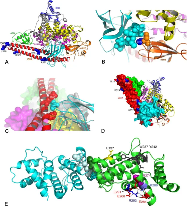
The positions of the missense mutated residues that are represented in the available crystal structure are shown in Figure β. Two mutations, Rγ58* and Nγ77K, were identified in the nSHβ domain. Recent findings suggest that this region of p85α acts as a scaffold for the p110α - p85α complex, with interactions with Cβ, helical and kinase domains of p110α and with p85α iSHβ [γβ]. Rγ58* truncates the protein at the nSHβ phosphopeptide binding site (FLVRDAS). This same mutation was recently reported in 5 endometrial cancer samples where it represented 5% of all mutations identified [β0]. Arginine γ58 has been identified as forming a salt bridge with E54β in p110α [γβ] and the engineered mutation Rγ58A was shown to abolish phosphopeptide binding [9]. Thus, loss of Rγ58 and truncation at this point is likely to result in disruption of phosphopeptide
binding and interaction of p85α with the helical domain of p110α and may mimic the effect of p110α helical domain mutations. We have shown previously that the helical domain mutations E54βK and E545K are by far the most common mutations found in PIK3CA in bladder cancer [4] and are more potent than H1047R in inducing signaling downstream of AKT and proliferation at confluence and under conditions of nutrient depletion when expressed in normal urothelial cells [β9]. As the Rγ58X truncation mutant removes both iSHβ and cSHβ regions of the protein, interaction with the Cβ domain of p110α is lost and effects may mimic those described for mutations and truncations affecting these domains [γγ]. From sequencing data, and knowledge that there was minimal contaminating normal tissue in the sample, this mutation appeared to be heterozygous. Thus it is predicted that in addition to the truncated protein, non-mutant protein was available for p110α binding and stabilisation in this tumour.
Nγ77, which was mutated to lysine, is adjacent to Gγ76, which was found previously to be mutated to arginine in γ cases of glioblastoma [16,γ4]. Both residues are within a region (γ74-γ77) that is predicted to interact with residues γ64-γ71 in the Cβ domain of p110α [γβ]. In the crystal structure of p110α in complex with p85niSHβ, both Gγ76 and Nγ77 are within hydrogen bonding distance of Eγ65 in p110α C Figure βB.
Six missense mutations and two in-frame deletions were identified in the iSHβ domain. This is the region where the majority of mutations have been found in other cancers, though none of the alterations found here are identical to those Figure 1. Distribution of PIK3R1 mutations identified in UC.
Figure 2. Position of mutations in the p85α protein. A. Ribbon diagram of the structure of the complex of p110α with the niSHβ region of p85α (PDB code βRD0) showing residues mutated in UC. B. Relationship of p85α nSHβ to p110α Cβ domain showing proximity of Nγ77 in nSHβ to Cβ domain residue Eγ65. C. Relationship of the iSHβ domain of p85α with the Cβ domain of p110α showing proximity of R557 to Nγ45 in Cβ. D. Space-filling model showing R481 and R557 residues in iSHβ of p85α in contact with Cβ of p110α. E. Structure of p85 dimer (PDB code 1PBW) showing position of PIKγR1 point-mutated residues E1γ7, Rβ6β and Kβ88 and the region deleted (Wβγ7-Yβ4β) in UC in relation to residues involved in p85 dimerization (M176, dark green/light cyan; L161, I177 and V181, light green/cyan). The position of residue Rβ74 (magenta), which is implicated in Rab-GAP activity is also shown. In addition, three glutamic acid residues (Eβ66, Eβ84 and Eβ91) that interact with Rβ6β are indicated, with Eβ91 also interacting with Kβ88.
reported previously [14,16,17,19,β0]. This region of the protein is involved in interaction with the adapter-binding region of p110α and in stabilisation and inhibition of its catalytic activity in the basal state via interaction with the Cβ domain. The predicted effect of some mutations in iSHβ is to disrupt the interface with p110α Cβ [16,γγ]. For example, p85α residue, N564, is within hydrogen-bonding distance of Nγ45 in p110α Cβ (Figure βC), a residue that has been found mutated in p110α [γ5]. D560Y reported in glioblastoma [16] is also within hydrogen bonding distance of Nγ45 and both of these are predicted therefore to mimic the oncogenic Nγ45 mutation. Here we found a novel mutation, R557P that is also close to Nγ45 of p110α (Figure βC). R481, which was found mutated to tryptophan, is also in close proximity to the Cβ domain of p110α Figure βD).
The two in-frame deletions identified in iSHβ (I566_D578del and Q579_Y580del) remove amino acid residues that have been found to be deleted in ovarian and colorectal cancers [14,17] and glioblastoma [16] and are predicted to disrupt the helical secondary structure in this region and disturb interaction with p110α Cβ. The mutation Q579_Y580del has been reported to bind and stabilise p110α but have reduced ability to regulate the lipid kinase activity of the p85α p110α holoenzyme [17]. Recently, nine of the nSHβ and iSHβ mutations found in other cancers were expressed in chicken embryo fibroblasts and all induced transformation, measured by focus-forming activity, increased proliferation and enhanced signaling via PIγK [γ6]. Specific inhibitors of p110α, but not inhibitors of p110 p110 , or p110δ abolished the phenotypic effects of two of these tumour-derived mutant forms (KS459delN, DKRMNS560del), indicating that the oncogenic activity of these mutants is uniquely mediated by p110α.
The most N-terminal iSHβ mutation identified, N441I, is in a linker region between nSHβ and iSHβ, and lies close to the end of the second alpha helix of iSHβ. This region is not resolved in the published crystal structures and its potential effect is not clear. T654I in the cSHβ domain is close to several residues that are mutated in colorectal cancers [17] and is immediately adjacent to the FLVRES motif (residues 646-651) required for phosphotyrosine engagement. A serine or threonine residue in this position is found in the SHβ domains of a wide range of proteins. Several mutations within this domain have been reported in colorectal cancers [17] but not in glioblastoma [16,γ4], raising the possibility that there may be tissue specific differences.
Interestingly, we found five mutations within the BH domain of PIKγR1 (β8% of mutations found) (Figure 1; Figure S1). Previous mutation screens have identified only seven mutations in this region (K14βfs, E160*, R16β*, I177N, Eβ17K, Nβ85H, Eβ97K) in more than 1550 samples of other tumours reported to date [14,16,17,18,19,β0,γ4] (< 0.5%). All missense mutations in this region (E1γ7K, Rβ6βT and Kβ88Q) are in highly conserved residues (Figure Sγ). The structure of this region of the protein (amino acids 105-γ19) as a homodimer has been reported [γ7]. The interface between BH domains was shown to involve interaction of M176 from one monomer with L161, I177 and V181 on the other. The three point
mutations and deletion identified here are remote from this region of interaction (Figure βE).
P85α BH domain mutants interact with and stabilise p110α
To determine whether BH domain mutant proteins have p110α dependent effects, we tested their ability to interact with and stabilise p110α. p85α, δ knockout (pan-p85 null) MEFs were transduced with retroviral expression vectors encoding N-terminus HA-tagged p85α cDNA encoding the BH domain mutant forms identified in UC (E1γ7K, Eβ18*, Δβγ7_β4β, Rβ6βT, Kβ88Q), other mutant forms as controls (R16β*, p85Δ, Rβ74A-bovine, N564D), wild-type (WT) or empty vector. R16β* mutant p85α is a BH domain mutation previously identified in cancer [17] and like p85Δ lacks the p110α binding region (n-iSHβ) [γ8]. Both have previously been shown not to interact with p110α [17]. N564D iSHβ mutant p85α was used as a positive control as it has previously been shown to bind p110α, increase PIγK and AKT activity, and induce cell survival, anchorage-independent growth and transformation-related phenotypes that were p110-dependent [17]. It was also used here to assess potential differences between BH and iSHβ domain mutant forms of p85. Rβ74A is an engineered BH domain mutant form of bovine p85α that has been previously shown to inhibit p85-GAP activity and was oncogenic [β1,γ9].
Expression of p85α was confirmed by western blotting (Figure γA). A truncated p85α protein was visible in the R16β*-MEF (18 kDa), but in the Eβ18*-R16β*-MEF, the truncated protein (β4 kDa) was expressed at a very low level. Three independent transductions with Eβ18* virus consistently induced low protein expression suggesting that this protein may be unstable. Interestingly, R16β*-MEF also expressed a small amount of full-length p85α protein.
Pan-p85 null MEFs have low levels of p110α, which can be restored by expression of wild-type p85α [40]. We confirmed this and showed that all mutant forms except Eβ18*, R16β* and p85Δ had the same effect (data not shown). Co-immunopreciptation of HA-tagged p85 proteins was used to assess p110α binding (Figure γB). Results indicated that all p85 proteins, except Eβ18*, R16β* and p85Δ, could bind p110α.
BH and iSH2 domain mutants of p85α show different effects on AKT activation and anchorage-independent growth
reflected in the proliferation rates of MEF and NIHγTγ cells when maintained in serum-containing medium and when assessing anchorage-independent growth of NIHγTγ cells (data not shown). Analysis of anchorage-independent growth of Rat1 cells expressing wild-type and mutant forms of p85α showed that whilst BH domain mutants induced some colony formation relative to wild-type p85α, N564D had the greatest effect (Figure S4).
Relationship to other mutations in PI3K pathway genes
Previously we screened the samples analyzed here for
PIK3CA mutation [[4] and unpublished data]. Only two samples
contained both PIK3R1 and PIK3CA (E545K) mutations and one of these was the tumour that had a BH domain mutation (E1γ7K) in PIK3R1. Overall, 61 of the tumours screened contain PIK3CA mutations (βγ%). Thus, PIK3CA mutation was underrepresented in the subset that had PIK3R1 mutation (1 observed; 5 expected). Thus PIK3R1 mutations not in the BH domain appear to be mutually exclusive with PIK3CA mutation in UC. There was no significant relationship of PIK3CA or
PIK3R1 mutation with tumour grade or stage. Four of the samples contain AKT1 mutations (E17K), none of which co-existed with PIK3R1 mutation. TSC1 mutation status is known for 148 tumours in the series, of which 14 contain a mutation Figure 3. Characterization of MEFs (KO p85α, β, δ) expressing WT or mutant p85α. A. Immunoblot showing p85α protein expression levels in transduced cells. B. Co-immunoprecipitation of HA-tagged proteins followed by immunoblot with p85α and p110α to determine p85-p110 binding. C. Immunoblot analysis of PIγK signaling (levels of pAKT (Ser47γ)) in serum-containing medium and bar chart displays the quantification of normalized pAKT to total AKT relative to control. D. Immunoblot analysis of pAKT (Ser47γ) in serum-containing medium and bar chart displays the quantification of normalized pAKT to total AKT relative to control.
[4]. One of these tumours had a mutation in the BH domain of
PIK3R1 (Wβγ7_Yβ4βdel). However the sample size and mutation frequencies are too small for any significant coincidence or mutual exclusivity to be identified.
PIK3R1 expression is reduced in UC and cell lines
As p85α mutations may alter protein:protein interactions of p85α, some of which, particularly those in the BH domain, might indicate a tumour suppressor role, we considered the possibility that downregulation of the protein might contribute to tumour development.
PIK3R1 is located on chromosome 5 (5q1γ.1). Loss of heterozygosity (LOH) and copy number loss of sequences on 5q have been reported in bladder cancer [41,4β]. By array CGH we have found that β9% of muscle invasive UC have copy number loss in the region of PIK3R1 [4γ]. Examination of publically available expression data indicated that UC has a significant reduction in PIKγR1 expression at the RNA level (Figure 4A).
We assessed p85α protein expression in 44 UC cell lines using western blotting. This revealed wide variation in expression levels with the majority of cell lines (80%) showing reduced p85α protein expression compared with normal urothelial cells (Figure 4B).
Discussion
In this study we found PIK3R1 mutations in 4.9% of UC. The higher frequency found here than in the previous study of UC [18] reflects our assessment of the entire gene rather than only exons 1β, 14 and 15. Our data for those three exons revealed 5 mutations (1.8% of tumours), compatible with this earlier study. The majority of mutations were located in the C-terminal region of p85α similar to findings in other human cancers [14,16,17,19,β0]. Mutations identified in the nSHβ and iSHβ domains may mimic the effect of p110α mutations by relieving the inhibitory regulation of p110α by p85α and the oncogenic activity of these mutants appears to be p110α dependent [γ6].
Interestingly, we found a higher frequency of BH domain mutations compared to previous mutation screens. Whether this is specific to UC is not yet clear due to the focus of many studies only on the p110α -interacting region of the gene. Recent data suggest that BH domain mutant forms of p85α may alter the stability of PTEN [β0]. p85α binds to unphosphorylated PTEN within the high molecular weight PTEN associated complex (PAC) [ββ]. This interaction involves the N-terminal region of p85 (SHγ-BH) and has been shown to positively regulate the lipid kinase activity of PTEN in a growth factor-dependent manner [βγ]. Expression of p85α with deletion of the BH domain alone significantly reduced interaction with PTEN and deletion of both domains abolished it, leading to increased amplitude and duration of AKT Figure 4. Expression of p85α in bladder tumours and cell lines. A. Analysis of p85α mRNA expression levels in 105 bladder tumour samples and 5β normal bladder samples. Data from Sanchez-Carbayo et al [γ0]. B. Quantitative analysis of p85α immunoblotted protein samples from bladder cancer cell lines normalized to tubulin and shown relative to pooled normal human urothelial cells (NHU-pool).
activation following growth factor stimulation [βγ]. Thus, PTEN: p85α interaction appears to potentiate the ability of PTEN to reduce PIPγ levels and terminate signaling to AKT. Indeed the mutation E160* identified in endometrial carcinoma has been shown to destabilise PTEN and result in increased AKT phosphorylation [β0].
The BH region also shows sequence homology to RhoGAP proteins and possesses GAP activity toward Rab5, Rab4, Cdc4β and Rac1 [β1]. The Rab proteins are regulators of endosome trafficking and influence vesicle fusion events during uptake, recycling or processing for degradation of growth factor receptors such as EGFR [44] and PDGFR [45]. Expression of a GAP-defective mutant of bovine p85α (Rβ74A) [γ9] led to sustained levels of PDGF receptor activation and downstream MAPK signaling in response to PDGF stimulation [β1]. Expression of this p85α-Rβ74A mutant in NIHγTγ cells induced focus formation in cell monolayers, anchorage-independent growth and tumorigenicity, demonstrating that disruption of p85α GAP function can contribute to cellular transformation [γ9]. Thus, the SHγ-BH region of p85α may be considered to exert a tumour suppressor function via positive regulation of PTEN activity or through its RabGAP activity that can impact receptor trafficking and signaling functions. Mutations that affect one or other of these functions may contribute to tumorigenesis in different ways.
All missense mutations that we identified in this region (E1γ7K, Rβ6βT and Kβ88Q) are in highly conserved residues. The three point mutations and deletion identified here are remote from the region of interaction between BH domains revealed by the structure of the homodimer of amino acids 105-γ19 [γ7]. The precise residues required for Rab5 and PTEN interaction and the multiple other proteins reported to interact with p85α remain to be defined. Rβ6β forms extensive contacts with three glutamic acid residues, one of which also interacts with Kβ88. Mutations of Rβ6β and/or Kβ88, may therefore destabilise the interhelix interactions mediated by these side chains that help maintain the structure of the BH domain. The truncating mutation R16β*, reported in a colorectal cancer [17], and Eβ18* found here, could exert their effect via deletion of the more C-terminal domains involved in p110α interaction, rather than loss of BH domain function(s), though the low levels of protein expression we achieved here for Eβ18* did not allow this to be properly assessed.
Our results indicated that all p85 BH mutant proteins, except Eβ18*, R16β* and p85Δ, could bind p110α. As these forms do not contain the p110α-binding domain, they are not predicted to activate, regulate or stabilise p110α. Recent work that characterized an E160* mutant of p85, showed that its expression enhanced PTEN ubiquitination and interfered with PTEN and wild-type p85 binding, thus destabilising PTEN [β0]. The large truncations of the p85 protein identified here (R16β* and Eβ18*) may function in a similar manner.
BH and iSHβ domain mutants of p85α show different effects on AKT activation, cell proliferation and anchorage-independent growth. p85α mutant forms that affect p110α activity normally result in activation of AKT [17,19,β0]. Here, only the iSHβ domain N564D mutant induced dramatically increased AKT activation, proliferation and
anchorage-independent growth. This suggests that BH domain mutants have p110α-independent functions and indicates that in contrast to i- and nSHβ mutants, the oncogenic activity of these mutants is not mediated by p110α. Further research is required to investigate the mechanisms of these BH domain mutants, particularly their effects on GAP activity and PTEN binding in urothelial cells.
In addition to mutational alterations in p85α that may interfere with protein:protein interactions, overall downregulation of the wildtype protein may contribute to tumour development. Some previous studies have suggested that levels of p85α and p110α in normal cells are tightly linked, arguing against a role for free p85α [46]. However, recent data indicate the potential importance of changes in overall levels of p85α in control of PIγK signaling and tumour development and suggest that it may exert a tumour suppressive role [βγ,47]. We found the majority of bladder cancer cell lines to have decreased levels of p85α expression. Reduced p85α has been shown to increase PIγK signaling in some tissues and decrease PIγK signaling in others, particularly in the context of heterozygous PTEN levels [48]. Thus, p85 has a critical role in PIγK signaling via effects on p110, but in addition it can also function in some tissues to inhibit PIγK signaling and suppress tumour formation, likely through effects on PTEN [ββ,49]. Overexpression of p110α has been shown to reduce p85α:PTEN heterodimers leading to the suggestion that p110α and PTEN form distinct complexes with p85α[β0]. Our finding of a novel class of PIK3R1 mutations in the region of p85α responsible for PTEN regulation in bladder cancer is consistent with this additional function for p85α. Taken together our data indicate that there are complex genomic imbalances affecting the PIK3R1 region in bladder cancer and major alterations in expression of PIKγR1.
Supporting Information
Figure S1. Sanger sequencing traces showing mutations identified in exons 2-6 of PIK3R1. Bracketed traces show mutations found in cis in tumor β.
(TIF)
Figure S2. Allele-specific PCR determination of phase of two mutations in the cell line LUCC3 and tumor sample 2. For each pair of mutations, c.1519 G>C (exon 1γ) / c.1670 G>C (exon 14) in LUCCγ (A) and c.65β G>T (exon 5) / c.86β A>C (exon 6) in tumor β (B), primers were used to amplify a single PCR product that spanned the two mutation sites. Electrophoresis analysis shows the PCR products from different DNA templates specific for the first 5’ mutant sequence. Electropherograms show the corresponding mutant (MT) nucleotide at the alternative mutation site on the same PCR product.
(TIF)
(TIF)
Figure S4. Anchorage-independent growth analysis of Rat1 cells expressing WT and mutant p85α. No colonies > 50 μm were seen in vector only cells.
(TIF)
Table S1. Urothelial carcinoma cell lines and their origins. (DOC)
Table S2. Primers used for high resolution melting analysis of PIK3R1.
(DOCX)
Table S3. Allele-specific PCR to determine phase of PIK3R1 mutations.
(DOCX)
Acknowledgements
We thank Joanne Brown for tissue collection and processing and staff and patients in the Pyrah Department of Urology St James’s University Hospital for their generosity in providing tissue samples. We also thank Helen McPherson for helping with the cloning of the p85 expression plasmids.
Author Contributions
Conceived and designed the experiments: RLR JEB CFT PM DHA MAK. Performed the experiments: RLR JEB CFT PM DHA. Analyzed the data: RLR JEB CFT PM DHA MAK. Wrote the manuscript: RLR JEB CFT PM DHA MAK.
References
1. Cantley LC (β00β) The phosphoinositide γ-kinase pathway. Science β96: 1655-1657. doi:10.11β6/science.β96.557γ.1655. PubMed: 1β040186.
β. Askham JM, Platt F, Chambers PA, Snowden H, Taylor CF et al. (β010) AKT1 mutations in bladder cancer: identification of a novel oncogenic mutation that can co-operate with E17K. Oncogene β9: 150-155. doi:10.10γ8/onc.β009.γ15. PubMed: 1980β009.
γ. Aveyard JS, Skilleter A, Habuchi T, Knowles MA (1999) Somatic mutation of PTEN in bladder carcinoma. Br J Cancer 80: 904-908. doi: 10.10γ8/sj.bjc.66904γ9. PubMed: 10γ6067γ.
4. Platt FM, Hurst CD, Taylor CF, Gregory WM, Harnden P et al. (β009) Spectrum of phosphatidylinositol γ-kinase pathway gene alterations in bladder cancer. Clin Cancer Res 15: 6008-6017. doi: 10.1158/1078-04γβ.CCR-09-0898. PubMed: 19789γ14.
5. López-Knowles E, Hernández S, Malats N, Kogevinas M, Lloreta J et al. (β006) PIKγCA mutations are an early genetic alteration associated with FGFRγ mutations in superficial papillary bladder tumors. Cancer Res 66: 7401-7404. doi:10.1158/0008-547β.CAN-06-118β. PubMed: 16885γγ4.
6. Cairns P, Evron E, Okami K, Halachmi N, Esteller M et al. (1998) Point mutation and homozygous deletion of PTEN/MMAC1 in primary bladder cancers. Oncogene 16: γβ15-γβ18. doi:10.10γ8/sj.onc. 1β01855. PubMed: 967140β.
7. Wang DS, Rieger-Christ K, Latini JM, Moinzadeh A, Stoffel J et al. (β000) Molecular analysis of PTEN and MXI1 in primary bladder carcinoma. Int J Cancer 88: 6β0-6β5. doi: 10.100β/1097-0β15(β0001115)88:4. PubMed: 11058880.
8. Knowles MA, Habuchi T, Kennedy W, Cuthbert-Heavens D (β00γ) Mutation Spectrum of the 9qγ4 Tuberous Sclerosis Gene TSC1 in Transitional Cell Carcinoma of the Bladder. Cancer Res 6γ: 765β-7656. PubMed: 146γγ685.
9. Yu J, Wjasow C, Backer JM (1998) Regulation of the p85/p110alpha phosphatidylinositol γ'-kinase. Distinct roles for the n-terminal and c-terminal SHβ domains. J Biol Chem β7γ: γ0199-γ0β0γ. doi:10.1074/ jbc.β7γ.46.γ0199. PubMed: 9804776.
10. Miled N, Yan Y, Hon W-C, Perisic O, Zvelebil M et al. (β007) Mechanism of two classes of cancer mutations in the phosphoinositide γ-kinase catalytic subunit. Science γ17: βγ9-β4β. doi:10.11β6/science. 11γ5γ94. PubMed: 176β688γ.
11. Jimenez C, Jones DR, Rodríguez-Viciana P, Gonzalez-García A, Leonardo E et al. (1998) Identification and characterization of a new oncogene derived from the regulatory subunit of phosphoinositide γ-kinase. EMBO J 17: 74γ-75γ. doi:10.109γ/emboj/17.γ.74γ. PubMed: 9450999.
1β. Shekar SC, Wu H, Fu Z, Yip SC, Nagajyothi et al. (β005) Mechanism of constitutive phosphoinositide γ-kinase activation by oncogenic mutants of the p85 regulatory subunit. J Biol Chem β80: β7850-β7855. doi: 10.1074/jbc.M506005β00. PubMed: 159γβ879.
1γ. Jücker M, Südel K, Horn S, Sickel M, Wegner W et al. (β00β) Expression of a mutated form of the p85alpha regulatory subunit of phosphatidylinositol γ-kinase in a Hodgkin's lymphoma-derived cell line (CO). Leukemia 16: 894-901. doi:10.10γ8/sj.leu.β40β484. PubMed: 1198695β.
14. Philp AJ, Campbell IG, Leet C, Vincan E, Rockman SP et al. (β001) The phosphatidylinositol γ'-kinase p85alpha gene is an oncogene in human ovarian and colon tumors. Cancer Res 61: 74β6-74β9. PubMed: 11606γ75.
15. Mizoguchi M, Nutt CL, Mohapatra G, Louis DN (β004) Genetic alterations of phosphoinositide γ-kinase subunit genes in human glioblastomas. Brain Pathol 14: γ7β-γ77. PubMed: 15605984. 16. Network. TCGAR (β008) Comprehensive genomic characterisation
defines human glioblastoma genes and core pathways. Nature 455: 1061-1068
17. Jaiswal BS, Janakiraman V, Kljavin NM, Chaudhuri S, Stern HM et al. (β009) Somatic mutations in p85alpha promote tumorigenesis through class IA PIγK activation. Cancer Cell 16: 46γ-474. doi:10.1016/j.ccr. β009.10.016. PubMed: 1996β665.
18. Sjödahl G, Lauss M, Gudjonsson S, Liedberg F, Halldén C et al. (β011) A systematic study of gene mutations in urothelial carcinoma; inactivating mutations in TSCβ and PIKγR1. PLOS ONE 6: e1858γ. doi:10.1γ71/journal.pone.001858γ. PubMed: β15γγ174.
19. Urick ME, Rudd ML, Godwin AK, Sgroi DC, Merino M et al. (β011) PIKγR1 (p85-alpha/p85{alpha}) is Somatically Mutated at High Frequency in Primary Endometrial. Cancer - Cancer Res 71: 4061-4067. doi:10.1158/0008-547β.CAN-11-0549.
β0. Cheung LW, Hennessy BT, Li J, Yu S, Myers AP et al. (β011) High Frequency of PIKγR1 and PIKγRβ Mutations in Endometrial Cancer Elucidates a Novel Mechanism for Regulation of PTEN Protein Stability. Cancer Discov 1: 170-185. Available online at: doi: 10.1158/β159-8β90.CD-11-00γ9 PubMed: β1984976.
β1. Chamberlain MD, Berry TR, Pastor MC, Anderson DH (β004) The p85alpha subunit of phosphatidylinositol γ'-kinase binds to and stimulates the GTPase activity of Rab proteins. J Biol Chem β79: 48607-48614. doi:10.1074/jbc.M409769β00. PubMed: 15γ7766β. ββ. Rabinovsky R, Pochanard P, McNear C, Brachmann SM, Duke-Cohan
JS et al. (β009) p85 Associates with unphosphorylated PTEN and the PTEN-associated complex. Mol Cell Biol β9: 5γ77-5γ88. doi:10.11β8/ MCB.01649-08. PubMed: 196γ5806.
βγ. Chagpar RB, Links PH, Pastor MC, Furber LA, Hawrysh AD et al. (β010) Direct positive regulation of PTEN by the p85 subunit of phosphatidylinositol γ-kinase. Proc Natl Acad Sci U S A 107: 5471-5476. doi:10.107γ/pnas.0908899107. PubMed: β0β1β11γ. β4. UICC (1978) TNM classification of malignant tumors, bladder. γ ed:
Union Internationale Contre le Cancer, Geneva. pp. 11γ-117
β5. WHO (197γ) Histological typing of urinary bladder tumours; Mostofi Fk, Geneva, editor. World Health Organisation.
β6. Chapman EJ, Williams SV, Platt FM, Hurst CD, Chambers P et al. (β009) Integrated genomic and transcriptional analysis of the in vitro evolution of telomerase-immortalized urothelial cells (TERT-NHUC). Genes Chromosomes Cancer 48: 694-710. doi:10.100β/gcc.β067β. PubMed: 19405089.
β7. Taylor CF (β009) Mutation scanning using high-resolution melting. Biochem Soc Trans γ7: 4γγ-4γ7. doi:10.104β/BST0γ704γγ. PubMed: 19β90876.
secreted isoform that inhibits fibroblast growth factor-induced proliferation and is repressed in urothelial carcinoma cell lines. Cancer Res 65: 10441-10449. doi:10.1158/0008-547β.CAN-05-1718. PubMed: 16β880γ5.
β9. Ross RL, Askham JM, Knowles MA (β01γ) PIKγCA mutation spectrum in urothelial carcinoma reflects cell context-dependent signaling and phenotypic outputs. Oncogene γβ: 768-776. PubMed: ββ4γ0β09. γ0. Sanchez-Carbayo M, Socci ND, Lozano J, Saint F, Cordon-Cardo C
(β006) Defining molecular profiles of poor outcome in patients with invasive bladder cancer using oligonucleotide microarrays. J Clin Oncol β4: 778-789. doi:10.1β00/JCO.β005.0γ.βγ75. PubMed: 164γβ078. γ1. Antonetti DA, Algenstaedt P, Kahn CR (1996) Insulin receptor substrate
1 binds two novel splice variants of the regulatory subunit of phosphatidylinositol γ-kinase in muscle and brain. Mol Cell Biol 16: β195-ββ0γ. PubMed: 86β8β86.
γβ. Mandelker D, Gabelli SB, Schmidt-Kittler O, Zhu J, Cheong I et al. (β009) A frequent kinase domain mutation that changes the interaction between PIγKalpha and the membrane. Proc Natl Acad Sci U S A 106: 16996-17001. doi:10.107γ/pnas.0908444106. PubMed: 19805105. γγ. Wu H, Shekar SC, Flinn RJ, El-Sibai M, Jaiswal BS et al. (β009)
Regulation of Class IA PI γ-kinases: Cβ domain-iSHβ domain contacts inhibit p85/p110alpha and are disrupted in oncogenic p85 mutants. Proc Natl Acad Sci U S A 106: β0β58-β0β6γ. doi:10.107γ/pnas. 090βγ69106. PubMed: 19915146.
γ4. Parsons DW, Jones S, Zhang X, Lin JC-H, Leary RJ et al. (β008) An integrated genomic analysis of human glioblastoma multiforme. Science γβ1: 1807-181β. doi:10.11β6/science.1164γ8β. PubMed: 1877βγ96.
γ5. Huang C-H, Mandelker D, Schmidt-Kittler O, Samuels Y, Velculescu VE et al. (β007) The structure of a human p110alpha/p85alpha complex elucidates the effects of oncogenic PIγKalpha mutations. Science γ18: 1744-1748. doi:10.11β6/science.1150799. PubMed: 18079γ94.
γ6. Sun M, Hillmann P, Hofmann BT, Hart JR, Vogt PK (β010) Cancer-derived mutations in the regulatory subunit p85alpha of phosphoinositide γ-kinase function through the catalytic subunit p110alpha. Proc Natl Acad Sci U S A 107: 15547-1555β. doi:10.107γ/ pnas.100965β107. PubMed: β071γ70β.
γ7. Musacchio A, Cantley LC, Harrison SC (1996) Crystal structure of the breakpoint cluster region-homology domain from phosphoinositide γ-kinase p85 alpha subunit. Proc Natl Acad Sci U S A 9γ: 14γ7γ-14γ78. doi:10.107γ/pnas.9γ.β5.14γ7γ. PubMed: 896β058.
γ8. Dhand R, Hara K, Hiles I, Bax B, Gout I et al. (1994) PI γ-kinase: structural and functional analysis of intersubunit interactions. EMBO J 1γ: 511-5β1. PubMed: 8γ1γ896.
γ9. Chamberlain MD, Chan T, Oberg JC, Hawrysh AD, James KM et al. (β008) Disrupted RabGAP function of the p85 subunit of phosphatidylinositol γ-kinase results in cell transformation. J Biol Chem β8γ: 15861-15868. doi:10.1074/jbc.M800941β00. PubMed: 18γ8794β. 40. Brachmann SM, Yballe CM, Innocenti M, Deane JA, Fruman DA et al.
(β005) Role of phosphoinositide γ-kinase regulatory isoforms in development and actin rearrangement. Mol Cell Biol β5: β59γ-β606. doi:10.11β8/MCB.β5.7.β59γ-β606.β005. PubMed: 15767666. 41. Zieger K, Wiuf C, Jensen KM, Ørntoft TF, Dyrskjøt L (β009)
Chromosomal imbalance in the progression of high-risk non-muscle invasive bladder cancer. BMC Cancer 9: 149. doi: 10.1186/1471-β407-9-149. PubMed: 19445696.
4β. Simon R, Burger H, Semjonow A, Hertle L, Terpe HJ et al. (β000) Patterns of chromosomal imbalances in muscle invasive bladder cancer. Int J Oncol 17: 10β5-10β9. PubMed: 110β9508.
4γ. Hurst CD, Platt FM, Taylor CF, Knowles MA (β01β) Novel tumor subgroups of urothelial carcinoma of the bladder defined by integrated genomic analysis. Clin Cancer Res 18: 5865-5877. doi: 10.1158/1078-04γβ.CCR-1β-1807. PubMed: ββ9γβ667.
44. Chen PI, Kong C, Su X, Stahl PD (β009) Rab5 isoforms differentially regulate the trafficking and degradation of epidermal growth factor receptors. J Biol Chem β84: γ0γβ8-γ0γγ8. doi:10.1074/ jbc.M109.0γ4546. PubMed: 197βγ6γγ.
45. Chamberlain MD, Oberg JC, Furber LA, Poland SF, Hawrysh AD et al. (β010) Deregulation of Rab5 and Rab4 proteins in p85Rβ74A-expressing cells alters PDGFR trafficking. Cell Signal ββ: 156β-1575. doi:10.1016/j.cellsig.β010.05.0β5. PubMed: β05707β9.
46. Geering B, Cutillas PR, Nock G, Gharbi SI, Vanhaesebroeck B (β007) Class IA phosphoinositide γ-kinases are obligate p85-p110 heterodimers. Proc Natl Acad Sci U S A 104: 7809-7814. doi:10.107γ/ pnas.0700γ7γ104. PubMed: 1747079β.
47. Taniguchi CM, Winnay J, Kondo T, Bronson RT, Guimaraes AR et al. (β010) The phosphoinositide γ-kinase regulatory subunit p85alpha can exert tumor suppressor properties through negative regulation of growth factor signaling. Cancer Res 70: 5γ05-5γ15. doi: 10.1158/0008-547β.CAN-09-γγ99. PubMed: β05γ0665.
48. Luo J, Sobkiw CL, Logsdon NM, Watt JM, Signoretti S et al. (β005) Modulation of epithelial neoplasia and lymphoid hyperplasia in PTEN+/-mice by the p85 regulatory subunits of phosphoinositide γ-kinase. Proc Natl Acad Sci U S A 10β: 10βγ8-10β4γ. doi:10.107γ/pnas.0504γ7810β. PubMed: 1600651γ.