*e-mail: [email protected]
In Vitro Studies of Bioactive Glass/polyhydroxybutyrate Composites
André Oliveira Paivaa*, Maria Gabriela Duarteb, Maria Helena Vaz Fernandesb,
Maria Helena Gilc, Necésio Gomes Costad*
a
Science Institute, Federal University of Itajubá, 37500-9002 Itajubá - MG, Brazil
b
Department of Ceramics and Glass Engineering, University of Aveiro,
Santiago Campus, 3810, Portugal
c
Department of Chemical Enginnering, University of Coimbra, 3030 Polo II,
Pinhal de Marrocos, Coimbra, Portugal
d
Mechanical Engineering Institute, Federal University of Itajubá,
37500-902 Itajubá - MG, Brazil
Received: September 4, 2006; Revised: October 24, 2006
Bioactive materials can help bone reparation and regeneration by offering support to bone growth. In vitro
studies of bioactive glass/polyhydroxybutyrate composites were carried out to evaluate the influence of the composition on the bioactivity through the presence of calcium phosphate (Ca-P) on the layer formed when the substrates were immerse in simulated body fluid (SBF). The in vitro tests were carried out by soaking the
composites bioactive glass/polyhydroxybutyrate 30/70 and 40/60 in SBF. The surface of the composites was analyzed by Scanning Electron Microscopy (SEM) with Energy Dispersive Spectroscopy (EDS) and also via x ray Diffraction (XRD). The solutions were analyzed by Inductively Couple Plasma (ICP). SEM images show a formation of a Ca-P rich layer on surface of composites. XRD results characterized the layer as calcium hydrogen phosphate. Ca/P ratios found via EDS results show a value close to 1.67. According to ICP results, the Ca e P ions are from SBF.
Keywords: bioactive glasses, polyhydroxybutyrate, composites and bioactivity
1. Introduction
The beginning of the 1980’s hydroxyapatite/polyethylene com-posite was the first bioactive comcom-posite to be investigated1. These biomaterials have as the main aim to help the bone reparation. They can act as a viable alternative to autogenous grafts, because they can eliminate the donor surgical sites by decreasing the post-surgical discomfort of patients. Nearly all polymeric matrix developed as bio-material is non-biodegradable; nevertheless biodegradable polymeric matrix has its importance such as2,3. The biodegradable composites have the advantage of allowing the natural growth of new tissue in order to maintain the load-bearing and function2.
According to Wang4 the quality of biomedical composites, e.g. mechanical properties, bioactivity, etc, are influenced by many factors, such as: size, shape and distribution of the reinforcement particles. The particle size will influence the reinforcement area. The particle shape can be spherical, irregular I, irregular II, acicular or parallel plates. The irregular I presents circular corners and the irregular II has shaped corners. Irregulars I and II have a lager surface area than the other shapes; lager it is, better the bioactivity will be. Glasses are produced having the irregular shape II, and therefore the glasses need to be milled to remove the shaped corners from the particles. The distribution factor of the reinforcement can be condensed – particles are hold together, dispersed – particles are spread out and interme-diate. The dispersed distribution avoids crack initiation sites4,5. The reinforcement size can be uniform (mono-modal) or with different size (multi-modal, also called hybrid). The multimodal distribution, also avoids starting point of fracture6.
Polyhydroxybutyrate (PHB) belongs to the group of polyhy-droxyalkanoates (PHA). This polymer is thermoplastic polyester
that occurs naturally and is synthesized by many species of bacteria under nutrient deficiency7. It uses a renewable feedstock such as glucose8. They need to respect some requirements to be used as biodegradable polymers, e.g. grafts. Among these requirements, the biocompatibility, the easy control of degradation, the non-toxicity of products from the degradation and easy excretion of these products stand out9. PHB has excellent properties of biocompatibility so much in tissue as in blood. The properties of biodegradability of PHB can be controlled and its degradation product (hydroxybutyric acid) is a common metabolite in human body10. Then, PHB attends to the requirements to be used as biomaterial.
The degradation of PHB and its copolymer hydroxyvalerate (HV) happen by hydrolysis in environments where extracellular enzymes from microorganisms are present; transforming the polymer in oli-gomers. These are finally transformed in carbon dioxide and water by intracellular enzymes11.
PHB was also used as reinforcement of hydroxyapatite2,7 and of tricalcium phosphate7. By reinforcing the PHB an increase of Young Modulus and micro-hardness were obtained. An increase of bioactiv-ity was also observed in these composites. It might have induced a formation of linking hydroxyapatite.
start with an exchange of ions from glass and from the solution and the formation of silanols (SiOH) on surface of glass; later there is a loss of soluble silica and more formation of silanols. They condense and form a SiO2-rich layer, where a calcium phosphate coating is deposited. Finally, the Ca-P layer crystallizes and forms the HCA. This HCA layer needs to be well adhered to the implant and later it will adhere to bone assuring the osteointegration14. The presence of some components, such as B2O3, CaF2, Fe2O3, in bioactive glass can inhibit the formation of HCA surface layer or yet the addition of Na2O, P2O5 can increase the formation of HCA layer12.
Hench et al.15 carried out in vitro studies using a tris-buffered solution to identify the sequence of reactions, which the HCA layer forms on bioactive glasses. In 1990, Kokubo et al.16 proposed a tris-buffered solution that simulates the body fluid, called as SBF-K9. It has similar ionic concentration found in human blood plasma. Since then, the SBF have been used as preliminary in vitro tests to
be undertaken in new biomaterials.
Generally, glasses present disadvantages in the mechanical fields, because they have low mechanical properties. Glasses are brittle and they have low shear strength and short critical crack propagation length. The limited amount of network crystalline structure formed in glasses provides these pour properties, however it provides a good compressive strength17.
In the present work a composite, bioactive glass/PHB, was pro-duced in order to have the bioactivity of the glass and structure of PHB. This novel composite will exhibit the capacity to form a Ca-P rich layer and to bond to the bone by the successive biodegradability of the bioactive glass and of the polymeric matrix. This composite will maintain the function of the bone while help the formation of a new tissue.
The main aim of this work was to evaluate the in vitro bioactivity
of composite bioactive glass/PHB. After the composite was immersed in SBF x ray Diffraction (XRD), Scanning Electron Microscopy (SEM), Energy Dispersive Spectroscopy (EDS) were used to char-acterize the Ca-P layer formed on composites and the solutions were analyzed by Inductively Couple Plasma (ICP).
2. Materials and Methods
A bioactive glass named VH30 was produced from the composi-tion 32.76 SiO2 – 40.44 3CaO.P2O5 – 26.8 MgO. The reagents SiO2, MgO, CaCO3 and Ca(H2PO4)2 were mixed manually with 96% ethylic alcohol and put into the planetary mill (Fritsh Pulveresette) using an agate lined jar and agate balls for 45 minutes to make a homogene-ous mixture. The mixture was put in the stove (Memmert) at 60 °C for 24 hours to be dried. Mixture batches were smashed to transform them into powder again. Batches with 80 g were melted in platinum crucible at a temperature of 1500 °C in a cylindrical oven (Termo-lab) for one hour. The melts were poured quickly to a bucket with water to have a fast cooling. The glass samples were dried in a stove (Memmert) for 24 hours. The samples were grinded in an agate mill (Retsh MR 100) for one hour and sieved manually in a 30 μm mesh. Particles retained on the sieve were separated. The final material obtained was a SiO2-CaO-MgO-P2O5 glass powder.
Coulter (LS Particle Size Analyzer) test was used to analyze the glass particles size using a Fraunhofer optical model in water as
fluid to obtain medium size of the glass particles. BET (Gemini 2370 V5.00) test was used in a sample of 0.7275 g of glass at a 300 mmHg/ min evacuation rate and under saturation pressure of 786 mm Hg to obtain the surface area of the powder. The glass was also characterized by x ray Diffraction to confirm its amorphous nature using CuKα
radiation (40 kV and 30 mA).
The poly(3-hydroxyburyrate) used was synthesized by
Burkholderia sacchari under nitrogen deficiency and present less than 3% of copolymer hydroxyvalerate (HV). The molar mass is about 250.000 g/mol and the melting point is 162 °C. The polymer was donated by Department of Chemical Engineering of University of Coimbra.
Composites bioactive glass/PHB in relations 30/70 and 40/60 wt. (%) was prepared following these steps: 300 or 400 mg of bioactive glass were mixture with acetone and 700 or 600 mg of PHB was added to the mixture, respectively, to perform 1 g of composite; after manual homogenization the samples were dried under 60 °C for 24 hours in a stove (Memmert); the samples were put in a cylindrical mold of 1 cm of diameter and taken to a hot press (Carvers Labora-tory Press C). The molding was undertaken under 150 °C and under a load of 3 ton for 30 min. After cooling the 30 samples of composite bioactive glass 30/70 and 40/60 were took in 2 different flasks.
Microhardness Vickers assay was carried out in 2 samples of each composite 30/70 and 40/60. They were polished with sandpapers in the sequence: 120, 500, 800 and 1200. The samples were cleaned by immersing them in distillated water and put them in ultrasonic bath for 20 minutes. The samples were dryed in a room temperature. Microhardness Vickers measurements were undertaken via micro-durometer Shimadzu Type M using a 300 g of load for 15 seconds. Twenty measurements were done for each composite.
SBF was prepared by mixing the reagents NaCl, NaHCO3, KCl, K2HPO4.3H2O, MgCl2.6H2O, HCl, CaCl2.6H2O, Na2SO4, NH2C(CH2OH)3 = C4H11NO3 in 500 mL of ultra pure water in a beaker according to the method proposed by Kokubo et al.16. The solution had a pH = 7.25 corrected by HCl 1N. The solution was transferred to a chemical flask and it was added ultra pure water to complete 1 L. The SBF ionic concentration simulates the human plasma, and has the chemical composition presented in Table 1.
The in vitro tests were undertaken immersing the circular tablets (5 x 10 mm) of composites bioactive glass/PHB 30/70 and 40/60 in 12 different flasks containing SBF. The samples were sterilized by UV radiation for 20 minutes for each side of the tablets. The samples were put in a stove under 37 °C for 2 and 7 hours and 1, 3, 7 and 14 days. After these periods the samples were withdrawn from the solution and dried in room temperature; the solution was kept in a refrigerator under 5 °C. The formation of a Ca-P rich layer in surface of both composites was observed via Scanning Electron Microscopy (SEM) (Hitachi S4100) and the characterization of composition of the layer was undertaken by Energy Dispersive Spectroscopy (EDS) (Rontec). The identification of the phase present in the Ca-P layer was carried out by x ray Diffraction (XRD) (Rigaku PMG-VH), at Cu Kα radiation (40 kV and 30 mA).
The chemical compositions of the solutions, that soaked the samples of composites, were analyzed by Inductively Couple Plasma (ICP) (Jobin-Yvon JY70 Plus Spectrometer), to evaluate the exchange of ions from the samples and the solutions.
Table 1. Ions concentration (mM) in plasma and in SBF16.
Na+ K+ Ca2+ Mg2+ Cl- HCO
3- HPO4- SO4
2-Plasma 142.0 5.0 2.5 1.5 103.0 27.0 1.0 0.5
3. Results and Discussion
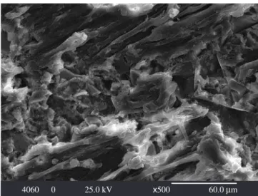
The average size of particles of bioactive glass was about 10.50 ± 9.4 μm. The particle size presents a high standard deviation value. It can provide an ideal distribution - hybrid distribution - of the re-inforcement particles in the composites, considering that the reinforce-ments present a great homogenization6. Figures 1-2 are SEM images of the composite bioactive glass/PHB, 30/70 and 40/60, respectively. As it can be observed the reinforcements are distributed randomly. Both samples present a hybrid distribution, however the sample bioactive glass/PHB 40/60 is the one that the particles present more size variation. The hybrid distribution increases the probability of the presence of the bioactive glass (reinforcement) in any part of the composite, decreasing the chance to crack propagation19. Its mechanical properties tend to be better than the bioactive glass/PHB 30/70. The average particle size results of this work are in agreement with the results found by others. The value 10.50 ± 9.4 μm is close to the average size of composites tricalcium phosphate/PHB (4.4 μm) used by Wang et al.2 and close to composites hydroxyapatite/PHB (24.5 μm) used by Ni and Wang20. They show bioactivity when immersed in SBF.
Jaakkola et al.21 concluded that the quantity of bioactive glass and the size of particles influence the bioactivity of composite. Larger particles and high quantity of particles presented less bioactivity than
composite with smaller particles and intermediate size particles. The authors used 40, 60 and 70 percent of bioactive glass with size particle of > 45 μm and 90-135 μm in composites ε-caprolactone-co
-DL-lac-tate/bioactive glass. However, Ni and Wang20, produced composites HA/PHB with less percentage of HA (10, 20 and 30%) and obtained a great formation of apatite layer, which indicates a higher bioactive with less quantity of hydroxyapatite.
The values of surface areas, BET results, for the bioactive glass were 0.7583 m2.g-1 and 0.6685 m2.g-1 in the multipoint mode and in the single point method, respectively. Zhong et al.22, affirm that only bioactive glasses with surface area larger than 40-80 m2.g-1 can induce HCA formation. However, Krajewski et al.23 produced bio-active glass and common glasses with surface area between 0.083 and 0.255 m2.g-1 and concluded that the bioactive glass with smaller surface areas also show some bioactivity. The surface area values of the bioactive glass, produced in the present work, are among the values obtained by Krajewski and Zhon and collaborators22,23. Regarding to the bioactivity, the surface area values of the samples produced in this work also exhibit bioactivity.
The microhardness Vickers results are presented in the Table 2. The results obtained for the bioactive glass/PHB 30/70 and 40/60 composites were superior to the results found by Chen and Wang7, when TCP/PHB and HA/PHB composites (30% of reinforcement) were studied, 10.18 ± 0.16 and 15.73 ± 0.44 (VHN) respectively. However, they were similar to the results obtained by Wang et al.3 in composite 30TCP/70PHB (24.66 ± 1.05 VHN). According to Wang4 and Doyler et al.9 higher is the quantity of ceramics higher will be the stiffness and there is a straight relationship between microhardness Vickers and Young Modulus. Therefore, the quantities of HA can also affect the bioactivity of mate-rial. The authors mentioned that the bioactivity is improved when the percentage of ceramic is between 20 and 40%. This amount of ceramic might not give the necessary mechanical properties to the composite; there is a need of increasing the amount of reinforcement. A balance between mechanical property and bioactivity needs to be found.
SEM results are presented in Figures 1-6. Composite bioactive glass/PHB 30/70 and 40/60, as prepared, are shown in Figures 1
Table 2. Microhardness Vickers.
Composite Microhardness (VHN)
40/60 24.6 ± 3.0
30/70 23.6 ± 2.5
3070 0 25.0 kV x500 60.0 Mm Figure 1. SEM image of composite bioactive glass/PHB 30/70 as prepared.
4060 0 25.0 kV x500 60.0 Mm Figure 2. SEM image of composite bioactive glass/PHB 40/60 as prepared.
307014 25.0 kV x600 50.0 Mm Figure 5. SEM image of composite bioactive glass/PHB 30/70 after 14 days of immersion in SBF.
COMP7B 15.0 kV x1.00 K 30.0 Mm Figure 4. SEM image of composite bioactive glass/PHB 40/60 after 7 days of immersion in SBF.
406014 25.0 kV x600 50.0 Mm Figure 6. SEM image of composite bioactive glass/PHB 40/60 after 14 days of immersion in SBF.
globular morphology. The globular morphology found by Jaakkola21 on the composite ε-caprolactone-co-DL-lactate/bioactive glass after
7 days of soaking in SBF is similar to the morphology found in this work. Chen and Wang7 also observed similar morphology formed on composites HA/PHB after 28 days immersed in SBF. Figure 4 show a layer formation on the composite bioactive glass/PHB 40/60 after 7 days of immersion in SBF and its morphology resembles the one found by Ni and Wang20 when composites HA/PHB were immersed in SBF during 7 days. Comparing the morphology of the layer formed on the composites used in this work, the layer formed on the bioac-tive glass/PHB 30/70 is more uniform and continuous than the layer formed on composite bioactive glass/PHB 40/60, Figures 3 and 4. It was observed a continuous formation of globular layers, however even more cohesive and uniform, when the in vitro studies were prolonged to 14 days on both composites, Figures 5 and 6. Roether et al.24 using porous composite poly(DL-lactide)/Bioglass® found similar morphology in layers formed in samples immersed in SBF. The authors observed the growth of HA on composite surface after 7 days of soaking in SBF, however the similar morphology was only observed after 28 days of immersion in SBF. It indicates a better bioactivity of the composites produced in this work than the one produced by Roether et al.24.
HA morphology can vary from acicular to equi-axed crystals and it depends on the concentration of ion carbonate (CO3-)25. All biologic apatites show a percentage of carbonate, CO3- and the globular morphology has about 12.5% of CO3-. The morphology observed via SEM is in agreement with the spherical format of HCA. The adherence of HCA layer on the composite will depend of the chemical interactions and thickness and it will influence the adhesion of the cells, orienting, for example, the formation of the conjunctive tissue26.
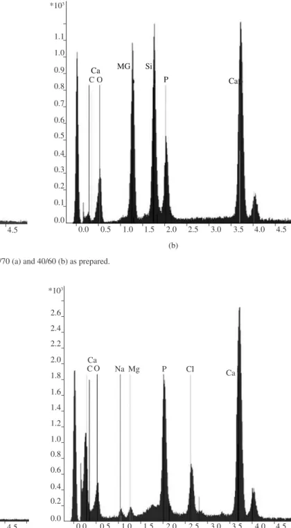
EDS results are presented in Figures 7-9. As it can be observed there is an intense formation of Ca-P rich layer. EDS spectrograms show clearly difference between intensity of Ca and P in the compos-ites as prepared (Figure 8) and in the composcompos-ites bioactive glass/PHB 30/70 and 40/60, after immersion in SBF during 14 days, Figures 8 and 9, respectively. The Ca/P ratio of the layer formed on bioactive glass/PHB 30/70 is near to the 1.67, which is the Ca/P ratio of HA of the human body25. The Ca/P ratio ≈1.67 of Ca-P layer deposited on the bioactive glass/PHB 30/70 was maintained constant for different time of immersion in SBF. Composite bioactive glass/PHB 40/60 did not show a constant Ca/P ratio for different time of immersion in SBF; however these values were between 1.0 to 2.0 that are typical of other phases present in human body.
In vitro studies of composite bioactive glass/PHB, carried out in the present work, showed that the composite with 30% of bioactive glass forms a Ca-P rich layer faster and more uniform than the com-posite with 40% of bioactive glass, Figures 3-4, respectively. There is a tendency in other works to use composites ceramics/polymers between 20 to 40% of ceramics reinforcements, because these percent-ages have shown a better bioactive behaviour5,7,9,20. It suggests that composite 30/70 is more bioactivity than composite 40/60.
XRD spectra of bioactive glass/PHB 30/70 and 40/60 after 3 days of immersion in SBF are presented in the Figures 10 and 11, respectively. Calcium hydrogen phosphate peaks are present in both samples; however they show up with low intensity due to the few days of immersion. As describe by Peitl14, initially an amorphous layer of calcium phosphate forms and after the incorporation of ions OH- and CO
3- the crystallization of HCA starts. These results suggest that 3 days of immersion in SBF is not enough to have the complete crystallization.
The change in the concentration of ions in SBF used in the in vitro studies of the samples of bioactive glass/PHB 30/70 and 40/60 and 2, respectively. The morphology of layer formed on the
Figure 7. EDS spectrogram of composites bioactive glass/PHB 30/70 (a) and 40/60 (b) as prepared. Ca
MG Si P Ca
0.0 0.5 1.0 1.5 2.0 2.5 3.0 3.5 4.0 4.5 2.2
2.0
1.8
1.6
1.4
1.2
1.0
0.8
0.6
0.4
0.2
0.0 *103
C O
(a)
Ca MG
P Ca
Si
C O
0.0 0.5 1.0 1.5 2.0 2.5 3.0 3.5 4.0 4.5 1.1
1.0
0.9
0.8
0.7
0.6
0.5
0.4
0.3
0.2
0.1
0.0 *103
(b)
Ca
C O Na Mg P Cl
Ca *103
4.5
4.0
3.5
3.0
2.5
2.0
1.5
1.0
0.5
0.0
0.0 0.5 1.0 1.5 2.0 2.5 3.0 3.5 4.0 4.5
Figure 8. EDS spectrogram of bioactive glass/PHB 30/70 after 7 days.
Ca
C O Na Mg P Cl Ca
*103
2.6
2.4
2.2
2.0
1.8
1.6
1.4
1.2
1.0
0.8
0.6
0.4
0.2
0.0
0.0 0.5 1.0 1.5 2.0 2.5 3.0 3.5 4.0 4.5
Figure 9. EDS spectrogram of bioactive glass/PHB 30/70 after 7 days.
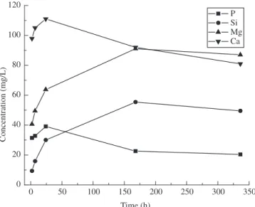
can be seen in the Figures 12 and 13, respectively. In the first stage, there is dissolution of all ions present in the glass structure. There-fore, after 1day, the concentration of ions Ca and P start to decrease. Concomitantly, Ca and P ions are being deposited on the composite as presented in the Figure 3-6. The concentration of Si and Mg ions in the SBF increasing continually; indicating the degradation of bioactive glass. The ICP results are similar to other works14,27,28. Jaakkola et al.21 state that the dissolution of Si from the bioactive glass is one the indicator of bioactivity and it is higher for composites that have smaller particles size of bioactive glass.
4. Conclusions
11.98 21.98 31.98 41.98 51.98 61.98
Intensity (cps)
2Q
600
500
400
300
200
100
0
Figure 10. XRD spectrum of bioactive glass/PHB 30/70 after 7 days of im-mersion in SBF (∇ Calcium Hydrogen Phosphate).
11.98 21.98 31.98 41.98 51.98 61.98
Intensity (cps)
2Q 600
500
400
300
200
100
0
Figure 11. XRD spectrum of bioactive glass/PHB 40/60 after 7 days of im-mersion in SBF (∇ Calcium Hydrogen Phosphate).
0 50 100 150 200 250 300 350
Concentration (mg/L)
Time (h) P
Si Mg Ca 140
130 120 110 100 90 80 70 60 50 40 30 20 10
Figure 12. ICP results of solution that soaks bioactive glass/PHB 30/70.
Figure 13. ICP results of solution that soaks bioactive glass/PHB 40/60.
0 50 100 150 200 250 300 350
Concentration (mg/L)
Time (h)
P Si Mg Ca 120
100
80
60
40
20
0
The Ca/P ratio found in the bioglass/PHB 40/60 did not present constant values; on the other hand they were between 1.1 and 1.9 that correspond to other phases of calcium phosphate. The HA layer formed on the bioglass/PHB 40/60 and bioglass/PHB 30/70 after being immersed in SBF during 3 days are not uniform. It presents some areas of cristalinity and others amorphous. The depletion of Ca and P in the SBF of ions Ca and P present in the calcium phosphate layer are from the SBF.
Acknowledgment
We are grateful to CAPES for the financial support and to Sandra Cachinho for the precious help.
References
1. Bonfield W, Grynpas MD, Tully AE, Bowman J, Abram J. Hydroxyapatite reinforced polyethylene – a mechanically compatible implant material for bone replacement. Biomaterials. 1981; 2(3):185-186.
2. Wang M. et al. Developing Tricalcium Phosphate/ Polyhydroxybutyrate composite as a new biodegradable material for clinical applications. Bioceramics. 2001; 1(3):193-195.
3. Ramakrishna S, Mayer J, Wintermantel E, Leong KW. Biomedical ap-plication of polymer composite material: a review. Composites Science and Technology. 2001; 61(3-4):1189-1224.
4. Wang M. Developing bioactive composite materials for tissue replace-ment. Biomaterials. 2003; 24(13):2133-2151.
5. Wang M, Bonfield W. Processing, characterization, and evaluation of hydroxyapatite reinforced polyethylene composites. Br. Ceram Trans. 1994; 93(3):91-95.
6. Anusavice KJ. Philips: Materiais Dentários. 10a ed. Rio de Janeiro:
Guanabara Koogan; 1998.
7. Chen L, Wang M. Production and evaluation of biodegradable composites based on PHB-PHV copolymer. Biomaterials. 2002; 23(13):2631-2639. 8. Owen AJ, Henzel J, Škrbić Ž, Divjaković. Crystallization and melt-ing behaviour of PHB and PHB/HV copolymer. Polymer. 1992; 33(7):1563-1567.
9. Doyle C, Tanner ET. Bonfiel W. In vitro and in vivo evaluation of poly-hydroxybutyrate and of polypoly-hydroxybutyrate reinforced with hydroxya-patite. Biomaterials. 1991; 12(9):841-847.
11. Nurbas NM, Kutsal T. Production of Polyhydroxyalkanoate (PHA) by Alcaligenes eutrophus. National Chemical Engineering Congress; Middle East Technical University, Ankara; 1994.
12. Oliveira JM, Correia RN, Fernades MH. Surface modifications of a glass and a glass-ceramic of the MgO-3CaO.P2O5-SiO2 system in a simulated body fluid. Biomaterials. 1995; 16(11):849-854.
13. Hench LL, Wilson J. An introduction to bioceramics. 1st ed. World
Sci-entific: Singapore; 1993.
14. Peitl O, Zanotto ED, Hench LL. Higly bioactive P2O5 -Na2O-CaO-SiO2 glass-ceramics. J. Non-Cryst Solids. 2001; 292(1-3):115-126. 15. Hench LL, Splinter RJ, Allen WC, Greenlee TK. Bonding mechanisms at
the interface of ceramic prosthetic materials. J Biomed Mater Res Symp. 1971; (2):117-141.
16. Kokubo T, Kushitani H, Ohtsuki C, Yamamuro T. Chemical reaction of bioactive glass and galss-ceramic with a simulated body fluid. J. Mater Sci: Materials in Medicine. 1992; 3(2):79-83.
17. Heikkilä JT, Aho AJ, Kangasniemi I, Yli-Urpo A. Polymethylmethacrylate composites: disturbed bone formation ate the surface of bioactive glass and hydroxyapatite. Biomaterials. 1996; 17(18):1755-1760.
18. Yoshie N, Saito M, Inoue Y. Effect of chemical compositional distribution on solid-state structures and properties of poly(3-hydroxybytrurte-co-3-hydroxyvalerate). Polymer. 2004; 45(6):1903-1911.
19. Schwartz MM, editor. Composite materials handbook. 2nd ed. New York:
McGraw-Hill; 1992.
20. Ni J, Wang M. In vitro evaluation of hydroxybutyrate reinforced polyhy-droxybutyrate composite. Mat. Sci and Eng. 2002; 20(1-2):101-109. 21. Jaakkola T, Rich J, Tirri T, Närhi T, Jokinen M, Seppälä J, Yli-Urpo A.
In vitro Ca-P precipitation on biodegradable thermoplastic composite of poly(e-caprolactone-co-dl-lactide) and bioactive glass (S53P4). Bioma-terials. 2004; 25(4):575-581.
22. Zhong JP, Greenspan DC, Feng JW. A microstructural examination of apatite induced by Bioglass® in vitro. J Mat Sci: Materials In Medicine. 2002; 13(3):321-326.
23. Krajewsk A, Malavolti R, Piancastelli A. Albumin adhesion on some bio-logical and non-biobio-logical glasses and connection with their Z-potentials. Biomaterials. 1996; 17(1): 53-60.
24. Roether J. et al. Development and in vitro characterization of novel bioresorbable and bioactive composite materials based on polylactide foams and Bioglass® for tissue engineering applications. Biomaterials. 2002; 23(18):3871-3878.
25. Le Geros RZ. Apatites in Biological Systems Prog. Crystal Growth Charact. 1981; 4(1-2):1-45.
26. Costa N, Maquis PM. Biomimetic processing of calcium phosphate coating. Medical Engineering & Physics. 1998; 20(8):602-606. 27. Leonor I. et al. In situ study of partially crystallized Bioglass® and
hy-droxyapatite in vitro bioactivity using atomic force microscopy. J Biomed Mater Res. 2002; 62(1):82-88.