SUMMARY
Objective: he authors show the clinical evaluation and follow-up results in 56 pa-tients diagnosed with a failed back surgery pain syndrome. Methods: Descriptive and prospective study conducted over a one-year period. In this study, 56 patients with a failed back surgery pain syndrome were assessed in our facility. he age ranged from 28 to 76 years (mean, 48.8 ± 13.9 years). he pain was assessed through a visual analog scale (VAS). Results: Postoperative pain was more severe (mean VAS score 8.3) than preoperative pain (7.2). Myofascial pain syndromes (MPS) were diagnosed in 85.7% of patients; neuropathic abnormalities associated or not with MPS were found in 73.3%. Drug therapy associated with physical medicine treatment provided ≥ 50% pain im-provement in 57.2% of cases; trigger point injection in 60.1%, and epidural infusion of morphine with lidocaína in 69.3% of refractory cases. Conclusion: In patients with a post-laminectomy syndrome, postoperative pain was more severe than preoperative pain from a herniated disk. A miofascial component was found in most patients.
Keywords: Low back pain; intervertebral disk displacement; post-laminectomy
syn-drome.
Study conducted at the Pain Center, Department of Neurology, Clinical Hospital of USP School of Medicine, São Paulo, SP, Brazil
Submitted on: 3/21/2008
Approved on: 3/21/2011
Correspondence to:
Wellingson Silva Paiva Divisão de Neurocirurgia Funcional e Centro de Dor Hospital das Clínicas da Faculdade
de Medicina da USP R. Dr. Eneas Aguiar, 255 5º andar – Cerqueira César
São Paulo – SP CEP: 05403-010 [email protected]
Conlicts of interest: None.
Failed back surgery pain syndrome: therapeutic approach descriptive
study in 56 patients
MANOEL JACOBSEN TEIXEIRA1, LIN TCHIA YENG2, OLIVER GARCIA GARCIA3, ERICH TALAMONI FONOFF4, WELLINGSON SILVA PAIVA5,
JOACI O ARAUJO6
1 Lecturer; Chair Professor in Neurosurgery, School of Medicine, Universidade de São Paulo (USP), São Paulo, SP, Brazil
2 Ph.D. in Science; Pain Center Coordinator, Division of Physical Medicine, Clinical Hospital of the School of Medicine, USP, São Paulo, SP, Brazil 3 M.D.; Residence in Neurosurgery; Medical Trainee, Pain Center, Clinical Hospital of the School of Medicine, USP, São Paulo, SP, Brazil 4 Ph.D. in Neurology; Pain Center Coordinator, Department of Neurology, Clinical Hospital of the School of Medicine, USP, São Paulo, SP, Brazil
INTRODUCTION
According to the International Association for the Study of Pain (IASP), the post-laminectomy syndrome is deined as a “low back pain of unknown origin, persisting at the same location as the original pain despite operative interven-tions or with a post-surgery onset. he low back pain may be associated with a referred or radiating pain”1. his dei-nition applies to all surgeries designed to treat pain arising from the low back spine, including those aiming to treat a herniated disk. Surgical management applied to herniated disks is a hemilaminectomy with a lavectomy, nerve root dislocation and a hernia excision. he many clinical mani-festations of the post-laminectomy pain oten overlap and have a low back pain as a common expression. he term “unknown origin” in the deinition should not be strictly used, as although the post-laminectomy syndrome is com-plex and the pain can arise from various nosological enti-ties afecting varied anatomical elements in the spine or away from the spine or even being a result from systemic conditions, its origin can be found in many cases2.
Low back pain causes are varied and diferential diag-nosis is wide. he structure causing the pain is identiied in less than 20% of cases3. he herniated disks are the most common indication for laminectomy to treat low back pain. Over 300 thousand laminectomies are estimated to be carried out in the United States, with a failure rate high-er than 40%4. Misinterpretation of pain origin as resulting from a herniated disk seen in imaging studies5, misidenti-ication of spine instability and other mechanical causes, including incomplete removal of the herniated disk6, and operative complications are blamed for the poor surgical results2. Pain can also result from articular facet instabil-ity or from reduced intervertebral space due to structure abnormalities or intervertebral disk removal, with conse-quent change in the articular facet angle7. Among non-me-chanical causes for the failed back surgery pain syndrome, disk infection, peridural “ibrosis”, arachnoiditis and psy-chosocial factors should be mentioned8. In this study, we aimed to assess the clinical features and the non-surgical management outcome in patients with a failed back sur-gery pain syndrome seen in a pain center.
METHODS
Fity-six patients diagnosed with a failed low back surgery pain syndrome were prospectively followed over one year at the Neurological Clinic Pain Center of the Clinical Hos-pital of Universidade de São Paulo in a descriptive study. he design was approved by the Institutional Ethics Board (213/05). Every patient signed an informed consent form. Patients with a persisting low back pain or an early relapse within less than three months of a surgical herniated disk procedure were selected. he patients were admitted into a speciic postoperative low back pain outpatient clinic according to a spontaneous demand in the unit. Patients
with evidence of metabolic, inlammatory, and oncologi-cal diseases or a radiologioncologi-cal segmental instability picture were excluded. No patients experienced a cognitive im-pairment. Out of 73 patients admitted into our unit, 14 were excluded by exclusion criteria and the follow-up was lost in three cases. We obtained data from history, general physical exam, neurological and physiatric evaluation in addition to pre- and postoperative imaging studies. Lab-oratory tests to rule out rheumatic or metabolic diseases (ANA, RF, ESR, CRP, blood cell count) were performed. he pain magnitude, characteristic, nature and location, as well as the radiating course in pre- (retrospectively) and postoperative periods. he pain intensity was assessed according to a visual analog scale (VAS) before and ater treatment. he physiatric exam aimed specially to assess low back spine and paravertebral, low back, gluteal and lower limb muscle groups, consisting of muscle power as-sessment, miofascial trigger point presence, spasms, sen-sitivity, cutaneous changes, and trophism. Data collection was performed by using a standard protocol. he statistical analysis used Sigmastat 4 sotware.
For all patients, a drug therapy with a rehabilitation program using kinesiotherapy and muscle stretching was adopted and if a rehabilitation refractory myofascial com-ponent (MPS) was associated, these patients underwent needling with a 1% lidocaine injection. MPS was identiied in 48 (85.7%) patients. Drug therapy consisted of amitrip-tyline 25 to 150 mg/day (mean, 64 mg/day), chlorproma-zine 20 to 100 mg/day (mean, 48 mg/day), naproxen 1000 to 1500 mg/day (mean = 1150 mg/day), acetaminophen 2 to 4 g/day (mean = 2.4 g/day) and codeine phosphate 120 to 240 mg/day (mean = 184 mg/day) according to the requirements and tolerability in each case. All patients un-derwent physiatric follow-up.
In case there was not ≥ 50% original pain improvement (VAS) all over the follow-up, the patients were considered frankly refractory, undergoing a 2 mL infusion containing morphine 1 mg/mL and 2% lidocaine by catheter place-ment into the low back peridural compartplace-ment bid over two weeks.
RESULTS
Overall, 37 (60.5%) patients were male and their ages ranged from 28 to 76 years (mean, 48.8 years ± 13.9 years). he patients’ mean age at the original pain onset ranged from 22 to 66 years (mean = 37.2 years). he patients had undergone from one up to four low back laminectomies (mean = 1.5) to treat low back pain or lumbosciatic pain. he mean symptomatology length was 96 months.
Muscle n %
Lumbar quadrate 33 69
Gluteus medius 9 19
Gluteus minimum 2 4
Piriform 3 6
Total 48 88
n, absolute numbers; %, percentage.
Table 1 – Patient disposition according to muscles affected more severely by the myofascial pain syndrome
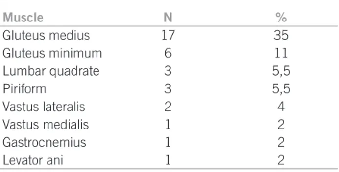
Muscle N %
Gluteus medius 17 35
Gluteus minimum 6 11
Lumbar quadrate 3 5,5
Piriform 3 5,5
Vastus lateralis 2 4
Vastus medialis 1 2
Gastrocnemius 1 2
Levator ani 1 2
MPS, myofascial pain syndrome
Table 2 – Affected muscle distribution by MPS satellite or secondary trigger points
with a postoperative root pain diagnosed had a mean score of 8.7, compared with 6.6 for those with no radiculopathy (p = 0.001).
In 17 (30.3%) patients, the preoperative pain history was consistent with a root origin, in 9 (16.1%) with re-ferred pain in musculoskeletal conditions in both lower limbs, in 22 (39,3%) with referred pain in one lower limb, in 2 (3.6%), the pain location was only the lower back re-gion and in 6 (10.7%) the pain had polyneuropathy char-acteristics. hree (5.4%) patients who did not experience pain with root characteristics preoperatively had under-gone a diskectomy and spinal ixation.
X-ray studies disclose a one- or two-lumbar-segment hemilaminectomy unilaterally (L4 or L5) in 53 (94.6%) pa-tients and bilateral lumbar laminectomy in 3 (5.4%). he lumbar spine dynamic study did not show instability in any patient. he computed tomography (CT) scan or mag-netic resonance (MR) imaging did reveal a periradicular scar at the operative site in 32 (57.1%) cases.
he pain intensity before the operations ranged from moderate to severe, according to the VAS, scoring from 5 to 10 (mean = 7.2); the pain intensity at the irst attendance to the Pain Center, Clinical Hospital, Universidade de São Paulo was severe, scoring from 7 to 10 (mean = 8.3).
hirty-six patients (64.9%) had undergone physical therapy, 53 (94.6%) had been on nonsteroidal anti-in-lammatory drugs (NSAIDs), and 17 (30.4%) had been on corticosteroids alone or in combination with B complex vitamins, four (7.1%) had been on opiates, and ive (8.9%) had been on tricyclic antidepressive agents before and af-ter the surgery. All of the patients had stayed at rest and those exerting an occupational activity had been put away from work.
In 38 (67.9%) patients, uniradicular (53.7%) or mul-tiradicular (14.3%) syndromes were shown. Ten (17.9%) patients had muscle pain and myofascial painful points in several body regions, sleep disturbances and depression, suggesting the ibromyalgia syndrome.
Trigger points characteristic of MPSs were identiied in 48 (85.7%) patients. Out of 17 (30.3%) patients with preoperative nerve root pain, 15 (88.2%) had lumbar or gluteal MPSs (Table 1).
Satellite or secondary trigger points were found in 29 (51.8%) patients (Table 2).
he treatment outcome was rated as excellent (> 75% improvement over the original pain according to the VAS), good (50% to 75% improvement), fair (25% to < 50% im-provement) and poor (< 25%).
Drug therapy combined with physical rehabilitation measures provided an excellent outcome in 5 (16.1%) pa-tients, a good outcome in 23 (41.1%), a fair outcome in 16 (28.6%) and a poor outcome in 12 (21.4%).
In 48 patients, MPS was found and trigger point in-jections were performed by using 1% lidocaine 0.5 mL.
By comparing the pain improvement scores, patients with MPS had worse outcomes over post-laminectomy syn-drome without MPS. he immediate results were excellent in 5 (10.4%), good in 17 (35.4%), fair in 18 (37.5%) and poor in 8 (17.8%). he results were satisfactory for 68.75% of patients with MPS versus 75% in patients without MPS, but the diference was not statistically signiicant (p = 0.2).
At the end of treatment, signiicant improvement (ex-cellent and good outcomes) occurred in 34 (60.1%) pa-tients, while 13 (23.2%) had a fair outcome. However, the outcome was considered poor in 8 (17.5%) patients. Re-garding the pain intensity at the inal follow-up, we found a reduced general mean in the visual analog scale from 7.2 to 4.7 (p = 0.01).
A peridural catheter for spinal infusion of a morphine and lidocaine solution was placed in thirteen patients; all of them had a MPS, being considered refractory ater a re-habilitative treatment attempt. he outcome was excellent in 4 (30.8%) patients, good in 5 (38.5%), and poor in 4 (30.8%), meaning the outcome was excellent or good in 9 (69.2%) patients treated with a peridural infusion.
DISCUSSION
In most cases, the course is favorable, even when no care measures are taken. However, low back pain becomes chronic in 15% to 20% of individuals9. In 13.8% of patients studied by Frymoyer10, the pain lasted more than two weeks and in 22% it was severe. In 21.2% of Deyo and Tsiu’s patients9, the pain was mild; in 43.4%, it was moderate and in 35%, severe; in 40% of cases, the low back pain irradi-ated to lower limbs and in only 1% a true sciatic pain could be found. In our series, preoperative pain was severe, with a 7.2 score according to the VAS; 45.4% had a low back pain and referred pain to lower limbs history before the surgery. In only 30.3%, the history suggested a true sciatic pain, and these indings indicate selection criteria for the surgery were likely inappropriate in most cases.
According to Hanley et al.11, the operative treatment outcome of herniated disks is poor in 14% of cases. he numbers of spine surgeries to relieve pain have steadily grown in the United States, with 170 thousand operations in 1974, 300,413 in 1994 reaching 392,948 in 200012, with 80 thousand cases of failed back surgery pain syndrome per year13. According to Deyo and Tsiu9, the main reason for an increasing number of laminectomies is the growing number of surgeons operating the spine in each country. In diferent countries and diferent regions, the frequency of operation indications is variable, and this is not ex-plained only by the diferent prevalence of low back pain or lumbosciatic pain; in 3% or 4% of individuals, herniated disk surgeries are indicated in the USA, but only in 1% of individuals in Sweden and Denmark.
Poor outcome of operative treatment might result from an incorrect diagnosis. Among the identiied causes for low back pain, the following could be highlighted: rheu-matic conditions, primary or secondary spine tumors, vascular conditions, hematological abnormalities, en-docrine conditions, pelvic or abdominal viscus diseases (endometriosis, ovarian cyst torsion, pelvic inlammatory disease, prostatitis, cystitis, pancreatopathy, nefropathy, kidney disease, peptic ulcer, urinary tract, biliary or duo-denal conditions), mechanical abnormalities (herniated in-tervertebral disk, articular facet injury, segmental instability or sacroiliac joint instability), systemic conditions (ibromy-algia, myositis, autoimmune or immune-allergic diseases), psychiatric diseases and other conditions (hip joint disease, trochanteric bursa injury, polyradiculoneuritis, meningeal irritation signs)14. Because of the great number of possibili-ties, the high surgical therapeutic failure rate is justiiable in care provided to these patients, but it also indicates there must be a more judicious semiologic evaluation.
Surgeries that do not meet the indication criteria to treat a herniated disk can result in maintenance or wors-ening of pain and preoperative deicits. A herniated disk misinterpreted as a cause for low back pain is the most common reason for indicating spine surgeries that prog-ress to a post-laminectomy chronic pain syndrome with
an early onset postoperatively. his is partly due to over-valuing the anatomical indings not related to the low back pain that are shown in imaging studies, but those usually do not warrant the pain and the surgical intervention14. In 35% of asymptomatic individuals studied by Hitselberg and Wihen15, the x-ray imaging revealed abnormalities suggesting a herniated disk. In 35% of asymptomatic in-dividuals studied by Wiesel et al.16, a spine CT scan found abnormalities; in 20.2% of cases, there was a herniated disk evidence. Boden et al.14 observed 60% of asymptomatic in-dividuals had a herniated disk on magnetic resonance im-aging. herefore imaging studies can conirm a herniated disk clinical diagnosis, but they are not the main determi-nants for indicating a surgery, since asymptomatic herni-ated disks are so commonly seen2.
Even in symptomatic conditions, there is a progressive absorption of the herniated disk fragment, a phenomenon accompanied by symptom improvement in most cases17. Hakelius18 observed 38% of patients with a herniated disk not undergoing a surgery, but having been on medical treatment, were clinically improved within a month, 52% within two months, and 73% within three months. Saal and Sall19 conducted a retrospective study involving 58 patients with a radiculopathy resulting from a herniated disk; 52 underwent a conservative treatment, resulting in improvement in over 90% of cases; only in three cases surgical ablation of extruded fragments was required. his means the indication criteria for diskectomy, represented by a cauda equina syndrome, marked acute or progressive motor deicit or lumbosciatic pain occurrence and evident radiculopathy, characterized by sensory, motor and deep tendon relexes deicits over one or more nerve root ter-ritory, nerve root irritation evidence, translated as a posi-tive straight leg raising maneuver and consistent imaging study indings20 in patients achieving no improvement ater symptomatic drug therapy with physical medicine measures during a period of over 6 to 12 weeks20,21, are not always met. Only 64.9% of patients included in the current casuistry had undergone physical medicine treatment and only 8.9% had undergone a tricyclic antidepressant thera-py before the operations, suggesting medical methods had not been adopted in most cases.
postop-erative imaging studies oten show similar abnormalities in individuals whether they are symptomatic or not24. Peridu-ral scar occurring ater a laminectomy is a frequent post-operative inding. Newly formed tissue can involve, distort and/or compress the nerve root. However, epidural ibrosis is oten shown on CT scan or MRI postoperatively in cases there is no pain8. In 57.1% of patients in this study, a perira-dicular scar was found.
he patients included in our study had undergone up to 4 surgical lumbar spine surgical procedures with no im-provement; the mean was 1.5 operations per patient. Many patients undergoing further operations to treat persisting or residual pain get frustrated. he improvement rate in reoperations is low, around 30% ater the second surgery, 15% ater the third surgery and 5% ater a fourth proce-dure with up to 20% of worsening13.
Out of 56 patients analyzed in the present study, 85,7% had MPS not found previously on physical exam. here is evidence that MPS is involved in low back pain genesis or maintenance23. However, MPS diagnosis is frequently dis-regarded25. Many lumbar muscles afected by MPS and the operative injury would result in pain worsening. Although physiatrically speaking lumbar and gluteal muscle MPS is considered the most important cause for low back pain, bone, tendinous, nerve, disk and bursa conditions are still valued as symptom causes26. he muscle iber injury is not necessarily a cause for pain, since in patients with primary degenerative conditions, as in Duchenne muscular dystro-phy, there is a disruption in a large amount of myoibrils and the sarcoplasmic reticulum, but there is no pain, sug-gesting MPS symptoms result from nonstructural muscle iber changes or dysfunctions26. he main electrophysio-logical abnormality seems to be a neuromuscular dysfunc-tion in the motor endplate.
he energy crisis theory postulates there is an increased calcium concentration in the sarcoplasm due to a sarco-plasmic reticulum, sarcolemma and or muscle cell mem-brane disruption. he sarcoplasmic reticulum function is storing and releasing ionized calcium, which activates contractile elements and causes sarcomere shortening. Sustained sarcomere contraction results in increased me-tabolism, causes localized ischemia and generates a local-ized energy crisis. he combination of electrophysiological and histopathological theories generated the neuromus-cular endplate multiple dysfunction concept. he poten-tials recorded as spontaneous activity or spikes in trigger points would result in abnormal acetylcholine release by the nervous ending. Acetylcholine release would accentu-ate depolarization and calcium release from the sarcoplas-mic reticulum, causing sarcomere contraction and small-caliber vessel compression. Increased depolarization due to acetylcholine release and sarcomere contraction would cause increased energy demand, which, if associated with hypoxia resulting from reduced muscle blood low, would
then cause the energy crisis. his energy crisis generates metabolites which sensitize nociceptores and referred pain from trigger points26. he abnormalities in nerve ibers re-sponsible for supplying the muscle could cause localized muscle contraction and MPS27. he referred pain from the trigger point is due to a sensory neuron sensitization in the spine cord posterior horn and may have a distribution similar to that in the radiculopathic pain. his referred pain associates with tingling and numbness26. In 88.2% of 17 patients with preoperative nerve root pain history, lum-bar and gluteal MPS was identiied.
he ages of patients included in the study when irstly seen at the Pain Center ranged from 28 to 76 years (mean age, 48.8 years). he mean symptom length was 96 months and the mean pain intensity was 8.3, showing the magni-tude and the extended distress the patients went through. he postoperative pain was also shown more severe than the preoperative pain.
he chronic pain treatment should involve a multi-disciplinary team and pharmacological, physiatric, psy-chotherapeutic, and neuroanesthesic procedures; func-tional neurosurgical procedures should be performed if required28. he treatment with analgesic drugs, whether they are anti-inlammatory drugs or not, psychotropic drugs and physical medicine provided > 50% original pain improvement in 57.2% of patients evaluated in this study. Myofascial trigger point treatment consists of using analgesic drugs, psychotherapeutic agents, muscle relax-ant drugs, refrigerrelax-ant vapor, dry needling, local anesthetic injection and stretching, as well as correction of causal or perpetuating factors29.
In 69.4% of patients undergoing administration of a morphine and lidocaine solution via peridural route in our study, the original pain had > 50% improvement.
he pain in patients with failed back surgery pain syn-drome is severe, afects individuals in the fullness of their activities, is oten found as a lumbar and/or gluteal MPS and, less frequently, has a neuropathic pattern alone or as-sociated with MPSs29.
CONCLUSION
he failed back surgery pain syndrome evaluation and management is challenging for the medical team. Analgesic drugs and physical medicine provide major improvement in most cases. he pain intensity in post-laminectomy syn-drome is worse than the herniated disk preoperative pain. he injection of myofascial trigger points and opiate infu-sion into the lumbar spine compartment can be required in refractory pain cases.
REFERENCES
2. Follett, KA, Dirks BA. Etiology and evaluation of the failed back sur-gery outcome. Neurosurg Q. 1993;3:40-59.
3. Saal JS, Franson RC, Dobrow R, Saal JA, White AH, Goldthwaite N. High levels of inlammatory phospholipase A2 activity in lumbar disc herniations. Spine 1990;15:674-8.
4. Martin BI, Mirza SK, Comstock BA, Gray DT, Kreuter W, Deyo RA. Are lumbar spine reoperation rates falling with greater use of fusion surgery and new surgical technology? Spine 2007;32:2119-26. 5. Deyo RA. Back surgery-who needs it? N Engl J Med.
2007;356:2239-43.
6. Deyo RA. Back pain revisited newer thinking on diagnosis and ther-apy. Consultant 1993;33:88-100.
7. Goupille P. Cause des echecs de la chirurgie discale. Rev Rheumat 1996;63:255-60.
8. Willberger RE, Wettenberg RH. Prostaglandin release from lumbar disc and facet joint tissue. Spine 1994;19:2068-70.
9. Deyo RA, Tsiu WU. Descriptive epidemiology of low-back pain and its related medical care in the United States. Spine 1987;12:164-8. 10. Frymoyer JW. An international challenge to the diagnosis and
treat-ment of disorders of the lumbar spine. Spine 1993;18:2147-52. 11. Hanley EN Jr, Shapiro DE. he development of low-back pain ater
excision of a lumbar disc. J Bone Joint Surg Am. 1989;71:719-21. 12. Hazard RG. Failed back surgery syndrome: surgical and nonsurgical
approaches, Clin Orthop Relat Res. 2006;443:228-32.
13. Ragab A, Deshazo RD. Management of back pain in patients with previous back surgery. Am J Med. 2008;121:272-8.
14. Boden SD, McCowin PR, Davis DO, Dina TS, Mark AS, Wiesel S. Abnormal magnetic-resonance scans of the cervical spine in asymp-tomatic subjects. A prospective investigation. J Bone Joint Surg Am. 1990;72:1178
15. Hitselberger WE, Witten RM. Abnormal myelograms in asympto-matic patients. J Neurosurg. 1968;28:204-6.
16. Wiesel SW, Tsourmas N, Fefer HL, Citrin CM, Patronas N. A study of computer assisted tomography. he incidence of positive cat scans in an asymptomatic group of patients. Spine 1984;9:549-51 17. Weber H. Lumbar disc herniation. A controlled, prospective study
with ten years of observation. Spine 1983;8:131-40.
18. Hakelius A. Prognosis in cciatica: a clinical follow-up of surgical and non surgical treatment. Acta Orthop Scand. 1970;129 (Suppl):1-76. 19. Saal JA, Saal JS. Non operative treatment of herniated lumbar
in-tervertebral disk with radiculopathy: An outcome study. Spine 1989;14:431-7.
20. Hurme M, Alaranta H. Factors predicting the results of surgery for lumbar intervertebral disc herniation. Spine. 1987;12:933-8. 21. Gibson JN, Grant IC, Waddell G. Cochrane Database Syst Rev.
2000;(3):CD001350.
22. Finnegan WJ, Fenlin JM, Marvel J. Results of surgical interven-tion in the symptomatic multiply-operated patient: analysis of sixty seven cases followed three to seven years. J Bone Joint Surg. 1979;611:1077-82.
23. Licber RL. Low back pain. A scientiic and clinical overview. Wash-inghton (DC): American Academy of Orthopaedics Surgeon Work-shop; 1995.
24. Fritsch EW, Heisel J, Rupp S. he failed back surgery syndrome: rea-sons, intraoperative indings and long term results: a report of 182 operative treatments. Spine 1996;21:626-33.
25. Imamura ST, Fischer AA, Imamura M, Teixeira MJ. Pain manage-ment using myofascial approach when other treatmanage-ment failed. Myo-fascial pain update in diagnosis and treatment. Phys Med Rehab Clin North Am. 1997;8:179-92.
26. Rachlin ES. Importance of trigger point management in orthopedic practice myofascial pain update in diagnosis and treatment. Phys Med Rehabil Clin North Am. 1997;8:171-7.
27. Cannon DE, Dillingham TR, Miao H, Andary MT, Pezzin LE. Mus-culoskeletal disorders in referrals for suspected lumbosacral radicu-lopathy. Am J Phys Med Rehabil. 2007;86:957-61.
28. Teixeira MJ. Princípios de tratamento da dor. In: Teixeira MJ, Figue-iró JAB, editors. Dor, epidemiologia, isiopatologia, avaliação, sín-dromes dolorosas e tratamento. São Paulo: Grupo Editorial Moreira Jr; 2001. p. 86-92.