RESUMO
JUSTIFICATIVA E OBJETIVOS: Corticosteróides fo-ram introduzidos no tratamento das infecções graves há várias décadas, Diversos estudos aleatórios e con-trolados com resultados negativos utilizando corticói-des em doses elevadas por curto período na sepse ou SARA trouxeram questionamentos acerca da eficácia deste tratamento. Recentemente, um elo entre a infla-mação sistêmica e a insuficiência adrenal foi estabele-cida. Este achado renovou o interesse na reposição de baixas doses de corticóides por períodos mais longos. O objetivo deste artigo é revisar o papel dos corticoste-róides na resposta do hospedeiro ao stress e atualizar o leitor com indicações novas e validadas de reposição de corticosteróides.
CONTEÚDO: Extensa revisão da fisiologia adrenal e das alterações fisiopatológicas e suas implicações clí-nicas em pacientes críticos.
CONCLUSÕES: Na vigência de sepse, instabilidade hemodinâmica e perpetuação da resposta inflamató-ria podem resultar de insuficiência adrenal (IA). Desta forma teste de estimulação com ACTH devem ser lizados precocemente para identificação de IA. À rea-lização do teste deve seguir a reposição de 50 mg de hidrocortisona em bolus e a cada 6 horas associado a
Adrenal Insufficiency in Sepsis*
Insuficiência Adrenal na Sepse
Andrea Polito1, Jérôme Aboab1 , Djillali Annane, PhD1
Faltou a titulação
*From Service de Reanimation Medicale, Hopital Raymond Poincare, Assistance Publique-Hopitaux de Paris, Faculte de Medecine Paris Ile de France Ouest, Universite de Versailles Saint-Quentin en Yveli-nes, Garches, France
Submitted to December, 22, 2005 Accepted to January, 27, 2006 Correspondence to:
Service de Reanimation, Hospital Raymond Poincaré (AP-HP) University of Versailles
SQ, 104 Boulevard Raymond Poincaré, Garches 92380.
Phone: 331 47 10 77 87 Fax: 331 47 10 77 83
E-mail: [email protected]
50 µg de fludrocortisona uma vez ao dia. Quando os resultados do teste com ACTH encontrarem-se dispo-níveis, a terapia deverá ser continuada por sete dias nos não-respondedores e descontinuada nos respon-dedores. A decisão acerca do tratamento de responde-dores ao ACTH com cortisol > 34 µg/dL com potencial resistência periférica ao cortisol deve ser avaliada em futuros ensaios clínicos.
Unitermos: ACTH, cortisol, disfunção orgânica múlti-pla, insuficiência adrenal, sepse
SUMMARY
BACKGROUND AND OBJECTIVES: Corticosteroids were introduced in the treatment of severe infection as early as in the nineteen forties. Several “negative” ran-domized controlled trials of high-dose of glucocorticoi-ds given for a short period of time in the early course of severe sepsis or acute respiratory distress syndrome raised serious doubts on the benefit of this treatment. Recently, a link between septic shock and adrenal insu-fficiency, or systemic inflammation induced glucocorti-coids receptor resistance had been established. This finding prompted renewed interest of a replacement therapy with low doses of corticosteroids during longer periods. The goal of this article is to review the key role of corticosteroids in the host response to stress and will update the reader with the new validated indica-tions of corticosteroids treatment in the ICU.
CONTENTS:Extensive review of the adrenal physiolo-gy and its pathophysiological derangements and clini-cal implications in criticlini-cally ill patients.
responders. Whether responders to ACTH with high baseline cortisol levels (> 34 µg/dL) have tissue resis-tance to cortisol and also should receive exogenous hormones remains to be evaluated in clinical trials.
Key Words: ACTH, adrenal insufficiency, cortisol, mul-tiple organ dysfunction syndrome, sepsis
INTRODUCTION
In the beginning of the twentieth century, observations of apoplectic adrenal glands in fatal meningococce-mia underlined their key role in host defense against infection. Thirty years after, cortisone was discovered and rapidly proven to have numerous and diversified physiological functions in the host response to stress. Corticosteroids were introduced in the treatment of se-vere infection as early as in the nineteen forties. Seve-ral “negative” randomized controlled trials of high-dose of glucocorticoids given for a short period of time in the early course of severe sepsis or acute respiratory distress syndrome raised serious doubts on the benefit of this treatment. Recently, a link between septic sho-ck and adrenal insufficiency, or systemic inflammation induced glucocorticoids receptor resistance had been established. This finding prompted renewed interest of a replacement therapy with low doses of corticosteroi-ds during longer periocorticosteroi-ds. We will review the key role of corticosteroids in the host response to stress and will update the reader with the new validated indications of corticosteroids treatment in the ICU.
REGULATION AND PRODUCTION OF CORTICOSTEROIDS
At Rest
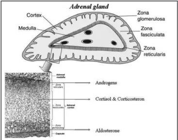
The adrenal gland is made of two functional units, the medulla which produces catecholamines and cortex composed of three zones: zone glomerulosa, superfi-cially located, producing mineralocorticoids (aldoste-rone and corticoste(aldoste-rone), zone reticularis deeper sited producing weak androgens and zone fasciculate pro-ducing corticosteroids (cortisol and corticosteron) (Fi-gure1).
Cortisol, the main corticosteroid, is a steroid hormo-ne of 19 carbon atoms derived from cholesterol by an enzyme of P450 cytochrome complex. Cortisol circu-lates in plasma either in its free and active form (whi-ch accounts only for 5% to 10% of total cortisol) or in its inactive form, reversibly bound to proteins. The two main binding proteins are the cortisol-binding globulin
(CBG) and albumin1.
Production of corticosteroids is regulated by the hypo-thalamic–pituitary-adrenal (HPA) axis. Cortisol produc-tion and secreproduc-tion is stimulated mainly by the adreno-corticotrophic hormone (ACTH). This is a 39 amino acids peptide produced in the anterior pituitary. It is obtained after the cleavage of a large precursor, the pro-opiome-lanocortin, which also liberates other peptides (β -en-dorphin, lipotropin, melanocyte-stimulating hormone). In the short term, ACTH stimulates cortisol production and secretion even if cortisol storage in adrenal glands being low; in the longer term, ACTH also stimulates the synthesis of enzymes that are involved in cortisol pro-duction, as well as their cofactors and adrenal recep-tors for low-density lipoprotein cholesterol. ACTH also stimulates the production of adrenal androgens and, to a lesser extent, mineralocorticoids1.
The half-life of ACTH is short and cortisol concentration in adrenal veins rises only a few minutes after ACTH secretion2. Secretion of ACTH is regulated by several
factors among which the corticotrophin-releasing hor-mone (CRH) and arginine vasopressin (AVP), which are both secreted by the hypothalamus. AVP stimulates ACTH secretion only weakly but it strongly promotes CRH action. Catecholamines, angiotensin II, seroto-nin and vasoactive intestinal peptide are also known stimulators of ACTH secretion. Finally, some inflam-matory cytokines influence ACTH secretion, exerting either a stimulatory action (IL-1, IL-2, IL-6, tumour ne-crosis factor TNF-α) or an inhibitory one (transforming growth factor-β)2-4.
CRH is a 41-amino-acid peptide secreted by the
pothalamus. Liberated in the hypothalamic–pituitary portal system, it stimulates the production and the se-cretion of pro-opiomelanocortin. Adrenergic agonists (noradrenaline) and serotonin stimulate its production whereas substance P, opioids and γ-aminobutyric acid inhibit it. Inflammation cytokines (IL-1, IL-2, IL-6,
TNF-α) also influence production of CRH2-5.
Finally, corticosteroids exert a negative feedback on the HPA axis, inhibiting ACTH production as well as pro-opiomelanocortin gene transcription, and CRH and AVP production. Secretion of the HPA axis hormo-nes (ACTH, CRH and AVP) follows a pulsatile course with a circadian rhythm. The amplitude of the secretory pulses varies throughout the day and is greatest in the morning between 6 and 8 AM, rapidly decreasing until noon and decreasing more slowly until midnight1.
However, the brain and the pituitary, which regulate the production of corticosteroid secretion through ne-gative feedback by sensing time-integrated tissue ex-posure to corticosteroids, are also targets of teroids. Thus, any generalized change in the corticos-teroid signaling system will be followed by corrective, compensatory, and generally protective changes in the activity of the HPA axis by adjusting target tissue effects to optimal levels2,6. Absence of complete
com-pensation - be it excessive or deficient - could result in chronically altered homeostasis or allostasis and tar-get tissue pathology, as occurs in chronically stressed or depressed individuals who have mild but persistent hyper- or hyposecretion of cortisol2,7. The
suprahypo-thalamic, hyposuprahypo-thalamic, and pituitary corticosteroid-sensing network, however, differs from the networks of corticosteroid signaling systems in the arousal, as-sociative, reward, metabolic, cardiovascular, and im-mune systems. Any change in one or more molecules that participate in the corticosteroid signaling system could potentially produce a discrepancy in the sensi-tivity of target tissues to corticosteroids and result in pathology2,8. For these reasons any polymorphism or
mutation of GR gene may affect corticosteroid signa-ling pathway and being responsible for different dise-ases according to corticosteroid tissue sensitivity that may be augmented (hypertension, diabetes mellitus II, visceral obesity-related insulin resistance)2,6,7 or
de-creased (asthma, ARDS)2,7. Moreover, tissue
sensitivi-ty to corticosteroids is affected by interactions of the host with viruses, such as human immunodeficiency viruses type 1, which increases corticosteroid sensiti-vity of host tissues, and the adenovirus, which decre-ases it19,10.
DURING STRESS
Corticosteroids are the main mediators of the stress response. During a stressing event it appears an imme-diate increment of ACTH secretion rapidly followed by increase in cortisol level11. Moreover CBG level
decre-ases and subsequently free cortisol rises12.
Pro-inflam-matory cytokines play a crucial role in the activation of HPA axis13. These events are associated with a loss of
the circadian rhythm of cortisol secretion secondary to an increase in CRH and ACTH production stimulated by inflammatory cytokines, vagal stimulation and reduc-tion in cortisol-negative feedback11,14. All the changes
during stress have the purpose to maintain homeosta-sis. Metabolic effects, especially hyperglycemia allow the redistribution of glucose toward insulin-dependant cells. Cardiovascular effects of corticosteroids aim to maintain normal vascular reactivity and almost all the component of the inflammatory cascade are prevented by cortisol. However, the ability of the HPA axis to corti-sol sustained inflammatory insult like one resulting from severe infection, is likely a determinant of the progres-sion from sepsis to septic shock and death.
About “normal” concentration of circulating cortisol in critical illness is not clear what the right value is. Has been proposed to consider the range from 15 to 20 µg/ dL as normal5,15-17. Even if is controversial, the higher
the cortisol level at admission on ICU, the higher the mortality rate [18]. Cortisol serum level seems an inde-pendent predictive factor for outcome and likely the se-verity of stress. However, it is more essential to deter-mine the ability of the endocrine system to respond to sustained stress and thus, dynamic testing of adrenal function is of paramount importance. The most conve-nient test in ICU is the standard ACTH stimulation test. Normally a rise of circulating cortisol concentration of 9 µg/dL or more is considered as a normal response19,20.
MECHANISM OF ADRENAL INSUFFICIENCY IN SEPSIS
The recognition of various disorders that cause adrenal insufficiency (AI), either at a clinical or molecular level, often has implications for the management of the pa-tient. Recent molecular-genetic analysis for the disor-der that causes AI gives valuable insights into adrenal organogenesis, regulation of steroid hormone biosyn-thesis, and the developmental and reproductive endo-crinology.
axis function, e.g. pre-existing defect of adrenal or pi-tuitary gland, inflammation of the endocrine tissues, anatomic damage of the gland, drugs inhibiting enzy-matic step of cortisol synthesis (e.g. etomidate, ketoco-nazole), or increasing cortisol metabolism (phenytoin, phenobarbital)11,21.
The various etiologies of AI can be subgrouped into three categories: 1) impaired steroidogenesis, 2) adre-nal dysgenesis/hypoplasia, and 3) adreadre-nal destruction. AI develops only when a large part of the function of the adrenal gland is lost. Primary AI is caused by proces-ses that damage the adrenal glands or by drugs that block cortisol synthesis. In contrast, secondary AI re-sults from processes that reduce the secretion of ACTH by the pituitary gland due to a pituitary or hypothalamic pathology. More often in sepsis necrosis or hemorrha-ge of the hypothalamus or of the pituitary gland have been reported, as a result of prolonged hypotension or severe coagulation disorders22. Sepsis may also
exa-cerbate chronic known or latent secondary AI, which may due to hypothalamic or pituitary tumors, chronic inflammation or congenital ACTH deficiency. Seconda-ry AI may follow drug therapy. Previous treatment with corticosteroids reduces secretion of CRH and ACTH, resulting in slow onset of secondary AI. This suppres-sion depends on duration of pre-treatment and on the type of corticosteroid used. Hydrocortisone is the least suppressive agent and dexamethasone the most23. The
opioids reduce cortisol serum level probably by acting at the level of the hypothalamus24. Infection diseases,
viral and fungal especially, may cause chronic primary AI, particularly in immunosuppressed patients, like HIV patients5. Certain drugs may decrease steroidogenesis,
like etomidate. This effect persists as long as 24 hours after a single dose in critically ill patients25,26. In addition
cyclosporine27, clarithromycin28 accelerate cortisol
me-tabolism.
DIAGNOSIS OF AI IN SEPSIS
Usually, the adrenal crisis is characterized by fever, nausea, vomiting, abdominal pain, impaired consciou-sness, hypotension which is refractory to fluids and vasopressor therapy, hypoglycemia, hyponatremia and hypereosinophilia. Unfortunately, these symptoms are very common during severe sepsis and thus they lack specificity to help in the decision making process. The-reafter, whenever AI is suspected endocrine tests are mandatory.
Nonstimulated Serum Cortisol: This test measure
plasma ACTH and serum cortisol in the morning, when the latter peaks. The test requires that cortisol circadian rhythm is normal. A serum cortisol level of less than 3 µg/dL probably indicates AI [29]. In the latter case, plasma ACTH level will distinguish between primary and secondary AI. In primary AI, the ACTH level is al-most invariably more than 100 pg/mL, while in secon-dary AI, plasma ACTH can be either low or inappropria-tely normal. In patients with severe stress, like sepsis, serum cortisol level should be more than 18 µg/dL at any time. One exception may the patient with severe hypoproteinemia from whom total cortisol levels are no more accurate30. However, in sepsis, high total
corti-sol levels may result from impaired corticorti-sol clearance or tissue resistance and thus dynamic tests of adrenal function are essential t rule out AI.
Insulin-Induced Hypoglycemia (Insulin Tolerance Test, ITT). This measures the patient’s cortisol res-ponse to hypoglycemia induced by the intravenous administration of insulin. This test is often considered as the gold standard because it assesses the ability of the entire HPA axis to respond to the stressful situation of hypoglycemia. Following insulin (0.1 IU/kg) adminis-tration, blood is drawn during symptomatic hypoglyce-mia (glucose should decrease to less than 40 mg/dL [< 2.22 mmol/L]). In obese patients with insulin resis-tance, the usual dose of insulin should be increased to 0.15 IU/kg31. A serum peak cortisol of more than 18
µg/dL is considered normal. ITT has some limitations in terms of reproducibility and clear cut-off levels32,33 and
is contraindicated in patients with unstable hemody-namic status. In addition, peripheral insulin resistance that characterizes sepsis, made this test less accurate in this context.
Metyrapone Test. The metyrapone test measures the ability of the HPA axis to respond to an acute reduc-tion in serum cortisol levels. Metyrapone inhibits 11-hydroxylase, the enzyme involved in the last step of cortisol synthesis. This inhibition causes a decrease in cortisol that results in a compensatory increase in ACTH and in the cortisol precursor, 11-deoxycortisol. The administration of metyrapone (30 mg/kg; maxi-mal dose, 3000 mg) occurs at midnight, and blood is drawn the following morning at 8 AM for cortisol and 11-deoxycortisol34 in response to metyrapone, serum
cautiously as it may cause adrenal crisis.
CRH Stimulation Test. The CRH stimulation test mea-sures the ability of the pituitary gland to secrete ACTH in response to CRH. Ovine CRH (1 µg/dL) is injected in-travenously, and cortisol is measured after 15, 30, and 60 minutes. The test has been proposed as a way to differentiate secondary (pituitary disease) from tertiary (hypothalamic disease) AI. However, the accuracy of this test remains very controversial35.
ACTH Stimulation Test. The ACTH stimulation test is based on the inability of a diseased adrenal gland to respond acutely to the injection of ACTH by secreting cortisol. ACTH is injected intravenously (or intramuscu-larly), and serum cortisol levels are measured at 30 or 60 minutes. In critical illness, cortisol levels less than 15 µg/dL or a cortisol increment of 9 µg/dL or less indica-tes AI and identifies patients that require a replacement therapy16,17,36. Finally, an increase in cortisol level more
than 9 µg/dL with the basal level above 34 µg/dL, sug-gests tissue resistance to corticosteroid.
As plasma ACTH levels following a bolus of 250 µg of ACTH exceed by several times the levels achieved during major surgery, it has been suggested that the “High Dose” that may miss “mild” secondary AI37.
Sub-sequently, the 1 µg ACTH test (LDST) was introduced as a more sensitive test38. However, 1 µg of ACTH
indu-ces supramaximal stimulation of the adrenals for only 30 min and may not appropriately evaluate the capacity of the adrenal glands to maintain maximal cortisol pro-duction in response to a major ongoing stress like sep-sis. In addition, interpretation of the results of the 1 µg dose ACTH testing is very dependent on determination of the lower limit of normality.
Given the uncertainty surrounding the reliability of the LDST in general, and the fact that in septic shock the benefit of cortisol replacement therapy has been sho-wn only in nonresponders to the “High Dose” test, we strongly recommend using the high dose ACTH test.
MOLECULAR MECHANISM OF ACTION OF CORTICOSTEROIDS
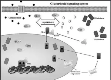
The human corticosteroid receptor (GR) is located in chromosome 5 and is responsible of the produc-tion of the subunit α and β. The classic receptor has three major distinct functional domains-the N-termi-nal or immunogenic domain, the DNA binding domain (DBD), and the ligand-binding domain (LBD). In its un-bound state, GRα is located primarily in the cytoplasm as part of a hetero-oligomeric complex that contains
heat shock proteins (HSPs) 90, 70, 50, and 20 and, possibly, other proteins as well7. After binding to an
agonist ligand, GR undergoes conformational chan-ges, dissociates from the HSPs and other chaperon proteins, and translocates into the nucleus through a nuclear pore as a monomer or dimer by means of an active adenosine triphosphate–dependent process mediated by its nuclear localization signals NL1 and NL2 7. Once in the nucleus, ligand-activated GRα
di-mers interact directly with specific DNA sequences in the promoter regions of target genes (the corticos-teroid response elements, GREs). Ligand-activated GRα monomers, interact with other transcription fac-tors also, at the cytoplasm level [e.g. nuclear factor
κB (NF-κB), activating protein 1 (AP-1), p53, CRE-bin-ding protein (CREB), signal transducer and activator of transcription 5 (STAT5)] through protein-protein interactions, indirectly influencing the activity of the latter on their own target genes [7]. GR contains two transactivation domains (activation function 1, AF-1 and AF-2) at its N-terminal and LBD, respectively. GR interacts through AF-1 and AF-2 with various protein complexes, including the nuclear receptor coactiva-tor complexes (p160, p300/CREB-binding protein, CBP, and p300/CBP-associated factor, p/CAF) and other factors that can alter the transcription rates of corticosteroid-responsive genes7 (Figure 2). The GR
isoform β does not itself bind corticosteroids, but
ther exerts dominant negative effects on GRα throu-gh several mechanisms, such as heterodimerization and competition with GRα for GREs or transcriptional nuclear receptor coactivators6,7,39-41. GRα translational
isoforms are differentially expressed in various cell li-nes42. There are multiple mechanisms through which
target cells alter the sensitivity and specificity of the response to corticosteroids. These take place at the levels of GR gene transcription, mRNA splicing, and mRNA translation, as well as through post-translatio-nal modifications and the inherent functiopost-translatio-nal activity of the expressed isoform monomer(s) or dimer(s) on responsive genes. N-terminal domain of GR subs-tantially contributes to this regulatory diversity42. The
mineralocorticoid (MR), estrogen (ER), and proges-terone43 receptors also contain potential alternative
translation initiation sites in their N-terminal domains. Thus, like the GR gene, the genes encoding these re-ceptors might produce N-terminal isoforms42.
There-fore, tissue-specific and regulated variable N-terminal isoform production may be a general mechanism that defines target tissue sensitivity to steroid hormones, further adding to the complexity of their own signal transduction systems.
It has been shown that corticosteroids upregulate and downregulate up to 2000 genes that are involved in re-gulation of the immune response44.
If cortisol is able to act in genomic manner it is also responsible for nongenomic interaction. Physioche-mical interactions occur in-between the cell’s mem-brane and corticosteriods inducing very rapid, nons-pecific, nongenomic effect45. For example during an
acute stress the loss in interaction with hypothalamic synaptosomes may partly explain the loss in cortisol circadian rhythm46. Moreover corticosteroids act in
this way about immediate catecholamine release from sympathetic cells, restoration of sympathetic modula-tion on heart and vessel47, sensitization to exogenous
catecholamine in septic shock48,49. In addition through
activation of endothelial nitric oxide synthase induces protective effects in ischemic brain injury50, and in
is-chemia/reperfusion injury51.
MAIN ACTIONS OF CORTICOSTEROIDS IN SEPSIS
Metabolic Effects
About metabolism of carbohydrates, corticosteroids play a major role in glucose metabolism stimulating liver gluconeogenesis and glycogenolysis also promoting the secretion of glucagon and adrenaline. They inhibit
cellular uptake of glucose by inducing peripheral insulin resistance. All these actions have as consequence a rise in blood glucose concentration52. About proteins
and fat metabolism, corticosteroids inhibit protein syn-thesis and activate proteinolysis in muscles, inhibit glu-cose uptake by adipocytes and activate lipolysis. Fi-nally they are involved in bone and mineral metabolism, activating osteoclasts, inhibiting osteoblasts, decrea-sing intestinal calcium uptake and increadecrea-sing calcium urinary secretion by decreasing its renal reabsorption1.
Immune Effects
Clinical relevance of corticosteroids in immune and inflammatory response is controversial; its affinity for immune cells is great. Its influence can realize through lymphocytes, natural killer (NK #0), monocytes, macro-phages, eosinophils, mast cells and basophils14.
Corti-costeroids administration is followed by a reduction of lymphocytes, which result in a passage of cells from blood to lymphoid organ. The opposite is true for granu-locytes. To the reduction of inflammatory reaction con-tribute the lowering of chemokines and subsequently of neutrophil migration. Macrophage secretion is inhibi-ted by the production of migration inhibitory factor [53], while eosinophils apoptosis is stimulated54.
Corticosteroids are involved in the immune response by inhibiting the production of IL-12 by macrophages and monocytes and influencing lymphocytes differen-tiations in favour of Th2 cells. All that would result in a rise of IL-4 and IL-13 secretion (which is normally inhibited by IL-12) and suppression of cellular immu-nity14. In patients with sepsis hydrocortisone does not
suppress IL-12 secretion, an effect that resulted from hydrocortisone induced inhibition of pro-inflammatory state55 (Figure 3).
Corticosteriods modulate the inflammatory respon-se also acting on the cellular level by the inhibition of proinflammatory cytokines (IL-1, IL-2, IL-3, IL-6, interferon-γ, TNF-α), chemochine, eicosanoids, bra-dykinins and inhibitory factor3,21,53. This is possible by
interaction of corticosteroid complex and DNA where it can active and inhibit transcriptional factors45.
In the last decade, in septic shock patients, corticos-teroids have been shown to attenuate the inflamma-tory response without causing immune suppression. Indeed, intravenous hydrocortisone decreased core temperature, heart rate and plasma levels of PLA2 and C-reactive protein56. Its decreased circulating
levels of IL-6 and IL-8, prevented the endothelial ac-tivation and also decreased circulating levels of TNF soluble receptors I and II, and IL-10. Hydrocortiso-ne is also able to favour the expansion of peripheral blood-derived CD56 (+) cells, allowing the differen-tiation of NK cells57.
Cardiovascular Effects
Corticosteroids regulate vascular reactivity to vaso-constrictors. Physiological concentrations of corti-costeroids play a key role in maintaining an appro-priate vascular reactivity to vasoconstrictors, hence the normal blood pressure. Multiple mechanisms are involved in corticosteroid-mediated vascular res-ponses. It is known that chronic excess in corticos-teroids induces hypertension, whereas AI induces hypotension. Corticosteroids act on vascular endo-thelial cells and inhibit the release of endoendo-thelial va-sodilators such as NO and PGI2 by downregulating enzyme expression and activity. In vascular smooth muscle cells, corticosteroids enhance agonist-me-diated pharmacomechanical coupling by increasing Ca++ mobilization and Ca++ sensitivity of myofilament.
The corticosteroid-induced vascular changes occur within minutes and thus are unlikely dependant on genomic interaction. The precise mechanism of cor-ticosteroids induced changes in the endothelium and vascular smooth muscle cells are not clear at pre-sent.
Administration of hydrocortisone simultaneously or just before infusion of lipopolysaccharide prevents vascular non responsiveness to noradrenaline in he-althy subjects, as well as hypotension, rise of both heart rate and plasma adrenaline levels. In septic shock, AI was associated with a marked hypores-ponsiveness to noradrenaline, which was fully rever-sed 1 h after 50 mg of intravenous hydrocortisone48.
Corticosteroids regulate response to noradrenaline and angiotensine II. The mechanism of action may be partially explained by iNOS pathways inhibition. Another study confirm ability of the corticosteroid to reverse vessels insensitivity within 1 h without rela-tionship with renin-angiotensin system or nitric oxi-de pathway49. Several placebo-controlled
randomi-zed studies emphasize the cardiovascular effects of low dose of corticosteroids (200-300 mg/day) for a prolonged period17,55,58-61. Hydrocortisone was
asso-ciated with increased vascular resistance with little effects on cardiac index and pulmonary hemodyna-mic. It may be possible that the very early hemody-namic effects are nongenomic, whereas subsequent ones are partially due to iNOS inhibition55.
These trials also showed that use of corticosteroids reduces duration of shock and time of varopressor weaning. The hemodynamic effect was confirmed by a phase III trial, that also demonstered survival benefit from a 7-day treatment with 50 mg of hydro-cortisone every 6 hrs and 50 µg of fludrohydro-cortisone per os daily30. The beneficial effect of
corticosteroi-ds is more pronounced in non responders to ACTH. In a more recent study, infusion of 50-mg bolus of hydrocortisone followed by a continuous infusion of 0.18 mg/kg /hr determined in 41 hyperdynamic sep-tic shock patients, was associated with a dramasep-tic reduction of vasopressor support (53 hrs vs. 120 hrs) ad duration of shock and of proinflammatory cytoki-nes. In this study the hemodynamic effects but not the immune effects of hydrocortisone were related to the adrenal status62.
Survival Effects
Data extracted from all more important trials (Table 1), shows mortality rate in control group and in tre-ated group not significantly different. But subgroup analysis on trials with long courses (≥ 5 days) of low dose corticosteroids (≤ 300 mg hydrocortisone or equivalent) showed a significant reduction in mortali-ty rate in favour of corticosteroids, at 28 days and at ICU and hospital discharge62.
Adverse Events
1.22, 0.84 to 1.78, p = 0.30)62. Only one trial reported
a rise in serum sodium concentration (>155 mmol/L) in 6/20 (30%) Patients in the treated group and in 1/20 (5%) in the placebo group60.
CONCLUSION AND RECOMMENDATIONS FOR CLINICAL PRACTICE
During sepsis, hemodynamic instability and perpetu-ation of inflammatory state may result from AI. Thus, an ACTH test should be performed as soon as possi-ble to identify non overt AI (Figure 4). It should be im-mediately followed by a replacement therapy with iv bolus of 50mg of hydrocortisone every 6 hours com-bined to 50 µg of fludrocortisone once daily. When the results of the ACTH test are available, treatment should be continued for 7 days in the non responders to ACTH and withdraw in the responders. Whether responders to ACTH with high baseline cortisol levels
(> 34 µg/dL) have tissue resistance to cortisol and also should receive exogenous hormones remains to be evaluated in clinical trials.
REFERENCES
01. Orth DN, De Bold CR – Williams Textbook of Endocrinology; em: Wilson JD, Foster DW - Williams Textbook of Endocrinology. Philadelphia: W.B. Saunders Company, 1992;489-531.
02. Chrousos G - The hypothalamic-pituitary-adrenal axis and immune-me-diated inflammation. N Engl J Med, 1995;332:1351-1362.
03. Cavaillon J - Action of glucocorticoids in the inflammatory cascade. Re-anim Urgences, 2000;9: 605-612.
04. Tsigos C, Chrousos G - Hypothalamic-pituitary-adrenal axis, neuroendo-crine factors and stress. J Psychosom Res, 2002;53:865-871. 05. Marik PE, Zaloga GP - Adrenal insufficiency in the critically ill: a new look
at an old problem. Chest, 2002;122:1784-1796.
06. Kino T, Chrousos G - Glucocorticoid and mineralocorticoid receptors and associated diseases. Essays Biochem, 2004;40:137-155.
07. Kino T, de Martino MU, Charmandari E et al - Tissue glucocorticoid resistance/hypersensitivity syndrome. J Steroid Biochem Mol Biol, 2003;85:457-467.
08. Chrousos GP, Charmandari E, Kino T - Glucocorticoid action networks-an introduction to systems biology. J Clin Endocrinol Metab, 2004;89:563-564. 09. Kino T, Mirani M, Alesci S et al - AIDS-related lipodystrophy/insuline
re-sistance syndrome. Horm Metab Res, 2003;35:129-136.
10. Kino T, Gragerov A, Slobodskaya O et al - Human immunodeficiency virus type 1 (HIV-1) accessory protein Vpr induces transcription of the HIV-1 and glucocorticoid-responsive promoters by binding directly to p300/CBP coactivators. J Virol, 2002;76:9724-9734.
11. Lamberts SW, Bruining HA, de Jong FH - Corticosteroid therapy in seve-re illness. N Engl J Med, 1997; 337:1285-1292.
12. Beishuizen A, Thijs LG, Vermes I - Patterns of corticosteroid--binding globulin and the free cortisol index during sepsic shock and multitrauma. Intensive Care Med, 2001;27:1584-1591.
13. Franchimont D, Martens H, Hagelstein MT et al - Tumor necrosis factor alpha decreases, and interleukin-10 increases, the sensitivity of human monocytes to dexamethasone: potential regulation of the glucocorticoid receptor. J Clin Endocrinol Metab, 1999;84:2834-2839.
14. Chrousos G - The stress response and immune function: clinical im-plications. The 1999 Novera H. Spector Lecture, Ann N Y Acad Sci, 2000;917:38-67.
15. McKee JI, Finlay WE - Cortisol replacement in severely stressed pa-tients. Lancet, 1983;1:(8322):484.
16. Cooper MS, Stewart PM - Corticosteroid insufficiency in acutely ill pa-tients. N Engl J Med, 2003;348: 727-734.
17. Annane D, Sebille V, Charpentier C et al - Effect of treatment with low
Table 1 – Corticosteroid Trials in Severe Sepsis and Septic Shock Patients
Reference Population Dose of Cortisone vs Placebo Results
Bollaert et al. (1998) Septic shock (41) 100 mg x 3 for 5 days of hydrocortisone
Reduction of mortality and shock reversal time Briegel et al. (1999) Septic shock (40) 100 mg + 0.18 mg/kg/hr of
hydrocortisone
Reduction of time of vasopressor infusion Chawla et al. (1999) Septic shock (44) 100 mg x 4 for 72 hr of
hydrocortisone
Reduction of shock reversal time and vasopressor infusion Annane et al. (2002) Septic shock (300) 50 mg x 6 of hydrocortisone +
fludrocortisone 50 µg for 7 days
Reduction of mortality and relative adrenal insufficiency Yildiz et al. (2002)63 Severe sepsis (40) Physiological dose of
prednisolone for 10 days
Reduction of mortality
Confalonieri et al. (2004)64 Severe sepsis (46) 200 mg + 10 mg/hr of
hydrocortisone
Reduction of mortality and delayed of septic shock Oppert et al (2005) Septic shock (41) 50mg + 0.18 mg/kg/hr of
hydrocortisone
Reduction of shock reversal time
Tandan et al (2005)65 Septic shock (28) Low doses of hydrocortisone Reduction of mortality
Biol, 2003;23:4319-4330.
42. Lu NZ, Cidlowski JA - Translational regulatory mechanisms generate N-terminal glucocorticoid receptor isoforms with unique transcriptional target gene. Mol Cell, 2005;18:331-342.
43. Bernard GR, Luce JM, Sprung CL et al - High dose corticosteroids in patients with the adult respiratory distress syndrome. N Engl J Med, 1987;317:1565-1570.
44. Galon J, Franchimont D, Hiroi N et al - Gene profiling reveals unknown enhancing and suppressive actions of glucocorticoids on immune cells. FASEB J, 2002;16:61-71.
45. Almawi WI - Molecular mechanisms of glucocorticoids effects. Mod Asp Immunobiol, 2001;2:78-82.
46. Edwardson JA, Bennett GW - Modulation of corticotropin-releasing fac-tor release from hypotalamic synaptosome. Nature, 1974;251:425-427. 47. Orlikowsky D, Castel M, Annane D - Acute effect of a single intravenous
bolus of 50 mg hydrocortisone on cardiovascular autonomic modulation in septic shock. Crit Care Med, 2003; 31:(Suppl):A124.
48. Annane D, Bellissant E, Sebille V et al - Impaired pressor sensitivity to noradrenaline in septic shock patients with and without impaired adrenal function reserve. Br J Clin Pharmacol, 1998;46:589-597.
49. Bellissant E, Annane D - Effect of hydrocortisone on phenylephrine - mean arterial pressure dose-response relationship in septic shock. Clin Pharmacol Ther, 2000;68:293-303.
50. Limbourg FP, Huang Z, Plumier JC et al - Rapid nontranscriptional ac-tivation of endothelial nitric oxide synthase mediates increased cere-bral blood flow and stroke protection by corticosteroids. J Clin Invest, 2002;110:1729-1738.
51. Hafezi-Moghadam A, Simoncini T, Yang Z et al - Acute cardiovascular protective effects of corticosteroids are mediated by non-transcriptio-nal activation of endothelial nitric oxide synthase Nat Med, 2002;8:473-479.
52. Pilkis SJ, Granner DK - Molecular physiology of the regulation of the he-patic gluconeogenesis and glycolysis. Annu Rev Physiol, 1992;54:885-909.
53. Beishuizen A, Thijs LG, Haanen C et al - Macrophage migration inhibitory factor and hypothalamo-pituitary-adrenal function during critical illness. J Clin Endocrinol Metab, 2001;86:2811-2816.
54. Beishuizen A, Vermes I, Hylkema BS et al - Relative eosinophilia and functional adrenal insufficiency in critically ill patients Lancet, 1999;353:1675-1676.
55. Keh D, Boehnke T, Weber-Cartens S et al - Immunologic and hemody-namic effects of “low-dose” hydrocortisone in septic shock: a double-blind, randomized, placebo-controlled, crossover study. Am J Respir Crit Care Med, 2003;167:512-520.
56. Briegel J, Kellermann W, Forst H et al - Low-dose hydrocortisone in-fusion attenuates the systemic inflammatory response syndrome. The Phospholipase A2 Study Group. Clin Investig, 1994;72:782-787. 57. Perez SA, Mahaira LG, Demirtzoglou FJ et al - A potential role for
hydro-cortisone in the positive regulation of IL-15-activated NK cell prolifera-tion and survival. Blood, 2005;106:158-166.
58. Oppert M, Schindler R, Husung C et al - Low-dose hydrocortisone im-proves shock reversal and reduces cytokine on early hyperdinamic sep-tic shock. Crit Care Med, 2005;33:2457-2464.
59. Bollaert PE, Charpentier C, Levy B et al - Reversal of late septic sho-ck with supraphysiologic doses of hydrocortisone. Crit Care Med, 1998;26:645-650.
60. Briegel J, Forst H, Haller M et al - Stress dose of hydrocortisone reverse hyperdynamic septic shock: a prospective, randomized, double-blind, single-center study. Crit Care Med, 1999;27:723-732.
61. Chawla K, Tessler S - Hydrocortisone reverses refractory septic shock. Crit Care Med, 1999;27:A33.
62. Annane D, Bellissant E, Bollaert PE et al - Corticosteroids for treating severe sepsis and septic shock: a systematic review and meta-analysis. BMJ,
2004;329:480-63. Yildiz O, Doganay M, Aygen B et al - Physiologic-dose steroid therapy in sepsis. Crit Care, 2002; 6:251-259.
64. Confalonieri M, Urbino R, Potena A et al - Hydrocortison infusion for se-vere community-acquired pneumonia: a preliminary randomized study. Am J Respir Crit Care Med, 2005;171:242-248.
65. Tandan SM, Gupta N – Low dose steroids and adrenocortical insuffi-ciency in septic shock: a double-blind randomised trial from India. Am J Respir Crit Care Med, 2005;[A24][Poster:326]
doses of hydrocortisone and fludrocortisone on mortality in patients with septic shock. JAMA, 2002;288: 862-871.
18. Rothwell PM, Lawler PG - Prediction of outcome in intensive care pa-tients using endocrine parameters. Crit Care Med, 1995;23:78-83. 19. Rothwell PM, Udwadia ZF, Lawler PG - Cortisol response to
corticotro-pin and survival in septic shock. Lancet, 1991;337:582-583.
20. Annane D, Sebille V, Troche G et al - A 3-level prognostic classification in septic shock based on cortisol levels and cortisol response to cortico-tropin. JAMA, 2000;283:1038-1045.
21. Soni A, Pepper GM, Wyrwinski PM et al - Adrenal insufficiency occurring during septic shock: incidence, outcome, and relationship to peripheral cytokine levels. Am J Med, 1995;98:266-271.
22. Sharshar T, Annane D, de la Grandmaison G et al - The neuropathology of septic shock. Brain Pathol, 2004;14:21-33.
23. Melby JC - Drug spotlight program: systemic corticosteroid therapy: pharmacology and endocrinologic considerations. Ann Intern Med, 1974;81:505-512.
24. Hall GM, Lacoumenta S, Hart GR et al - Site of action of fentanyl in inhibiting the pituitary-adrenal response to surgery in man. Br J Anaesth, 1990;65:251-253.
25. Absalom A, Pledger D, Kong A - Adrenocortical function in criti-cally ill patients 24 h after a single dose of etomidate. Anaesthesia, 1999;54:861-867.
26. Annane D - ICU physicians should abandon the use of etomidate! Inten-sive Care Med, 2005; 31:325-326.
27. Abel SM, Back DJ – Cortisol metabolism in vitro – III. Inhibition of mi-crosomal 6 beta-hydroxylase and cytosolic 4-ene-reductase. J Steroid Biochem Mol Biol, 1993;46:827-832.
28. Ushiama H, Echizen H, Nachi S et al - Dose-dependent inhibition of CYP3A activity by clarithromycin during Helicobacter pylori eradication therapy assessed by changes in plasma lansoprazole levels and par-tial cortisol clearance to 6 beta-hydroxycortisol. Clin Pharmacol Ther, 2002;72:33-43.
29. Oelkers W - Adrenal insufficiency. N Engl J Med, 1996;335:1206-1212. 30. Bernier J, Jobin N, Emptoz-Bonneton A et al - Decreased
corticosteroid-binding globulin in burn patients: Relationship with interleukin-6 and fat in nutritional support. Crit Care Med, 1998;26:452-460.
31. Cordido F, Alvarez-Castro P, Isidro ML et al - Comparison between in-sulin tolerance test, growth hormone (GH)-releasing hormone (GHRH), GHRH plus acipimox and GHRH plus GH-releasing peptide-6 for the diagnosis of adult GH deficiency in normal subjets, obese and hypopi-tuitary patients. Eur J Endocrinol, 2003;149:117-122.
32. Pfeifer M, Kanc K, Verhovec R et al - Reproducibility of the (ITT) for as-sessment of GH and cortisol responses in normal and hypopituitaric adult man. Clin Endocrinol (Oxf), 2001;54:17-22.
33. Nye E, Grice JE, Hockings GI et al - The insulin hypoglycemia test: hypo-glycemic criteria and reproducibility. J Neuroendocrinol, 2001;13:524-530. 34. Berneis K, Staub JJ, Gessler A et al - Combined stimulation pf
adreno-corticotropin and compound-S by single dose metyrapone test as an outpatient procedure to assess hypotalamic-pituitary-adrenal function. J Clin Endocrinol Metab, 2002;87:5470-5475.
35. Schmidt IL, Lahner H, Mann K et al - Diagnosis of adrenal insufficiency: evaluation of the corticotropin-releasing hormone test and basal se-rum cortisol in comparison to the insuline tolerance test in patients with hypotalamic-pituitary-adrenal disease. J Clin Endocrinol Metab, 2003;88:4193-4198.
36. Doe R, Fernandez R, Seal US - Measurement of corticosteroid-binding globulin in man. J Clin Endocrinol Metab, 1964;24:1029-1039. 37. Graybeal ML, Fang VS - Physiological dosimg of exogenous ACTH Acta
Endocrinol, 1985;108:401-406.
38. Dickstein G, Shechner C, Nicholson WE et al - Adrenocorticotropin sti-mulation test: effect of basal cortisol level, time of day and suggested new sensitive low dose test. J Clin Endocrinol Metab, 1991;72:773-778. 39. Bamberger CM, Bamberger AM, de Castro M et al - Glucocorticoid re-ceptor beta, a potential endogenous inhibitor fo glucocorticoid action in humans. J Clin Invest, 1995;95:2435-2441.
40. Charmandari E, Chrousos GP, Ichijo T et al - The human glucocorticoid receptor (hGR) beta-isoform suppresses the transcriptional activity of HGRalpha by interfering with formation of active coactivator complexes. Mol Endocrinol, 2005;19:52-64.