Abstract
Picrorhiza kurroais an important medicinal herb valued for iridoid glycosides, Picroside-I (P-I) and Picroside-II (P-I(P-I), which have several pharmacological activities. Genetic interven-tions for developing a picroside production platform would require knowledge on biosyn-thetic pathway and key control points, which does not exist as of today. The current study reports that geranyl pyrophosphate (GPP) moiety is mainly contributed by the non-mevalo-nate (MEP) route, which is further modified to P-I and P-II through phenylpropanoid and iri-doid pathways, in total consisting of 41 and 35 enzymatic steps, respectively. The role of the MEP pathway was ascertained through enzyme inhibitors fosmidomycin and mevinolin along with importance of other integrating pathways using glyphosate, aminooxy acetic acid (AOA) and actinomycin D, which overall resulted in 17%-92% inhibition of P-I accumulation. Retrieval of gene sequences for enzymatic steps from NGS transcriptomes and their expression analysis vis-à-vis picrosides content in different tissues/organs showed ele-vated transcripts for twenty genes, which were further shortlisted to seven key genes, ISPD, DXPS, ISPE, PMK, 2HFD, EPSPS and SK, on the basis of expression analysis between high versus low picrosides content strains ofP.kurroaso as to eliminate tissue type/ devel-opmental variations in picrosides contents. The higher expression of the majority of the MEP pathway genes (ISPD, DXPS and ISPE), coupled with higher inhibition of DXPR enzyme by fosmidomycin, suggested that the MEP route contributed to the biosynthesis of P-I inP.kurroa. The outcome of the study is expected to be useful in designing a suitable genetic intervention strategy towards enhanced production of picrosides. Possible key genes contributing to picroside biosynthesis have been identified with potential implications in molecular breeding and metabolic engineering ofP.kurroa.
Introduction
Picrorhiza kurroaRoyle ex. Benth is an important medicinal herb valued for its hepatoprotec-tive activity as well as other medicinal properties like malarial, inflammatory,
anti-OPEN ACCESS
Citation:Shitiz K, Sharma N, Pal T, Sood H, Chauhan RS (2015) NGS Transcriptomes and Enzyme Inhibitors Unravel Complexity of Picrosides Biosynthesis inPicrorhiza kurroaRoyle ex. Benth. PLoS ONE 10(12): e0144546. doi:10.1371/journal. pone.0144546
Editor:Keqiang Wu, National Taiwan University, TAIWAN
Received:September 4, 2015
Accepted:November 19, 2015
Published:December 11, 2015
Copyright:© 2015 Shitiz et al. This is an open access article distributed under the terms of the
Creative Commons Attribution License, which permits unrestricted use, distribution, and reproduction in any medium, provided the original author and source are credited.
Data Availability Statement:All relevant data are within the paper and its Supporting Information files.
Funding:The research work has been funded by Department of Biotechnology, Ministry of Science and Technology, Government of India. The funders had no role in study design, data collection and analysis, decision to publish, or preparation of the manuscript.
Competing Interests:The authors have declared that no competing interests exist.
oxidant, anti-bacterial, immune modulator, etc., which are attributed to the presence of iridoid glycosides, Picroside-I (P-I) and Picroside-II (P-II) [1]. The reckless collection of plant material from wild along with unorganized cultivation and low seed viability has led to the endangered status of this important medicinal herb.
Herbal drug formulations have been an integral part of Ayurvedic system of medicine for centuries. With an ever-increasing global demand for herbal medicine, there is not only a demand for large quantity of raw material of medicinal plants, but also of appropriate quality where active compounds are present in desired concentrations [2].P.kurroais used in a num-ber of commercially available drug formulations like livocare, livomap, livplus, katuki, arogya, etc. for different disorders containing combinations of P-I and P-II in different concentrations [1]. P-I and P-II possess different medicinal properties individually as well as in combination and are, therefore, two major constituents ofP.kurroahaving therapeutic importance in sev-eral herbal drug formulations [3]. P-I is reported to be antimicrobial [4] and used against hepa-titis B [5]. P-II possess different pharmacological activities such as antiapoptotic [6],
neuroprotective [7], anti-inflammatory [8], anti-oxidant [9] and prevents myocardial ischemia reperfusion injury [10]. The proper concentration and ratio of P-I and P-II are, therefore, important in determining the quality and efficacy ofP.kurroa-based herbal drug formulations.
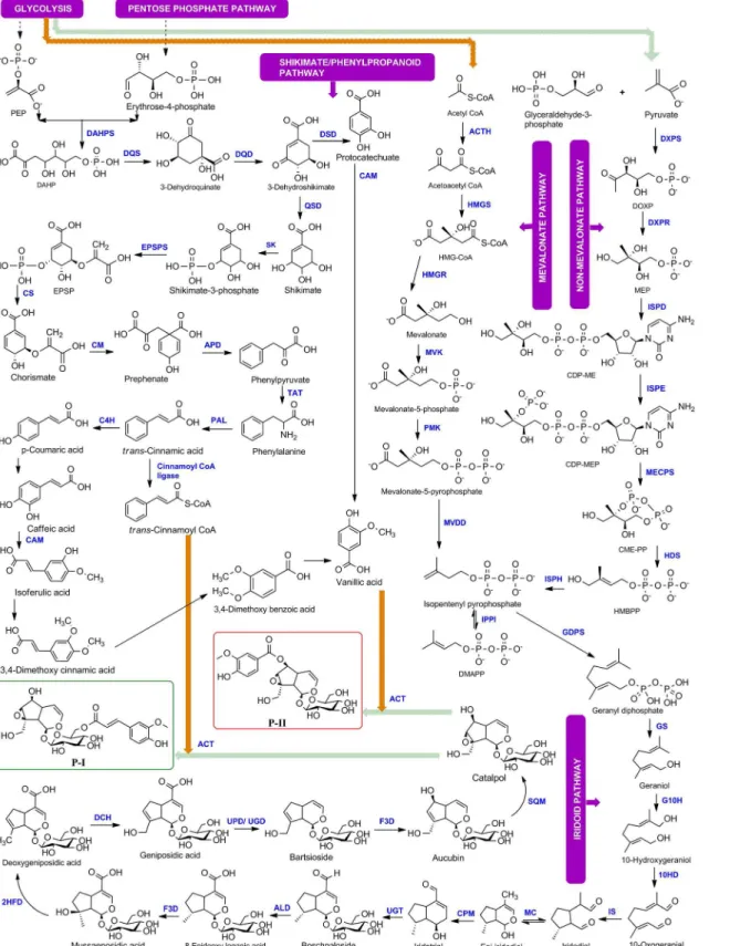
The biosynthesis and accumulation of P-I and P-II occur differentially in different tissues of
P.kurroa. The biosynthesis of P-I occurs in shoots and P-II in roots or stolons whereas both accumulate in rhizomes [11,12]. Biosynthesis of picrosides occurs through a combined biosyn-thetic route involving non-mevalonate (MEP), mevalonate (MVA), phenylpropanoid and iri-doid pathways. Picrosides are monoterpenoids with iriiri-doid backbone and glycoside moiety. Picrosides are classified as P-I and P-II based on functional group moieties; P-I having cinn-mate moiety and P-II having vanillate moiety [13], both derived from phenylpropanoid path-way. Iridoid backbone is derived from geranyl pyrophosphate (GPP) which is synthesized by head to tail condensation of isopentenyl pyrophosphate (IPP) and its allelic isomer dimethylal-lyl diphosphate (DMAPP) via cytosolic mevalonate (MVA) and/or plastidic non-mevalonate (MEP) pathway [14,15]. Biosynthesis of picrosides involves the synthesis of iridoid moiety from GPP through series of oxidation and cyclization steps followed by condensation of glu-cose moiety and cinnamate/vanillate from phenylpropanoid pathway [16,17]. The complete biosynthetic pathway of picrosides has been deciphered for all possible intermediates [17], however, the role and contribution of corresponding genes catalysing the enzymatic steps are not known. There are 41 steps (15 from MEP and MVA pathway, 14 from iridoid pathway, 11 from phenylpropanoid pathway, 1 involved in esterification of catalpol) and 35 steps (15 from MEP and MVA pathway, 14 from iridoid pathway, 5 from phenylpropanoid pathway, 1 involved in esterification of catalpol) involved in the biosynthesis of P-I and P-II, respectively. The cinnamate/vanillate moieties are first CoA activated and then transferred to the catalpol for the formation of respective iridoids. Out of 35 steps, 32 till 3-dehydroshikimate are com-mon for both P-I and P-II. After that P-II pathway is diverted for production of vanillic acid (4-hydroxy-3-methoxybenzoic acid) and P-I pathway is diverted for production of cinnamic acid. The final step involved in the esterification of catalpol (iridoid backbone) for the forma-tion of P-I and P-II is common. An alternative route for the formaforma-tion of vanillic acid by degra-dation of ferulic acid has also been reported inVanilla planifolia[18] andPseudomonas fluorescens[19]. Various studies have reported the partial biosynthetic pathway for picrosides along with few enzymatic steps. Kawoosa et al [16] reported 15 steps of MEP and MVA path-way with their corresponding enzymes but intermediate steps from GPP till the formation of picrosides were missing. Two genes of phenylpropanoid pathway (4-CH and 3-CH) and involvement of CYPs and glycosyltransferases in picrosides biosynthesis was also reported [20]. Singh et al [13] cloned 8 genes of the MEP and MVA pathways and reported two reductase; MVK, Mevalonate kinase; PMK,
way is the main source of universal terpenoid precursor isopentenyl diphosphate (IPP) for biosynthesis of taxanes inTaxus baccata. The major involvement of mevalonate pathway for shikonins biosynthesis inArnebia euchromahas also been proved through inhibitor assays [24]. However, any such studies has not been taken up inP.kurroafor identification of major contributing pathway for picrosides among various integrating pathways.
Present work reports on ascertaining the contribution of MVA and/or MEP route in the biosynthesis of P-I through enzyme inhibitor experiments. Also, genes catalysing the enzy-matic steps were mapped to iridoid branch of the picrosides biosynthetic pathway which were not known inP.kurroa. The availability of NGS transcriptomes of differentP.kurroatissues enabled the selection of appropriate paralogs for pathway genes. Expression analysis of all genes involved in the complete biosynthetic pathway was carried out in four different tissues of
P.kurroawith varying contents of P-I (0.0% and 2.7%) and P-II (0.0% and 0.4%) to associate key genes involved in picrosides biosynthesis. Further, to ascertain the involvement of genes in picrosides biosynthesis as well as to eliminate the effect of tissue type/developmental stage vis-à-vis picrosides contents, expression analysis of pathway genes was also assesed in high versus low P-I content strains ofP.kurroa.
Results
Mapping genes to enzymatic steps of iridoid branch of the pathway
The first seven enzymatic steps GS, G10H, 10HGO (10HD), IS, MC, CPM, UGT of iridoid pathway have recently been identified during this study in medicinal plant species such asP.
reaction is involved in the conversion of geniposidic acid to bartsioside. The thirteenth step is the conversion of bartsioside to aucubin and this reaction is similar to ninth step, which is being catalyzed by a similar enzyme flavanone 3-dioxygenase/hydroxylase (F3D). Fourteenth step is conversion of aucubin to catalpol which is catalyzed by an epoxidase or monooxygenase enzyme. We searched for all the monooxygenases and epoxidases inP.kurroatranscriptomes and identified two enzymes squalene monooxygenase (SQM) and squalene epoxidase (SQE) involved in secondary metabolism but squalene epoxidase did not show significant expression w.r.t. P-I or P-II contents. The last step in the formation of P-I and P-II from catalpol involves acyl group transfer reaction and we predicted this reaction to be catalyzed by anthocyanin acyl-transferase (ACT). The complete biosynthetic pathway with corresponding enzymatic steps is shown inFig 1.
Identification of appropriate paralogs/isoforms
NGS transcriptomes were generated from differentP.kurroatissues varying in picrosides con-tent mentioned in our previous work [27]. The assembly statistics for transcriptome datasets is given inS1 Table. Mining of transcriptomes for picrosides biosynthetic pathway genes showed the existence of multiple paralogs of these genes. The appropriate paralog for each gene was identified and selected for expression analysis using the methodology described in methods section. The paralogs selected for pathway genes in different shoot transcriptomes ofP.kurroa
are given inS2 Table.
Expression analysis of P-I and P-II biosynthetic pathway genes vis-
à
-vis
picrosides content
The results are in continuation to our previous work which reported the expression status of 15 genes of MEP and MVA pathway in different tissues ofP.kurroa[21]. Expression analysis of remaining genes of the biosynthetic pathway (15 genes from iridoid pathway and 12 from phe-nylpropanoid pathway) was done in four tissues varying for P-I and P-II contents. Majority of the genes showed higher expression in relation to picrosides content. Four genes of iridoid pathway (GS, 2HFD, DCH and SQM) and two genes of phenylpropanoid pathway (DQS and TAT) showed elevated levels of transcripts (~12–130 folds) in FGS having 2.7% P-I content w. r.t. TCS having negligible (0.01%) P-I. Two genes of iridoid pathway (F3D and ACT) and six genes of phenylpropanoid pathway (DQD, QSD, CAM, SK, EPSPS and PAL) showed ~16–96 folds expression in FGR having 0.4% P-II content wr.t. TCR having no P-II content. Four genes of iridoid pathway (G10H, CPM, ALD and UPD/UGD) and two genes of phenylpropa-noid pathway (CM and APD) showed ~12–42 folds higher expression in FGS as well as FGR w.r.t. their tissue cultured counterparts. The difference in fold expression in field grown tissues in comparison to tissue culture grown for iridoid and phenypropanoid pathways is given inFig 2a, 2b, 2c and 2d. The overall expression status of pathway genes w.r.t. P-I and P-II is given in Fig 3. The genes which showed above 10 folds transcript abundance in initial screening w.r.t. either P-I or P-II or both (including MEP/MVA pathway genes from our previous study) were checked for their expression status on shoot tissues of high versus low P-I content strains ofP.
kurroa. A high P-I content strain, PKS-1 (2.7%) and a low content strain, PKS-4 (0.3%) were used in comparative expression analysis. This resulted in the identification of seven genes, ISPD, DXPS, ISPE, PMK, 2HFD, EPSPS, SK from the complete biosynthetic pathway with ~5–
57 folds higher transcript abundance in high content strain compared to low content strain of
Fig 1. Complete biosynthetic pathway for Picroside-I and Picroside-II ofPicrorhiza kurroa.
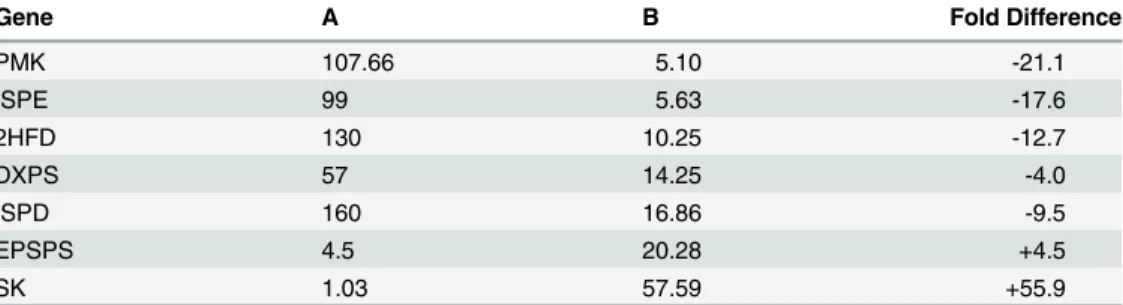
different tissue samples. The expression level of three genes of MEP pathway ISPD, DXPS and ISPE decreased from 160, 57 and 99 folds to 16.8, 14.2 and 5.6 folds, respectively in high con-tent strain. The expression ofPMKgene of MVA pathway decreased from 107.6 fold to 5.1 fold. 2HFD of iridoid pathway decreased from 130 to 10.2 fold (Table 1). The relative expres-sion status of pathway genes between high versus low content strains thus provided a realistic association with the biosynthesis of picrosides.
Fig 2. Expression status of iridoid (a, b) and phenylpropanoid (c, d) pathway genes in field grown tissues (FGS: Field grown shoots having 2.7% P-I and FGR: Field grown roots having 0.4% P-II) w.r.t. tissue cultured shoots (TCS having 0.01% P-I) and roots (TCR having 0.0% P-II).
doi:10.1371/journal.pone.0144546.g002
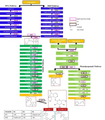
Fig 3. Expression status of all genes depicted as fold increase, for P-I and P-II biosynthetic pathways inPicrorhiza kurroa: MVA and MEP pathway genes data from Pandit et al. (2012).
Effect of enzyme inhibitors on biosynthesis of Picroside-I
The inhibition profile of five inhibitors was assessed at 15thand 30thday of culturingP.kurroa
shoots by quantifying P-I content (Fig 5) because cultured shoots are reported to accumulate only P-I [11]. Out of five inhibitors, mevinolin, fosmidomycin, glyphosate and AOA are com-petitive inhibitors which are highly specific for their target enzymes while actinomycin D is a transcriptional inhibitor. The best inhibitory concentrations were 5μM for mevinolin and
acti-nomycin D, 150μM for fosmidomycin, 4 mM for glyphosate and AOA. Mevinolin was found
to be least effective in inhibiting P-I biosynthesis as it showed maximum inhibition of only 17% and 11.6% at 15thand 30thday, respectively. Other inhibitors showed upto 92% inhibition in P-I content in comparison to control. All inhibitors showed higher inhibition at 30thday in comparison to 15thday, except mevinolin which unlike others showed higher inhibition at 15th day. At their best inhibitory concentrations, fosmidomycin, glyphosate and AOA showed a drastic reduction of 85.3%, 87.3% and 64.6% respectively in P-I accumulation at 15thday and reduction of 90.6%, 92.9% and 63.4%, respectively at 30thday. Fosmidomycin, glyphosate and AOA were thus found to be potent inhibitors of P-I biosynthesis. Actinomycin D moderately inhibited P-I biosynthesis inP.kurroaas it showed 47.9% and 61.2% inhibition at 15thand 30th day, respectively.
Gene expression in response to inhibitors
To see the effect of inbitor treatments on target gene transcripts, expression analysis was car-ried out on inhibitor treated shoot tissues in comparison to untreated control for related genes Fig 4. Expression pattern of key genes in shoot tissues of high (PKS-1) versus low (PKS-4) Picroside-I content accessions ofPicrorhiza kurroa.
doi:10.1371/journal.pone.0144546.g004
Table 1. Folds difference in expression status of key genes between A (FGS vs TCS) and B (PKS-1 vs PKS-4).
Gene A B Fold Difference
PMK 107.66 5.10 -21.1
ISPE 99 5.63 -17.6
2HFD 130 10.25 -12.7
DXPS 57 14.25 -4.0
ISPD 160 16.86 -9.5
EPSPS 4.5 20.28 +4.5
SK 1.03 57.59 +55.9
(DXPR, HMGR, EPSPS and PAL) alongwith upstream and downstream genes (1 each). The expression analysis was carried out at the best inhibitory concentrations of 150μM
(fosmido-mycin), 5μM (mevinolin), 4 mM (glyphosate and AOA), and 5μM (actinomycin D) at 15th
and 30thday. The inhibitors fosmidomycin, mevinolin, glyphosate and AOA did not show sig-nificant reduction in the transcript levels of their target steps or upstream/downstream genes. Fosmidomycin showed 1.25–2.5 folds reduction in transcript levels in comparison to control at both 15thand 30thday. Mevinolin, glyphosate and AOA also showed similar gene expression pattern with mevinolin (1–3.5 folds), glyphosate (1–4.7 folds) and AOA (1–4 folds) reduction in transcript levels as compared to control (Fig 6). Actinomycin D showed somewhat higher reduction in transcripts from 1 to 17.5 folds (Fig 7).
Discussion
Through this study MEP pathway has been identified as the major contributor of geranyl pyro-phosphate (GPP), the iridoid backbone for picrosides biosynthesis inP.kurroa. After GPP bio-synthesis, iridoid and phenylpropanoid pathways play equally important role in picrosides biosynthesis by providing catalpol and cinnamate/vanillate moieties, respectively. The impor-tance of iridoid and phenylpropanoid pathways has been justified by precursor feeding studies inP.kurroa, where exogenous feeding of catalpol and cinnamic acid showed that both path-ways act in conjunction for increased production of P-I inP.kurroa[Kumar et al. 2015, unpublished].
Current study is an initial endeavor towards deciphering the complete biosynthetic pathway including corresponding enzymatic steps for picrosides biosynthesis inP.kurroa. The identifi-cation of enzymatic steps of iridoid branch of the pathway, not known earlier has been done, however functional characterization is required to support their proposed functions inP. kur-roa. The availability of NGS transcriptomes of differentP.kurroatissues enabled the selection of appropriate paralogs for pathway genes as multiple paralogs were present for these genes. The higher expression of gene transcripts for corresponding enzymes vis-à-vis P-I and P-II contents and their involvement in secondary metabolism in other plant species suggested their Fig 5. Inhibition profiles of enzyme inhibitors on Picroside-I biosynthesis inPicrorhiza kurroa.
possible role in catalysing the required enzymatic reactions in picrosides biosynthesis. Alde-hyde dehydrogenase is reported to be involved in the biosynthesis of ferulic acid and sinapic acid inArabidopsis thaliana[28]. Flavanone 3-dioxygenase/hydoxylase (F3D) is involved in flavanoids biosynthesis in many plant species likeGinkgo biloba,Camellia sinensis[29,30]. 2-hydroxyisoflavanone dehydratase is involved in isoflavone biosynthesis inLotus japonica
[31]. Deacetoxycephalosporin-C hydroxylase is involved in cephalosporin biosynthesis [32]. Uroporphyrinogen decarboxylase is associated with the activity of enzymes involved in tetra-pyrrole biosynthesis and pathogen defense response inNicotiana tabacum[33,34]. Squalene epoxidase/monooxygenase is involved in ginsenoside biosynthesis inPanax ginseng[35,36] and triterpenoids inUncaria tomentosa[37]. Anthocyanin acyltransferases catalyze regiospeci-fic acyl transfer from acyl-CoA to the sugar moiety of anthocyanins [38] and are involved in anthocyanin biosynthesis in plant species [39]. The identified enzymes belong to cytochrome P450 family which possess broad substrate specificity i.e. same enzyme can bind to different substrates but enzyme kinetics vary considerably from one substrate to another. Therefore, these enzymes inspite of preferably using other substrates can also catalyse the similar reactions for picrosides biosynthesis using pathway intermediates as their substrates.
The expression analysis revealed that most of the genes of picrosides biosynthetic pathway had relatively higher expression in field grown tissues ofP.kurroacontaining higher amounts of P-I or P-II contents compared to tissue culture grown plants with negligible or no picrosides content at all. Genes, GS, G10H, CPM, ALD, F3D, 2HFD, DCH, UPD/UGD, SQM, ACT from Fig 6. Effect of inhibitors, fosmidomycin, mevinolin, glyphosate and AOA on transcript levels of target as well as upstream and downstream genes in Picroside-I biosynthesis inPicrorhiza kurroa.
iridoid pathway and DQS, DQD, QSD, SK, EPSPS, CM, APD, TAT, PAL from shikimic acid/ phenylpropanoid pathway showed significantly higher folds expression vis-à-vis picrosides content. Various studies have reported expression of multiple genes of biosynthetic pathways to be positively correlated with the terpenoid biosynthesis; shikonins inArnebia euchroma
[24], artemisinin inArtemisia annua[40], MIAs inCatharanthus roseus[41], flavonoids bio-synthesis inFagopyrumspecies [42], lignin biosynthesis inArabidopsis thaliana[43]. Geraniol synthase (GS) is an important enzyme which initiates monoterpenoid branch of the pathway inCatharanthus roseus[44] and G10H is reported to be a rate-limiting enzyme for biosynthesis of TIAs inCatharanthus roseus[45] alongwith its importance in iridoid monoterpenoid swer-tiamarin biosynthesis inSwertia mussotii[46]. EPSPS, SK and PAL are also reported to be important regulatory enzymes of shikimate/phenylpropanoid pathway [47–49]. In initial anal-ysis, multiple genes showed higher expression for P-I or P-II or both. But the differential expression in field grown tissues in comparison to tissue cultured plants might be due to tissue type or environmental variations. Therefore, to ascertain whether the elevated levels of tran-scripts of pathway genes are only affecting the biosynthesis of picrosides uniquely, the expres-sion status of genes was further studied between shoots ofP.kurroagenotypes that were varying for Picroside-I. High and low Picroside-I content genotypes ofP.kurroa(one each) grown for 3 years in the controlled environment in greenhouse were used for comparative expression analysis so as to reflect genetic differences contributing to the increase or decrease of gene transcripts rather than tissue type or developmental stage. It was observed that most of the genes did not show significant difference in expression between high versus low content Fig 7. Effect of actinomycin D on expression of significant genes of Picroside-I biosynthetic pathway inPicrorhiza kurroa.
key role in the regulation of isoflavone biosynthesis as its overexpression resulted in accumula-tion of daidzein and genistein inLotus japonicas[31]. Majority of these key genes belonged to MEP pathway. Also higher expression of two genes of shikimic acid/phenylpropanoid pathway and one gene of iridoid pathway, among seven genes, highlights the importance of these mod-ules of the biosynthetic pathway. This suggests that each module of the pathway is important in contributing to the structures of parental compounds, P-I and P-II inP.kurroa.
Five inhibitors targeting important enzymatic steps of the picrosides biosynthetic pathway were selected to assess their effect on picrosides accumulation. The inhibitor concentrations were chosen based on previous reports [56–59], [23]. Mevinolin and fosmidomycin are highly specific inhibitors of MVA (HMGR) and MEP (DXPR) pathways, respectively [60,61]. These two inhibitors were selected to rule out whether MVA and/or MEP pathway contributes in picrosides biosynthesis. Fosmidomycin produced drastic inhibition of upto 90.6% in P-I accu-mulation whereas mevinolin resulted in slight (17%) inhibition. This suggested that the MEP pathway plays major role in the production of GPP, the precursor for iridoid backbone biosyn-thesis. Picrosides are monoterpenoids and our results are in accordance with the previous find-ings that monoterpenoids have non-mevalonate (plastidial) origin and monoterpenoid synthases are localized to plastids [62]. It has also been reported by Eisenreich et al. [63] that MEP pathway is a predominant contributor for monoterpenoid biosynthesis, however, cross-talk occurs between two pathways [64]. Our previous reports also suggested the predominant role of MEP pathway in picrosides biosynthesis as majority of the MEP pathway genes were highly expressed in relation to picrosides content [21]. Sood and Chauhan [11] have also highlighted the importance of plastids (chloroplasts) by showing that the biosynthesis of Picro-side-I occurs only inin vitrocultured leaf and stem segments but not in undifferentiated callus cultures.
The other inhibitors, glyphosate and AOA were selected targeting the shikimate/phenylpro-panoid pathway enzymes whereas actinomycin D is a common transcriptional inhibitor. Glyphosate, a broad spectrum herbicide which competitively inhibits EPSPS [65] and AOA acts as an inhibitor of PAL [58] which is an important regulatory enzyme in phenylpropanoid pathway. Shikimic acid/phenylpropanoid pathway has a major contribution in picrosides bio-synthesis, thereby, providing cinnamate and vanillate moieties for P-I and P-II, respectively. The decrease in P-I biosynthesis by inhibiting shikimic acid/phenylpropanoid pathway enzymes confirms major contribution of this component of the pathway in picrosides biosyn-thesis inP.kurroa. Glyphosate is reported to inhibit secondary metabolites content in soyabean and buckwheat [57,66]. AOA treatment resulted in decreased accumulation of phytoalexins in banana [59] and 2-hydroxy-4-methoxybenzaldehyde inHemidesmus indicusroots [67].
our case. Therefore, most of it is getting used up till 15thday, hence not showing significant inhibition at 30thday.
The inhibitor treatment thus resulted in decrease in picrosides biosynthesis. We further looked at whether the expression of biosynthetic pathway genes, which are targets of corre-sponding inhibitors, is getting affected or not. Competitive inhibitors did not show significant decrease in expression of transcripts because these might be affecting the genes only at enzy-matic level but are not producing any effect at the transcriptional level. It has been reported in
Arabidopsisthat inhibitor treatment did not show significant effect on the expression of genes involved in sterol, chlorophyll and carotenoid metabolism, thereby indicating that posttran-scriptional processes might be playing an important role in regulating the flux through isopren-oid metabolic pathways [68].
Conclusion
The knowledge of complete pathway and corresponding genes would be helpful in understand-ing molecular basis of picrosides biosynthesis as well as plannunderstand-ing genetic improvement strate-gies for enhancing picrosides content inP.kurroa. Inhibitor experiments revealed that MEP pathway is a major contributor of GPP for picrosides inP.kurroaand picrosides biosynthesis is regulated at various control points in different modules of the biosynthetic route. Therefore, common regulatory elements for multiple genes need to be identified so as to ease genetic interventions for enhancing picrosides content. This is the first report wherein possible key genes have been identified in picrosides biosynthetic pathway which after further functional validation can have potential implications in molecular breeding and metabolic engineering.
Materials and Methods
Plant material
P.kurroaplants were procured from the nursery of Himalayan Forest Research Institute, Jagat-sukh, Manali, H.P., India (1,900 m altitude, 20°35.60–32°6.10N and 78°57.80–77°33.70E) and
maintained in the greenhouse of Jaypee University. These plants were cultured and maintained in an optimized Murashige and Skoog (MS) medium [69] supplemented with 3 mg/L indole-3-butyric acid and 1 mg/L kinetin in a plant tissue culture chamber maintained at 25±2°C with 16 h photoperiod provided by cool white fluorescent light (3,000 lux) (Sood and Chauhan 2010). FourP.kurroatissues with varying picrosides content growing in field conditions and tissue culture were taken for the study (Table 2, [21]). These included field grown shoots hav-ing 2.7% P-I (FGS), tissue cultured shoots havhav-ing negligible P-I content (TCS), field grown roots having 0.4% P-II content (FGR) and tissue cultured roots with no P-II content (TCR). Two strains ofP.kurroawith high and low content viz. PKS-1 (P-I 2.7%) and PKS-4 (P-I 0.3%) maintained in controlled environment in green house were also taken.
Identification of unknown enzymatic steps
qRT-PCR in different tissues ofP.kurroawith varying contents of P-I and P-II. The nucleotide sequences for phenylpropanoid and iridoid pathway genes were extracted from whole genome transcriptomes ofP.kurroaand primers were designed using online tool Primer3.
RNA isolation and cDNA synthesis
Total RNA was isolated fromP.kurroatissues by using RaFlex™total RNA isolation kit (GeNei™) by following manufacturer’s instructions. The quality of RNA was checked by 1% (w/ v) ethidium bromide-stained agarose gel. RNA was quantified in a NanoDrop spectrophotom-eter (Thermo Scientific) by measuring absorbance at 260nm and 280nm and 1μg RNA was
used for cDNA synthesis. cDNA was synthesized using Verso cDNA synthesis kit (Thermo Sci-entific) according to manufacturer’s protocol. Concentration of each cDNA sample was adjusted to 100ng/μl for qRT-PCR.
Identification of appropriate paralogs/isoforms for pathway genes in
transcriptomes
NGS transcriptomes forP.kurroawere generated and analysed using the methodology repre-sented inS1 FigMultiple paralogs of the genes were present inP.kurroatranscriptomes, therefore appropriate paralogs/isoforms were identified for each gene. For selection of suitable paralogs of genes, initially multiple sequence alignment of the selected transcripts using Clus-talW was carried out to see the homology within paralogs for each gene. Further, shortlisting of transcripts was done through BLAST analysis by looking for highest similarity with func-tionally characterized sequences (retrieved from the NCBI databases which were funcfunc-tionally characterized in the same plant or in different plant species). Thereafter, the transcripts were selected on the basis of transcript abundance (FPKM values), in different transcriptomes gen-erated fromP.kurroatissues varying in picrosides contents. The selected paralogs were fur-ther validated through qRT-PCR analysis under differential conditions of picrosides biosynthesis.
Expression analysis using quantitative real time PCR (qRT-PCR)
Table 3. Gene specific primers used for qRT-PCR.
Genes FP RP Annealing Temp (°C) Fragment size (bp)
26S CACAATGATAGGAAGAGCCGAC CAAGGGAACGGGCTTGGCAGAATC 58 500
GAPDH TTGCCATCAATGACCCCTTCA CGCCCCACTTGATTTTGGA 56 215
ACTH AGTGTTACTAGAGAGGAGCAGGACA CCTAGACCTTCATCCTTATCAACAA 50 110
HMGS GATGGTGCAAGAAAAGGCAACTAGA GGATATTCACTGGCAAGATTGGGCT 54 110
HMGR CGTTCATCTACCTTCTAGGGTTCTT GACATAACAACTTCTTCATCGTCCT 60 100
MVK ATTAACTCTGAGTATGACGGGTCTG GAGAGCCCATTTATTTAGCAACTC 50 110
PMK TGGATGTTGTCGCATCAGCACCTGG GTAATAGGCAGTCCACTCGCTTCAA 58 100
MVDD GTAACTCTGGATCCTGACCACCT TAATACCCCCTCTTTTTCATCCTC 54 100
IPPI TCTCCTATTCACTGTAAGGGATGTT ACCACTTAAACAAGAAGTTGTCCAC 54 110
GDS GATATATGTTCTGAGGGAATGGATG ATACACCTAGCGAAATTCCTCAACT 55 110
DXPS ACATTTAAGTTCAAGTCTGGGAGTG ATGTGCACTCTCTTCTCTTTTAGGA 55.9 110
DXPR GGAGGAACTATGACTGGTGTTCTT CAGGTCATAGTGTACGATTTCCTCT 54.9 110
ISPD GAGAAAAGTGTATCTGTGCTTCTTAG AATAACCTGCGGTGTATGCATTTCC 56 150
ISPE TTCATCTAGATAAGAAGGTGCCAAC CCTCTACCAGTACAATAAGCAGCTC 55 110
MECPS ATCTATAGCGGCAAACCTACAC ACTTTAGAGAGGGATGGAGGG 57.1 110
HDS ATGCCATTTAAGGAACTTGCAACAG GGAGCACCACCAACATATCCAAATT 58 110
ISPH CATACTGGGTTGACAGTGATGTAAG TAAGGACATCTTCAACAGCCTTATC 57.2 110
GS TGGGTAGATTAGAAGCCAGA CTGGTGATTTCTACCAGCTC 52 139
G10H TATCGAGCTTTTCAGTGGAT GATGTGAGTCCTGTCGATTT 52 136
10HD GGTAGTGTTTATTGGTGCAG GATCAACTGATCAAGGTCAA 54 172
IS AATAAGGCCTTGGTTTATCC TTAGCCTTAGGATCAACTGC 49 116
MC AAGGCTGCTCGTACCGATAA AATGGGGCAGTAGAGTGGTG 58.5 132
CPM ACCTGAGAGATGGCTAGATG AGTTCACAGGTTTGTGTTCA 53 188
UGT AATGGTTGGACTCACAAGAG ACAAGAAGGTCTGGTTGCTA 55 126
ALD ACTTTGCTGTGGGATTAAGT TTCCACAAACTAGAGCAATG 57 174
F3D AAGAATATGGCTTCTTCCAG ATCCCTCCAATTATGAACAG 55 197
2HFD ATTTACTCGACAGGATGTGG AGCACAAATCAACACCTTCT 52 134
DCH CATTTCACATCTCCACTGAG ATGGACAAAGCTTCTTTAGC 53 194
UPD CTGACGGCGTTATTATTTTC TCTTCAGAACGGATAGGAGA 51 115
UGD CATGTTGGACCTTTCAATCT TGGGCCTGAACTCTATCTTA 51 109
SQM GTTGATATACCCGGTCAGAA TTTCTCAACAGCAGACATGA 51 126
ACT TATGTCCAAGAATCCAGTGT AAGAACTCGTCTCGTTCAA 55 111
DAHPS ACACCATTAAAGCTCCTTGT TAACAGTCTGAGATCCACCA 59 171
DQS TGTTCAAAGTGTTGGTGTCT GTGACATGTGTGGATATGCT 54 192
DQD AAAAACAACCAGCTTCAAAT TCTTTGAAACACAATCGAAA 55 193
CAM GAAGATGCTCCTTCTTATCC AACACTCGACCAGAATCAC 53 187
QSD CATAGTTTGGTTCCTCCTCT ACTGTCTTTCTAGCCCAGAT 52 186
SK TATGGTGAGAGCTTCTTCAG AGTTCCGACAGCTGTAATC 56 195
EPSPS CACAGAACTCAGAAAGTTGG AGTAATCAGGGAAGGTCTTG 48.5 203
CS CTCGCTACCAGTCATAAAAG GAGTGTGTGGTGATTCTGTT 49 185
CM GTCTACACACCTGCCATTAG GTACAAATCAGCAACTAGGC 52 198
APD GCGTATTGGCTATAGAACTG CGACCTCTGAGAGTAGTTGA 50 192
TAT CTACATTGAGATCGACTTCC ACTATCTCCTGAGGACGTTT 53 206
PAL GCAAGATAGATACGCTCTAA GTTCCTTGAGACGTCAAT 49 136
C4H GCAACATTGATGTTCTCAAC TCCAGCTCTTCAAGGACTAT 53 169
Five inhibitors Mevinolin (HMGR), Fosmidomycin (DXPR), Glyphosate (EPSPS), Actinomy-cin D (transcriptional inhibitor) and Aminooxyacetic acid-AOA (PAL) inhibiting different enzymatic steps (mentioned above in braces) of MEP, MVA and phenylpropanoid pathways were purchased from Sigma-Aldrich, USA. To prepare stock solutions mevinolin and actino-mycin D were dissolved in DMSO, rest of the inhibitors were dissolved in autoclaved distilled water and filter sterilized using 0.22μm sterile filters (Millipore). Shoot apices of length 1 cm
were cut from six weeks old cultures ofP.kurroashoots raisedin vitroat 25°C and cultured in test tubes containing 10 ml agar gelled MS media containing different concentrations of inhibi-tors. Inhibitors were added separately at three different concentrations as follows: Mevinolin and actinomycin D (2.5μM, 5μM, 10μM), fosmidomycin (100μM, 150μM, 200μM),
glypho-sate (2 mM, 4 mM, 6mM) and AOA (2 mM, 4 mM, 8 mM). Two controls were also used, one having only agar gelled MS media and hormones another having equivalent concentration of DMSO as used for dissolving mevinolin and actinomycin D. After inoculation cultures were incubated at 15±2°C. Shoots were harvested at 15thand 30thday for quantification of picro-sides. Estimation of picrosides was done using RP-HPLC following the method described by Sood and Chauhan (2010). The experiment was done in triplicates.
Supporting Information
S1 Fig. Workflow ofde novowhole transcriptome analysis forP.kurroatissues.
(TIF)
S1 Table. Assembly statistics forP.kurroatissue datasets.PKS-25 (Tissue culture grown
shoots at 25°C), PKSR (Field grown root), PKSTS (Field grown stolon), PKR-25 (Tissue culture grown roots at 25°C), PKSS (Field grown shoot), PKS-15 (Tissue culture grown shoots at 15°C).
(DOCX)
S2 Table. The paralogs selected for pathway genes in different transcriptomes generated fromP.kurroashoot tissues.
(DOCX)
Acknowledgments
Author Contributions
Conceived and designed the experiments: RC KS HS. Performed the experiments: KS NS. Ana-lyzed the data: KS TP. Wrote the paper: KS. Revised the manuscript critically and finally approved the manuscript to be published: RC HS.
References
1. Bhandari P, Kumar N, Singh B, Gupta AP, Kaul VK, Ahuja PS. Stability indicating LC-PDA method for determination of picrosides in hepatoprotective Indian herbal preparations of Picrorhiza kurroa. Chro-matographia. 2008; 69:221–227.
2. Shahzad A, Saeed T.In vitroconservation protocols for some rare medicinal plant species. Recent Trends in Biotechnology and Therapeutic Applications of Medicinal Plants. 2013;263–291.
3. Dutt S, Kiddle G, Singh B, Khambay B, Foyer CH. Differential accumulation of picrosides in Picrorhiza kurroa Royle ex Benth plants. Available:www.rothamstedinternational.org/files/posters/Posters/ Sondutt/pdf. 2004; Accessed 25 March 2015.
4. Rathee D, Thanki M, Bhuva S, Anandjiwala S, Agrawal R. Iridoid glycosides-Kutkin, Picroside I and Kutkoside from Picrorrhiza kurroa Benth inhibits the invasion and migration of MCF-7 breast cancer cells through the down regulation of matrix metalloproteinases: 1st Cancer Update. Arab J Chem 2011; doi:10.1016/j.arabjc.2011.01.011
5. Mehrotra R, Rawat S, Kulshreshtha DK, Patnaik GK, Dhawan BN.In vitrostudies on the effect of cer-tain natural products against hepatitis B virus. Indian J Med Res. 1990; 92:133–138. PMID:2370093 6. Wang L, Liu X, Chen H, Chen Z, Weng X, Qiu T, et al. Effect of picroside II on apoptosis induced by
renal ischemia/reperfusion injury in rats. Exp Ther Med. 2014; doi:10.3892/etm.2015.2192
7. Zhao L, Guo Y, Ji X, Zhang M. The neuroprotective effect of picroside II via regulating the expression of myelin basic protein after cerebral ischemia injury in rats. BMC Neurosci. 2014; 15:25. doi:10.1186/ 1471-2202-15-25PMID:24524292
8. Guo Y, Xu X, Li Q, Li Z, Du F. Anti-inflammation effects of picroside 2 in cerebral ischemic injury rats. Behav Brain Funct. 2010; 6:43. doi:10.1186/1744-9081-6-43PMID:20618938
9. Zhang M, Fangfang P, Zhang R, Zhao L, Wu Y. The antioxidant effect of picroside II and the optimizing of therapeutic dose and time window in cerebral ischemic injury in rats,Merit Research Journal of Phar-macy and Pharmaceutical Sciences. 2013; 1:001–007.
10. Wu N, Li W, Shu W, Jia D. Protective effect of picroside II on myocardial ischemia reperfusion injury in rats. Drug Des Dev Ther. 2014; 8:545–554.
11. Sood H, Chauhan RS. Biosynthesis and accumulation of a medicinal compound, Picroside-I, in cultures ofPicrorhiza kurroaRoyle ex Benth. Plant Cell Tiss Org. 2010; 100:113–117.
12. Pandit S, Shitiz K, Sood H, Chauhan RS. Differential biosynthesis and accumulation of picrosides in an endangered medicinal herb Picrorhiza kurroa. J Plant Biochem Biot. 2012; doi: 10.1007/s13562-012-0136-z
13. Singh H, Gahlan P, Kumar S. Cloning and expression analysis of ten genes associated with picrosides biosynthesis inPicrorhiza kurroa. Gene. 2012;320–328.
14. Mahmoud SS, Croteau RB. Metabolic engineering of essential oil yield and composition in mint by alter-ing expression of deoxyxylulose phosphate reductoisomerase and menthofuran synthase. P Natl Acad Sci USA. 2001; 98:8915–8920.
15. Hampel D, Mosandl A, Wust M. Biosynthesis of mono and sesquiterpenes in strawberry fruits and foliage: 2H labelling studies. J Agr Food Chem. 2006; 54:1473–1478.
16. Kawoosa T, Singh H, Kumar A, Sharma SK, Devi K, Dutt S, et al. Light and temperature regulated ter-pene biosynthesis: hepatoprotective monoterter-penepicroside accumulation in Picrorhiza kurroa. Funct Integr Genomic. 2010; 10:393–404.
17. Kumar V, Sood H, Chauhan RS. A proposed biosynthetic pathway of picrosides linked through the detection of biochemical intermediates in the endangered medicinal herb Picrorhiza kurroa. Phytochem Analysis. 2013; 24:598–602.
18. Funk C, Brodelius PE. Phenylpropanoid metabolism in suspension cultures ofVanilla planifolia. Andr Plant Physiol. 1992; 99:256–262. PMID:16668858
24. Singh RS, Gara RK, Bhardwaj PK, Kaachra A, Malik S, Kumar R, et al. Expression of 3-hydroxy-3-methylglutaryl-CoA reductase, p-hydroxybenzoate-m-geranyltransferase and genes of phenylpropa-noid pathway exhibits positive correlation with shikonins content in arnebia [Arnebia euchroma (Royle) Johnston]. BMC Mol Biol. 2010; 11:88. doi:10.1186/1471-2199-11-88PMID:21092138
25. Miettinen K, Dong L, Navrot N, Schneider T, Burlat V, Pollier J, et al. The seco-iridoid pathway from Catharanthus roseus. Nat Commun. 2014: 5:3606. doi:10.1038/ncomms4606PMID:24710322 26. Nagatoshi M, Terasaka K, Nagatsu A, Mizukami H. Iridoid-specific Glucosyltransferase fromGardenia
jasminoides. J Biol Chem. 2011; 286:32866–32874. doi:10.1074/jbc.M111.242586PMID:21799001 27. Vashisht I, Mishra P, Pal T, Chanumolu S, Singh TR, Chauhan RS. Mining NGS transcriptomes for
miRNAs and dissecting their role in regulating growth, development, and secondary metabolites pro-duction in different organs of a medicinal herb, Picrorhiza kurroa. Planta. 2015; 241:1255–1268. doi: 10.1007/s00425-015-2255-yPMID:25663583
28. Nair RB, Bastress KL, Ruegger MO, Denault JW, Chapple C. TheArabidopsis thalianaREDUCED EPI-DERMALFLUORESCENCE1 gene encodes an aldehyde dehydrogenase involved in ferulic acid and sinapic acid biosynthesis. Plant Cell. 2004; 16:544–554. PMID:14729911
29. Shen G, Pang Y, Wu W, Deng Z, Zhao L, Cao Y, et al. Cloning and characterization of a flavanone 3-hydroxylase gene fromGinkgo biloba. Bioscience Rep. 2006; 26:19–29.
30. Singh K, Rani A, Kumar S, Sood P, Mahajan M, Yadav SK, et al. An early gene of the flavonoid path-way, flavanone 3hydroxylase, exhibits a positive relationship with the concentration of catechins in tea (Camellia sinensis). Tree Physiol. 2008; 28:134956.
31. Shimamura M, Akashi T, Sakurai N, Suzuki H, Saito K, Shibata D, et al. 2-Hydroxyisoflavanone dehy-dratase is a critical determinant of isoflavone productivity in hairy root cultures ofLotus japonicas. Plant Cell Physiol. 2007; 48:1652–1657. PMID:17921150
32. Dotzlaf JE, Yeh WK. Copurification and characterization of deacetoxycephalosporin C synthetase/ hydroxylase fromCephalosporium acremonium. J Bacteriol. 1987; 169:1611–1618. PMID:3558321 33. Mock HP, Grimm B. Reduction of uroporphyrinogen decarboxylase by antisense RNA expression
affects activities of other enzymes involved in tetrapyrrole biosynthesis and leads to light-dependent necrosis. Plant Physiol. 1997; 113:1101–1112. PMID:12223662
34. Mock HP, Keetman U, Kruse E, Rank B, Grimm B. Defense responses to tetrapyrrole-induced oxidative stress in transgenic plants with reduced uroporphyrinogen decarboxylase or coproporphyrinogen oxi-dase activity. Plant Physiol. 1998; 116:107–116.
35. Han JY, In DG, Kwon YS, Choi YE. Regulation of ginsenoside and phytosterol biosynthesis by RNA interferences of squalene epoxidase gene inPanax ginseng. Phytochemistry 2010; 71:36–46. doi:10. 1016/j.phytochem.2009.09.031PMID:19857882
36. Yendo AC, de Costa F, Gosmann G, Fett-Neto AG. Production of plant bioactive triterpenoid saponins: elicitation strategies and target genes to improve yields. Mol Biotechnol. 2010; 46:94–104. doi:10. 1007/s12033-010-9257-6PMID:20204713
37. Flores-Sánchez IJ, Ortega-López J, del Carmen Montes-Horcasitas M, Ramos-Valdivia AC. Biosynthe-sis of sterols and triterpenes in cell suspension cultures ofUncaria tomentosa. Plant Cell Physiol. 2002; 43:1502–1509. PMID:12514247
38. Nakayama T, Suzuki H, Nishino T. Anthocyanin acyltransferases: specificities, mechanism, phyloge-netics, and applications. J Mol Catal B-Enzym. 2003; 23:117–132.
40. Olofsson L, Engström A, Lundgren A, Brodelius PE. Relative expression of genes of terpene metabo-lism in different tissues ofArtemisia annuaL. BMC Plant Biol. 2011; 11:45. doi: 10.1186/1471-2229-11-45PMID:21388533
41. Peebles CA, Sander GW, Hughes EH, Peacock R, Shanks JV, San KY. The expression of 1-deoxy-D-xylulose synthase and geraniol-10-hydroxylase or anthranilate synthase increases terpenoidindole alkaloid accumulation inCatharanthus roseushairy roots. Metab Eng. 2011; 13:234–240. doi:10.1016/ j.ymben.2010.11.005PMID:21144909
42. Gupta N, Sharma SK, Rana JC, Chauhan RS. Expression of flavonoid biosynthesis genes vis-à-vis
rutin content variation in different growth stages ofFagopyrumspecies. J Plant Physiol. 2011; 168:2117–2123. doi:10.1016/j.jplph.2011.06.018PMID:21872967
43. Ali MB, McNear DH. Induced transcriptional profiling of phenylpropanoid pathway genes increased fla-vonoid and lignin content inArabidopsisleaves in response to microbial products. BMC Plant Biol. 2014; 14:84. doi:10.1186/1471-2229-14-84PMID:24690446
44. Simkin AJ, Miettinen K, Claudel P, Burlat V, Guirimand G, Courdavault V, et al. Characterization of the plastidial geraniol synthase from Madagascar periwinkle which initiates the monoterpenoid branch of the alkaloid pathway in internal phloem associated parenchyma. Phytochemistry 2013; 85:36–43. doi: 10.1016/j.phytochem.2012.09.014PMID:23102596
45. Cui L, Ni X, Ji Q, Teng X, Yang Y, Wu C, et al. Co-overexpression of geraniol-10-hydroxylase and stric-tosidine synthase improves anti-cancer drug camptothecin accumulation in Ophiorrhiza pumil. Sci Rep UK 2015; 5:8227.
46. Wang J, Liu Y, Cai Y, Zhang F, Xia G, Xiang F. Cloning and functional analysis of geraniol 10-hydroxy-lase, a cytochrome P450 from Swertia mussotii Franch. Biosci Biotech Bioch. 2010; 74:1583–1590. 47. Klee HJ, Muskopf YM, Gasser CS. Cloning of anArabidopsis thalianagene encoding
5-enolpyruvylshi-kimate-3-phosphatesynthase: sequence analysis and manipulation to obtain glyphosate-tolerant plants. Mol Genet Genomics. 1987; 210:437–442.
48. Herrmann KM. The shikimate pathway: early steps in the biosynthesis of aromatic compounds. Plant Cell 1995; 7:907–919. PMID:12242393
49. Bauer N, Fulgosi H, Jelaska S. Over expression of phenylalanine ammonia-lyase in transgenic roots of Coleus blumeialters growth and rosmarinic acid synthesis. Food Technol Biotech. 2011; 49:24–31. 50. Lois LM, Rodríguez-Concepción M, Gallego F, Campos N, Boronat A. Carotenoid biosynthesis during
tomato fruit development: regulatory role of 1-deoxy-D-xylulose 5-phosphate synthase. Plant J. 2000; 22:503–513. PMID:10886770
51. Estévez JM, Cantero A, Reindl A, Reichler S, León P. 1-deoxy-D-xylulose-5-phosphate synthase, a lim-iting enzyme for plastidicisoprenoid biosynthesis in plants. J Biol Chem. 2001; 276:22901–22909.
PMID:11264287
52. Ye GN, Hajdukiewicz PT, Broyles D, Rodriguez D, Xu CW, Nehra N, et al. Plastid-expressed 5-enolpyr-uvyl shikimate-3-phosphatesynthase genes provide high level glyphosate tolerance in tobacco. Plant J. 2001; 25:261–270. PMID:11208018
53. Cao G, Liu Y, Zhang S, Yang X, Chen R, Zhang Y, et al. A novel 5-enolpyruvylshikimate-3-phosphate synthase shows high glyphosate tolerance in Escherichia coli and tobacco plants. PLoS ONE. 2012; 7: e38718. doi:10.1371/journal.pone.0038718PMID:22715408
54. Garg B, Vaid N, Tuteja N. In-silico analysis and expression profiling implicate diverse role of EPSPS family genes in regulating developmental and metabolic processes. BMC Res Notes. 2014; 7:58. doi:
10.1186/1756-0500-7-58PMID:24450620
55. Cabe MC, Gopalan C. Transformation of tobacco (Nicotianatabaccum) with ESPS (5-enolpyruvlshiki-mate 3-phosphatesynthase)-a key enzyme in the shiki(5-enolpyruvlshiki-mate pathway, SACNAS National Conference, Los Angeles Convention Center, October 16–18, 2014
56. Brecke BJ, Duke WB. Effect of glyphosate on intact bean plants (Phaseolus vulgaris L.) and isolated cells. Plant Physiol. 1980; 66:656–659. PMID:16661497
57. Hoagland RE. Effects of glyphosate on metabolism of phenolic compounds: VI. Effects of glyphosine and glyphosate metabolites on phenylalanine ammonia-lyase activity, growth, and protein, chlorophyll, and anthocyanin levels in Soybean (Glycine max) seedlings. Weed Sci. 1980; 28:393–400.
58. Peiser G, Lopez-Galvez G, Cantwell M, Saltveit ME. Phenylalanine ammonia lyase inhibitors control browning of cut lettuce. Postharvest Biol Tec. 1998; 14:171–177.
59. Kamo T, Hirai N, Tsuda M, Fujioka D. Changes in the content and biosynthesis of phytoalexins in banana fruit. Biosci Biotech Bioch. 2000; 64:2089–2098.
32955.
66. Hollander H, Amrhein N. The site of the inhibition of the shikimate pathway by glyphosate: I. Inhibition by glyphosate of phenylpropanoid synthesis in buckwheat (Fagopyrum esculentum moench). Plant Physiol. 1980; 66:823–829. PMID:16661534
67. Chakraborty D, Sircar D, Mitra A. Phenylalanine ammonia-lyase-mediated biosynthesis of 2-hydroxy-4-methoxybenzaldehyde in roots ofHemidesmus indicus. J Plant Physiol. 2008; 165:1033–1040. PMID: 18023917
68. Laule O, Furholz A, Chang H, Zhu T, Wang X, Heifetz PB, et al. Crosstalk between cytosolic and plas-tidial pathways of isoprenoid biosynthesis in Arabidopsis thaliana. P Natl Acad Sci USA. 2003; 100:6866–6871.