OR
IGI
N
A
L
R
E
S
E
A
R
C
H
Corresponding address: Guilherme Augusto de Freitas Fregonezi – Avenida Senador Salgado Filho, 3000, Lagoa Nova, Natal (RN) – CEP 59072-970 – E-mail [email protected] – Finance source: CAPES PROCAD NF 764/2010 UFRN/UFMG/UFPE – Conlict of interests: Nothing to declare – Presentation:Dec. 5, 2016– Accepted for publication: June 7, 2017 – Approved by the Research Ethics Committee of the University Hospital under protocol no. 497/10.
This study was partially presented in the XIV European Respiratory Annual Congress – 2011 – Amsterdam.
1Master’s degree and Reseacher in the Laboratório de Desempenho PneumoCardioVascular & Músculos Respiratórios of the Physiotherapy Department at the Universidade Federal do Rio Grande do Norte (UFRN) – Natal (RN), Brazil.
2PhD Student and Reseacher in the Laboratório de Desempenho PneumoCardioVascular & Músculos Respiratórios of the Physiotherapy Department at the Fisioterapia da Universidade Federal do Rio Grande do Norte (UFRN) – Natal (RN), Brazil. 3Professor of the Faculdade de Ciências da Saúde do Trairí at the Universidade Federal do Rio Grande do Norte (UFRN) – Natal (RN), Brazil.
4Coordinator and Professor of the Laboratório de Desempenho PneumoCardioVascular & Músculos Respiratórios of the Physiotherapy Department at the Universidade Federal do Rio Grande do Norte (UFRN) – Natal (RN), Brazil. Professor of the PneumoCardioVascular Lab do Hospital Universitário Onofre Lopes at the Empresa Brasileira de Serviços Hospitalares (EBSERH) of the Universidade Federal do Rio Grande do Norte Norte (UFRN) – Natal (RN), Brazil.
5Professor of the Dipartimento di Elettronica, Informazione e Bioingegneria – Politecnico di Milano, Milano, Itália. 6Professor of the Physiotherapy Department at the Universidade Federal de Pernambuco (UFPE) – Recife (PE), Brazil. 7Professor of the Laboratório de Desempenho PneumoCardioVascular & Músculos Respiratórios of the Physiotherapy Department at the Universidade Federal do Rio Grande do Norte (UFRN) – Natal (RN), Brazil. Coordinator and Professor of the PneumoCardioVascular Lab do Hospital Universitário Onofre Lopes at the Empresa Brasileira de Serviços Hospitalares (EBSERH) of the Universidade Federal do Rio Grande do Norte Norte (UFRN) – Natal (RN), Brazil.
ABSTRACT | Positive Expiratory Pressure (PEP) improves lung function, however, PEP-induced changes are not fully established. The aim of this study was to assess the acute efects of diferent PEP levels on chest wall volumes and the breathing pattern in children with Cystic Fibrosis (CF). Anthropometric data, lung function values, and respiratory muscle strength were collected. Chest wall volumes were assessed by Optoelectronic plethysmography at rest and during the use of diferent PEP levels (10 and 20 cm H2O), randomly chosen. Eight subjects with CF (5M, 11.5±3.2 years, 32±9.5 kilograms) and seven control subjects (4M, 10.7±1.5 years, 38.2±7.8 kilograms) were recruited. The CF group showed signiicantly lower FEF values 25-75% (CF: 1.8±0.8 vs. CG: 2.3±0.6) and FEV1/FVC ratio (CF: 0.8±0.1 vs. CG: 1±0.1) compared with the control group (p<0.05). Diferent PEP levels increased the usual volume in chest wall and its compartments in both groups; however, this volume was signiicantly higher in the control group compared
311
with the CF group during PEP20 (CW: 0.77±0.25 L vs.
0.44±0.16 L; RCp: 0.3±0.13 L vs. 0.18±0.1 L; RCa: 0.21±0.1 L
vs. 0.12±0.1 L; AB: 0.25±0.1 L vs. 0.15±0.1 L; p<0.05 for all variables). Minute ventilation was signiicantly higher during PEP compared with breathing at rest in both groups (p<0.005). End-expiratory volume was also higher during PEP compared with breathing at rest for chest wall and pulmonary rib cage in both groups (p<0.05). Diferent PEP levels may increase chest wall volumes in CF patients.
Keywords | Cystic Fibrosis; Respiratory Therapy; Respiratory System; Thoracic Wall.
RESUMO | Pressão Expiratória Positiva (PEwP) melhora a função pulmonar, entretanto, as mudanças induzidas pela PEP não estão totalmente estabelecidas. O objetivo do estudo foi avaliar os efeitos agudos de diferentes intensidades de PEP nos volumes da parede torácica (PT) e padrão respiratório em crianças com Fibrose Cística (FC). Dados antropométricos,
Comparison of diferent levels of positive expiratory
pressure on chest wall volumes in healthy children
and patients with ibrosis
Comparação de diferentes níveis de pressão expiratória positiva em volumes de parede
torácica em crianças saudáveis e pacientes com ibrose
Comparación de los diferentes niveles de la presión espiratoria positiva en los volúmenes de la
pared torácica en niños saludables y pacientes con ibrosis
Silvia Angélica Brilhante1, Rêcio Bento Florêncio2, Lucien Peroni Gualdi3, Vanessa Regiane Resqueti4,
função pulmonar e força da musculatura respiratória. Os volumes da PT foram avaliados através da Pletismograia Optoeletrônica (POE) em repouso e durante o uso de diferentes intensidades de PEP (10 e 20 cm H2O). Foram recrutados 8 sujeitos com FC (5H; 11,5 ± 3,2 anos; 32 ± 9,5 kg) e 7 sujeitos (4H; 10,7 ± 1,5 anos; 38,2 ± 7,8 kg). Grupo FC mostrou valores signiicativamente menores para FEF 25-75% (FC: 1,8 ± 0,8 vs. GC: 2,3 ± 0,6) e relação VEF1/CVF (FC: 0,8 ± 0,1
vs. GC: 1 ± 0,1) comparado ao grupo controle (p>0,05). Diferentes intensidades de PEP levaram a um aumento do volume corrente da PT e seus compartimentos em ambos os grupos, entretanto, este volume aumentou de forma signiicativa no grupo controle quando comparado ao grupo FC durante PEP20 (CW: 0,77 ± 0,25 L vs. 0,44 ± 0,16 L; RCp: 0,3 ± 0,13 L vs. 0,18 ± 0,1 L; RCa: 0,21 ± 0,1 L vs. 0,12 ± 0,1 L; AB: 0,25 ± 0,1 L
vs. 0,15 ± 0,1 L; p>0,05 para todas as variáveis). A ventilação minuto aumentou de forma signiicativa durante a PEP em comparação a respiração em repouso para ambos os grupos (p>0,005). Volume expiratório inal também foi maior durante a PEP em comparação a respiração em repouso para PT e caixa torácica pulmonar em ambos os grupos (p>0,05). Diferentes intensidades de PEP podem induzir aumentos nos volumes da parede torácica em pacientes com FC.
Descritores | Fibrose Cística; Terapia Respiratória Sistema Respiratório; Parede Torácica.
RESUMEN | La Presión Espiratoria Positiva (PEP) mejora la función pulmonar, mientras tanto, los cambios inducidos por la PEP no están totalmente establecidos. El objetivo del estudio fue evaluar los
efectos agudos de distintas intensidades de PEP en los volúmenes de la pared torácica (PT) y patrón respiratorio en niños con Fibrosis Cística (FC). Datos antropométricos, función pulmonar y fuerza de la musculatura respiratoria. Los volúmenes de la PT fueron evaluados a través de la Pletismografía Optoelectrónica (POE) en reposo y durante el uso de distintas intensidades de PEP (10 y 20 cm H2O). Fueron reclutados 8 sujetos con FC (5H; 11,5 ± 3,2 años; 32 ± 9,5 kg) y 7 sujetos (4H; 10,7 ± 1,5 años; 38,2 ± 7,8 kg). Grupo FC mostró valores signiicativamente menores para FEF 25-75% (FC: 1,8 ± 0,8
vs. GC: 2,3 ± 0,6) y relación VEF1/CVF (FC: 0,8 ± 0,1 vs. GC: 1 ± 0,1) comparado al grupo control (p>0,05). Distintas intensidades de PEP conllevaron a un incremento del volumen corriente de la PT y sus compartimentos en ambos los grupos, mientras tanto, este volumen incrementó de manera signiicativa en el grupo control cuando comparado al grupo FC durante PEP20 (CW: 0,77 ± 0,25 L
vs. 0,44 ± 0,16 L; RCp: 0,3 ± 0,13 L vs. 0,18 ± 0,1 L; RCa: 0,21 ± 0,1 L
vs. 0,12 ± 0,1 L; AB: 0,25 ± 0,1 L vs. 0,15 ± 0,1 L; p>0,05 para todas las variables). La ventilación minuto incrementó de manera signiicativa durante la PEP en comparación a la respiración en reposo para ambos grupos (p>0,005). El volumen espiratorio inal también fue más grande durante la PEP en comparación a la respiración en reposo para PT y la caja torácica pulmonar en ambos los grupos (p>0,05). Las distintas intensidades de PEP pueden inducir incrementos en los volúmenes de la pared torácica en pacientes con FC.
Palabras clave | Fibrosis Quística; Terapia Respiratoria; Sistema Respiratorio; Pared Torácica.
INTRODUCTION
Cystic Fibrosis (CF) is a multi-system, autosomal recessive genetic disease characterized by chromosomal alteration that leads to ionic imbalance, promoting changes in exocrine glands secretion and resulting in abnormal functioning of several organs and systems1. CF patients show lung disorders such as mucosal secretion dehydration and viscosity increase, which lead to the obstruction of the small airways and triggering of a chronic inlammatory process2. Several respiratory complications can occur in CF patients as bronchiolitis, bronchitis, atelectasis, bronchiectasis, pneumothorax, hemoptysis, recurrent pneumonia, cor pulmonale, and respiratory failure3. herefore, due to the pathophysiological process of CF, these patients require daily respiratory therapy, aiming to improve lung ventilation and mucociliary clearance through secretion removal4,5.
motion between chest wall and abdomen during breathing12-14. his alteration is related to disease severity, increased risk of respiratory failure, and poor prognosis in patients with obstructive disease15,16.
herefore, the aim of this study was to compare the changes of diferent intensities of PEP on chest wall volumes and breathing pattern in children with CF and healthy controls. Chest wall volume analysis was performed by Optoelectronic Plethysmography (OEP), which can detect variations in motion and volume of the chest wall and its compartments during breathing, allowing the analysis and evaluation of these changes during rest and/or exercise17.
METHODOLOGY
Subjects
Subjects with CF diagnosis were recruited at the Cystic Fibrosis Multidisciplinary Clinic of a University Hospital. Age-matched healthy controls without previous history of cardiopulmonary disease were recruited in the community. Individuals of both genders, aged 7 years, able to perform acceptable evaluation exams, lung function, and respiratory muscle strength tests and with no postural disorders were included in the study. Subjects who presented disease exacerbation, such as hospitalization three weeks prior to data collection due to respiratory infection, used medication that could interfere in the exam results, such as short-term bronchodilator, and that did not complete all exams, quit or missed an appointment during the evaluation period were excluded from the study. his study was submitted and approved by the Research Ethics Committee of the University Hospital (number 497/10). All participants and their guardians were informed about the study and signed an Informed Consent form in accordance with the principles of the Helsinki declaration18.
Study design
his is a cross-sectional study performed in a single day at the Laboratory of PneumoCardioVascular
and Respiratory Muscle Performance. Before sample
collection, the study was explained to the individuals, who were then interviewed regarding their medical history and medication use. Anthropometric
characteristics were assessed before data collection. Next, subjects underwent spirometry and respiratory muscle strength assessment. Lastly, chest wall volumes were assessed by Optoelectronic Plethysmography using two diferent intensities of PEP (10 and 20 cm H2O). PEP intensity order randomization was performed manually by using a brown paper envelope. Room temperature during data collection was set between 22 and 24°C, with relative air humidity between 50 and 60%.
Anthropometric evaluation
Weight and height were determined using an anthropometric scale (Welmy, Santa Bárbara D’Oeste, São Paulo, Brazil). he values obtained were used to calculate Body Mass Index (BMI) (weight (kg)/ height2 (m)). Percentile values of BMI were used for anthropometric characterization according to the cutof points recommended by the World Health Organization19.
Spirometry
he technical procedure, acceptability and reproducibility criteria, reference and interpretative values, as well as standardization and equipment followed the recommendations of the American horacic Society (ATS)/European Respiratory Society (ERS)20. Reference forced expiratory volume in the irst second (FEV1), forced vital capacity (FVC), and FEV1/ FVC ratio were obtained by derivations from the pre-established equations21. All procedures were performed in seated position. A DATOSPIR® 120 (Sibelmed, Barcelona, Spain) device, calibrated daily, was used.
Respiratory muscle strength
position. A nozzle with a hole of approximately 1mmwas used to dissipate facial and oropharynx muscles pressure. Five to eight tests were performed until maximal values were reproducible.
Assessment of chest wall volumes and PEP
Optoelectronic pletysmography (OEP) (OEP® system, BTS, Milan, Italy) was used to evaluate chest wall and its compartments (Pulmonary Rib Cage – Rcp, Abdominal Rib Cage – Rca and Abdomen – Ab), volumes, and their variations during rest and diferent levels of PEP in both groups. All individuals were positioned in seated position on a backless bench and centralized in a system of six cameras previously calibrated according to the manufacturer’s recommendations and previously published studies24-26. Subjects were requested to remain motionless breathing freely for 180 seconds. After quiet breathing (QB), chest wall volumes was evaluated using two diferent PEP intensities (10 and 20 cm H2O), randomly chosen, using the hreshold PEP® independent low device (Health Scan Products Inc. Cedar Grove, USA) for the same period of QB (180 seconds). A minimal resting period of 20 minutes between diferent intensities of PEP was given to subjects. After the data was acquired the most stable period of 30 seconds of each period was analyzed. We considered the following variables for analysis: total tidal volume, percentage contribution of the diferent chest wall compartments (RCp, RCa and AB) to tidal volume, minute ventilation, respiratory rate, inspiratory time (Tinsp), expiratory time (Texp), total respiratory cycle time, total and compartmental chest wall operating volumes, namely end-expiratory (EEV), and end-inspiratory (EIV ) volumes. he changes in length of rib cage inspiratory muscle can be estimated by the relationship among Pulmonary Rib Cage volume variation and Inspiratory Time (VTrcp/Ti). he relationship can be used as a shortening velocity index of the rib cage inspiratory muscle 25.
Statistical analysis
Shapiro-Wilk normality test was applied to the variables. Two-way analysis of variance (ANOVA) with Bonferroni post hoc was performed to verify the diferences between the variables’ means during quiet breathing and diferent PEP levels in the study groups. GraphPad Prism 5 software (GraphPad Software Inc.
San Diego, California, USA) was used for the analysis, with signiicance level set at p>0.05. he efect size was calculated using the G*Power software (G*Power 3.1.9.2, Kiel, Germany).
RESULTS
Fourteen subjects with CF and 12 healthy individuals were enrolled. Six subjects from CF group were excluded: three due to disease exacerbation prior to sample recruitment, two due to irregularities and artifacts originated during data collection, and one due to study withdrawal). As for the healthy controls, ive were excluded: three due to irregularities and artifacts originated during data collection and two due to study withdrawal.
he mean, standard deviation and standard deviation diference of respiratory rate were considered to calculate the efect size. We found a Cohen’s d of 1.4 considering a α error probability of less than 0.05 (p>0.05) with 0.70 of power. he result found indicated a large efect size27. Ideal sample size calculated to the study was eight subjects per group.
Anthropometric characteristics
No signiicant diference was found regarding anthropometric characteristics between CF group and controls for age, gender, weight, and height (p>0.05). According to body mass index (BMI) percentile cutof points, two subjects with CF (25%) showed values below those recommended for the age. In contrast, three (43%) controls showed BMI values above those recommended for the age at the moment of sample collection. All anthropometric characteristics are shown in Table 1.
Spirometry and maximal inspiratory/expiratory muscles pressures
Table 1. Anthropometric characteristics and lung function values Cystic Fibrosis group
(n = 8)
Control group
(n = 7) p-value
Anthropometric characteristics
Gender (M/F) 5/3 4/3 1
Age (years) 11.5 ± 3.2 10.7 ± 1.5 0.55
Weight (kg) 32.1 ± 9.6 38.2 ± 7.8 0.20
Height (cm) 140 ± 12.8 143 ± 8.5 0.48
Lung function
FVC (l/s) 1.9 ± 0.5 2 ± 0.5 0.60
FVC (% pred) 84.3 ± 17.5 82.8 ± 11.9 0.84
FEV1 (l/s) 1.5 ± 0.4 2 ± 0.5 0.06
FEV1 (%pred) 72.7 ± 12.8 84.6 ± 14.4 0.11
FEF25-75% (l/s) 1.8 ± 0.8 2.3 ± 0.6 0.007
FEV1/FVC 0.8 ± 0.1 1 ± 0.1 0.01
FEV1/FVC (%) 81.5 ± 5.7 96.9 ± 13.5 0.01
MIP (cm H2O) 78.9 ± 25.6 85.3 ± 21.4 0.66
MIP (% pred) 85.5 ± 23.5 91.1 ± 21.2 0.39
MEP (cm H2O) 85.1 ± 25.9 109 ± 21.5 0.05
MEP (% pred) 84.4 ± 27.9 110.3 ± 24.57 0.15
*absolute values, values expressed in predicted percentage, p-value calculated using Fisher exact test for gender and unpaired t test for the other variables; FVC: Forced Vital Capacity; FEV1: Forced
Expiratory Volume in the irst second; FEF25-75%: Forced Expiratory Flow between 25 and 75% of the spirometric curve; VEF1/CVF% ratio expressed in percentage; MIP: Maximal inspiratory pressure;
MEP: Maximal expiratory pressure.
Chest wall variations and breathing pattern during QB and PEP
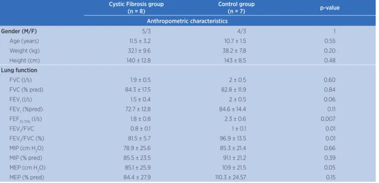
Intragroup analysis showed that tidal volume values in chest wall and its compartments were higher during the use of diferent PEP levels compared to quiet breathing in both groups (p>0.05). Regarding PEP10, no signiicant diference was found inintergroup analysis. However, total and compartmental tidal volumes were signiicantly higher in controls compared to CF patients during PEP20 (CW: 0.77 ± 0.25 L vs. 0.44 ± 0.16 L; RCp: 0.3 ± 0.13 L
vs. 0.18 ± 0.1 L; RCa: 0.21 ± 0.1 L vs. 0.12 ± 0.1 L; AB: 0.25 ± 0.1 L vs. 0.15 ± 0.1 L; p>0.05) (Figure 1).
Respiratory rate (RR, breaths/min) was signiicantly higher in the CF group (36.3±7, 33.8±14 and 37.8 ± 14.1 during QB, PEP10 and PEP20, respectively) compared to controls (28.2 ± 5.1, 30.6 ± 17.1 and 24.3 ± 14.6, p>0.005). In both groups, minute ventilation (MV, L/min) was signiicantly higher during PEP10 (15.6 ± 7.6 vs 19.5 ± 9.2) and PEP20 (16.6 ± 8.1 vs 17.8 ± 9.6) in CF patients and controls, when compared to quiet breathing (QB: 10.4 ± 3.3
vs. 8.1 ± 1.2,p>0.005). he increase in minute ventilation when using diferent intensities of PEP was obtained, however, by adopting diferent breathing patterns in the two groups (Figure 2A). While controls achieved increased ventilation with higher tidal volume (QB: 0.289 ± 0.062 L; PEP10: 0.719 ± 0.279 L; and PEP20: 0.755 ± 0.259 L)
and lower respiratory rate during PEP20, CF children showed an inverse pattern with lower tidal volume (QB: 0.292 ± 0.094 L; PEP10: 0.492 ± 0.169 L; and PEP20: 0.442 ± 0.160 L) and higher respiratory rate (Figure 2A).
Correspondingly, during QB, PEP10 and PEP20, total respiratory cycle time (Ttot, seconds) was lower in CF patients compared to controls (p>0.02, igure 2B). In CF patients, inspiratory time (Tinsp, seconds) was signiicantly lower than controls during PEP20 (0.74 ± 0.2
Figure 1. Volumes of chest wall and its compartments during quiet breathing and diferent levels of PEP in control group and CF
Values represent mean ± standard deviation. QB: quiet breathing; PEP10: positive expiratory pressure 10 cm H2O; PEP20: positive expiratory pressure 20 cm H2O. *p>0.05 PEP20 vs. QB (two-way
ANOVA).
Total and compartmental chest wall operating volumes during diferent intensities of PEP
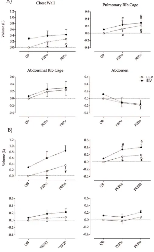
End-expiratory volume (EEV) of total chest wall and pulmonary rib cage compartment signiicantly increased during the use of PEP (both intensities)
compared to resting conditions (p>0.05) in both groups. Intergroup analysis did not show signiicant diferences regarding EEV. End-inspiratory volume (EIV) was signiicantly increased in the rib cage pulmonary compartment during PEP compared to QB (p>0.01) (Figure 3).
Figure 3. Operating total and compartmental chest wall volumes during diferent intensities of PEP
Values represent mean ± standard deviation. A) CF group; B) Control group. QB: quiet breathing; PEP10: Positive expiratory pressure 10 cm H2O; PEP20: Positive expiratory pressure 20 cm H2O; EEV:
When considering total and compartmental EIVs, intergroup analysis showed signiicant diference between CF and controls only at PEP20 for abdomen compartment (p>0.01, Figure 4).
Figure 4. End-inspiratory volume (EIV) in abdomen during diferent intensities of PEP
Values represent mean ± deviation. QB: Quiet breathing; PEP10: Positive expiratory pressure 10
cmH2O; PEP20: Positive expiratory pressure 20 cmH2O. *p>0.01 (two-way ANOVA). Bonferroni’s
post hoc between groups
DISCUSSION
Results showed that chest wall tidal volume increased during the use of PEP in comparison to quiet breathing. Moreover, the increase was signiicantly higher in controls compared to CF during PEP20. We also found that, compared to controls, children with CF are characterized by a more rapid and shallow breathing pattern, both during spontaneous quiet breathing at rest and during the use of PEP, as showed in Figure 2A. In addition, we have shown that minute ventilation increases in CF children during the use of PEP due to an increase of tidal volume. End-expiratory volume also increases compared to quiet breathing.
PEP is one of the most common airway clearance techniques used in diferent countries, such as Canada5. A recent study with 6,372 CF patients performed in the United Kingdom, showed that PEP was the third most popular technique used to manage patients’ conditions28. Despite its wide dissemination and use, little is known about its efects in ventilation. his feature is not related only to PEP, but all airway clearance techniques. he lack of knowledge about the physiological responses of airway clearance techniques was observed in the
conclusion of ive Cochrane systematic reviews that were inconclusive regarding the best airway clearance technique for CF patients5.
he acute efects of PEP have been previously studied in the literature. However, in the previous studies the possible dynamic changes in ventilation and breathing pattern were not assessed. Van Winden et al. (1998) studied the efects of lutter and PEP mask in symptoms and lung function in 22 CF patients29. he authors did not ind any signiicant changes in lung function parameters after a single session or 2 weeks of PEP or lutter use. A recent study, by McIlwaine et al. 30, analyzed long-term eicacy of high frequency chest wall oscillation (HFCWO) in comparison to theraPEP in patients with CF30. he primary outcome was the number of pulmonary exacerbations. he authors found no signiicant diference in quality of life and lung function between the groups. PEP showed to be more eicient in terms of time of use and number of exacerbations (1.14 for PEP vs 2.0 for HFCWO). A great number of CF studies aim to compare airway clearance techniques. On the other hand, we consider that is more important to irst understand the mechanisms and how patients respond to each technique, in this case, PEP.
To our knowledge, this is the irst study that evaluated the acute efects of PEP in the volumes of chest wall and its compartments in CF children. For this propose, we have used Optoelectronic Plethysmography, which provides continuous dynamic measurements of volume variations of the chest wall, divided into compartments17. Our results showed a rapid and shallow breathing pattern at rest and during use of diferent intensities of PEP in children with CF. Even though minute ventilation was similar between groups, CF patients showed less eicient breathing patterns compared to controls, as observed by the increased respiratory rate (at rest and during PEP use) and a decreased tidal volume during PEP use.
our indings is, therefore, that high levels of PEP should not be used in children with CF, to avoid overloading respiratory muscles to overcome the load imposed by the use of PEP.
here is still a lack of studies showing respiratory strength impairment in subjects with CF in the literature. Published data in the same topic are also controversial, however, we may speculate that individuals with CF show diiculty in overcoming a pressure load of 20 cmH2O due to reduction of the strength of expiratory muscles, as shown by the lower values of maximal expiratory pressure (MEP: 83.1 ± 25.9 vs. 109 ± 21.5 for CF and control groups, respectively), compared to controls, found in this study.
We may hypothesize that the increase in end-expiratory volume observed during PEP may be beneicial, particularly in those children in which the restrictive pattern is prevalent. On the other hand, in those children in which the obstructive pattern is prevalent, the EEV increase induced by PEP could not be clinically interesting as these subjects present pulmonary hyperinlation due to air trapping in the lungs, which was maintained.
We believe that the main limitation of our study is the small sample of participants and extrapolation of the results should be done carefully. However, CF is not a common lung disease, therefore, its low prevalence makes the recruitment of individuals even more limited.
CONCLUSION
In conclusion, diferent levels of PEP induce an increase of chest wall volumes in CF children with diferent mechanisms compared to controls. Even with the improvement caused by PEP, CF children still show shallow breathing characteristics. PEP levels above 10 cm H2O should be used with caution in CF children.
REFERENCES
1. Ramsey BW. Management of pulmonary disease in patients with cystic ibrosis. N Engl J Med. 1996;335(3):179-88. doi: 10.1056/NEJM199607183350307.
2. Knowles MR, Stutts MJ, Yankaskas JR, Gatzy JT, Boucher RC Jr. Abnormal respiratory epithelial ion transport in cystic ibrosis. Clin Chest Med. 1986;7(2):285-97.
3. Phelan PD, Olinski A, Robertson CF. Respiratory ilness in children. Oxford: Blackwell; 1994.
4. Lannefors L, Button BM, McIlwaine M. Physiotherapy in infants and young children with cystic ibrosis: current practice and future developments. J R Soc Med [Internet]. 2004 [acesso em 17 ago. 2017];97(Suppl. 44):S8-25. Disponível em: https:// goo.gl/GZ63FG
5. Bradley JM, Moran FM, Elborn JS. Evidence for physical therapies (airway clearance and physical training) in cystic ibrosis: an overview of ive Cochrane systematic reviews. Respir Med. 2006;100(2):191-201. doi: 10.1016/j. rmed.2005.11.028.
6. Pisi G, Chetta A. Airway clearance therapy in cystic ibrosis patients. Acta Biomed [Internet]. 2009 [acesso em 17 ago. 2017];80(2):102-6. Disponível em: https://goo.gl/bqEz1s 7. Main E, Prasad A, Schans C. Conventional chest physiotherapy
compared to other airway clearance techniques for cystic ibrosis. Cochrane Database Syst Rev. 2005;(1):CD002011. doi: 10.1002/14651858.
8. Warnock L, Gates A, van der Schans CP. Chest physiotherapy compared to no chest physiotherapy for cystic ibrosis. Cochrane Database Syst Rev. 2013;(2):CD001401. doi: 10.1002/14651858.CD001401.pub2.
9. Prasad SA, Tannenbaum E-L, Mikelsons, C. Physiotherapy in cystic ibrosis. J R Soc Med [Internet]. 2000 [acesso em 17 ago. 2017];93(Suppl. 38):27-38. Disponível em: https://goo. gl/V7TfVo
10. Laube BL, Geller DE, Lin TC, Dalby RN, Diener-West M, Zeitlin PL. Positive expiratory pressure changes aerosol distribution in patients with cystic ibrosis. Respir Care [Internet]. 2005 [acesso em 17 ago. 2017];50(11):1438-44. Disponível em: https://goo.gl/cFtL78
11. Hunter JM, Sperry EE, Ravilly S, Colin AA. Thoracoabdominal asynchrony and ratio of time to peak tidal expiratory low over total expiratory time in adolescents with cystic ibrosis. Pediatr Pulmonol. 1999;28(3):199-204.
12. Allen JL, Wolfson MR, McDowell K, Shafer TH.
Thoracoabdominal asynchrony in infants with airlow obstruction. Am Rev Respir Dis. 1990;141(2):337-42. doi: 10.1164/ajrccm/141.2.337.
13. Mayer OH, Clayton RG Sr, Jawad AF, McDonough JM, Allen JL. Respiratory inductance plethysmography in healthy 3- to 5-year-old children. Chest 2003;124(5): 1812-9. doi: 10.1378/ chest.124.5.1812.
14. Reber A, Geiduschek JM, Bobbià SA, Bruppacher HR, Frei FJ. Efect of continuous positive airway pressure on the measurement of thoracoabdominal asynchrony and minute ventilation in children anesthetized with sevolurane and nitrous oxide. Chest. 2002;122(2):473-8. doi: 10.1378/ chest.122.2.473.
15. Capria ME, D’Nedri C, De Vito EL. Relationship between Hoover sign, functional and variables, and curvature radius in patients with obstructive pulmonary disease. Medicina (B Aires). 2003;63(5):369-76.
16. Garcia-Pachon E, Padilla-Navas I. Frequency of
Hoover’s sign in stable patients with chronic obstructive pulmonary disease. Int J Clin Pract. 2006;60(5):514-7. doi: 10.1111/j.1368-5031.2006.00850.x.
18. World Health Organization. Declaration of Helsinki: ethical principals for research involving human subjects. Bull World Health Organ [Internet]. 2001 [acesso em 17 ago. 2017];79(4):373-4. Disponível em: https://goo.gl/4rCKZp 19. World Health Organization. Child growth standards based on
lenght/height, weight and age. Acta Paediatr. 2007;95:76-85. doi: 10.1111/j.1651-2227.2006.tb02378.x.
20. Miller MR, Hankinson J, Brusasco V, Burgos F, Casaburi R, Coates A, et al. Standardisation of spirometry. Eur Respir J. 2005;26(2):319-38. doi: 10.1183/09031936.05.00034805. 21. Mallozi MC. Valores de referência para espirometria em
crianças e adolescentes, calculados a partir de uma amostra da cidade de São Paulo [Tese]. São Paulo: Escola Paulista de Medicina; 1995.
22. American Thoracic Society, European Respiratory Society. ATS/ERS Statement on respiratory muscle testing. Am J Respir Crit Care Med. 2002;166(4):518-624.. doi: 10.1164/ rccm.166.4.518.
23. Lanza FC, Santos MLM, Selman JPR, Silva JC, Marcolin N, Santos J, et al. Reference equation for respiratory pressures in pediatric population: a multicenter study. PLoS One. 2015;10(8):e0135662. doi: 10.1371/journal.pone.0135662. 24. Cala SJ, Kenyon CM, Ferrigno G, Carnevali P, Aliverti A,
Pedotti A, et al. Chest wall and lung volume estimation by optical relectance motion analysis. J Appl Physiol [Internet].
1996 [acesso em 17 ago. 2017];81(6):2680-9. Disponível em: https://goo.gl/CyGkv3
25. Aliverti A, Cala SJ, Duranti R, Ferrigno G, Kenyon CM, Pedotti A, et al. Human respiratory muscle actions and control during exercise. J Appl Physiol [Internet]. 1997 [acesso em 17 ago. 2017];83(4):1256-69. Disponível em: https://goo.gl/HW7vgc 26. Aliverti A, Uva B, Laviola M, Bovio D, Lo Mauro A, Tarperi
C, et al. Concomitant ventilatory and circulatory functions of the diaphragm and abdominal muscles. J App Physiol. 2010;109(5):1432-40. doi: 10.1152/japplphysiol.00576.2010. 27. Cohen J. Statistical power analysis for the behavioral
sciences. 2. ed. Hillsdale: Lawrence Erbaum; 1988.
28. Hoo ZH, Daniels T, Wildman MJ, Teare MD, Bradley JM. Airway clearance techniques used by people with cystic ibrosis in the UK. Physiotherapy. 2015;101(4):340-8. doi: 10.1016/j. physio.2015.01.008.
29. van Winden CMQ, Visser A, Hop W, Sterk PJ, Beckers S, Jongste JC. Efects of lutter and PEP mask physiotherapy on symptoms and lung function in children with cystic ibrosis. Eur Respir J. 1998;12(1):143-7. doi: 10.1183/09031936.98.12010143. 30. McIlwaine MP, Alarie N, Davidson GF, Lands LC, Ratjen F,