Comparison between irradiated lung volumes
with two-dimensional and three-dimensional conformal
radiotherapy techniques for locally advanced lung
cancer*
Comparação entre os volumes pulmonares irradiados com técnica bidimensional e tridimensional conformada na radioterapia de pacientes com tumores de pulmão localmente avançados
Heloisa de Andrade Carvalho1, Camila Pessoa de Sales2, Silvia Radwanski Stuart3, Erlon Gil3, André Costa Navega Nunes4, Debora Cartelle Ferauche5
OBJECTIVE: To compare and quantify irradiated lung volumes using two-dimensional (2D) and three-dimensional (3D) conformal planning for radiotherapy in the treatment of lung cancer. MATERIALS AND METHODS: 2D and 3D conformal radiotherapy plannings were performed for 27 patients with lung cancer. Prescribed doses ranged from 45 to 66 Gy. The analysis covered the doses to planning target volume (PTV), gross tumor volume (GTV) and lungs (lung volume receiving 20 Gy or 30 Gy – V20 and V30, respectively, and mean dose). The doses to adjacent organs at risk (spinal cord, esophagus and heart) were maintained below the tolerance limits. RESULTS: GTV ranged from 10.5 to 1,290.0 cm3
(mean, 189.65 cm3
). On average, a total of 59.33 fields were utilized in the 2D planning and 75.65 fields in the 3D planning. Lung volumes were significantly preserved (P < 0.05) with the 3D conformal planning in all the evaluated cases, with
about 15% decrease in the irradiated lung volumes. Lungs without tumor were most benefited from this technique. CONCLUSION: 3D radiotherapy allowed a better sparing of the lungs, both in cases of early and advanced tumors. 3D radiotherapy should be used in the treatment of patients with lung cancer, even in cases of large tumors.
Keywords: Lung cancer; Radiotherapy; Conformal radiotherapy; Organs at risk; Lung volumes.
OBJETIVO: Comparar e quantificar os volumes pulmonares irradiados utilizando planejamentos bidimensio-nal (2D) e tridimensiobidimensio-nal (3D) conformado na radioterapia de tumores de pulmão. MATERIAIS E MÉTODOS: Em 27 pacientes portadores de câncer de pulmão foi feito planejamento 3D e outro correspondente em 2D. As doses prescritas variaram de 45 a 66 Gy. Foram avaliadas as doses no volume alvo planejado (PTV), volume tumoral macroscópico (GTV) e pulmões (volume de pulmão que recebe 20 Gy ou 30 Gy – V20 e V30, respectivamente, e dose média). Os órgãos de risco adjacentes (medula espinhal, esôfago e coração) receberam doses abaixo dos limites de tolerância. RESULTADOS: O GTV variou de 10,5 a 1.290,0 cm3
(média de 189,65 cm3
). Nos planejamentos 2D foi utilizado, em média, um total de 59,33 campos, e nos planeja-mentos 3D, 75,65 campos. Em todas as situações analisadas houve significante (P < 0,05) preservação dos volumes pulmonares com o planejamento 3D, com diminuição de cerca de 15% dos volumes irradiados. O pulmão sem tumor foi mais beneficiado. CONCLUSÃO: A radioterapia 3D permitiu maior preservação dos pulmões, tanto para tumores iniciais quanto avançados. A radioterapia 3D deve ser utilizada nos pacientes com tumores de pulmão, mesmo que volumosos.
Unitermos: Câncer de pulmão; Radioterapia; Radioterapia conformada; Órgãos em risco; Volumes pulmonares. Abstract
Resumo
* Study developed at Unit of Radiotherapy, Department of Radiology – Hospital das Clínicas da Faculdade de Medicina da Universidade de São Paulo (HC-FMUSP), São Paulo, SP, Brazil.
1. PhD, Assistant Physician at Unit of Radiotherapy, Depart-ment of Radiology – Hospital das Clínicas da Faculdade de Me-dicina da Universidade de São Paulo (HC-FMUSP), São Paulo, SP, Brazil.
2. Medical Physicist, Physicist at Unit of Radiotherapy,
Depart-Carvalho HA, Sales CP, Stuart SR, Gil E, Nunes ACN, Ferauche DC. Comparison between irradiated lung volumes with two-dimensional and three-two-dimensional conformal radiotherapy techniques for locally advanced lung cancer. Radiol Bras. 2009;42(5):303–308.
ment of Radiology – Hospital das Clínicas da Faculdade de Me-dicina da Universidade de São Paulo (HC-FMUSP), São Paulo, SP, Brazil.
3. MDs, Radiotherapists, Assistant Physicians at Unit of Radiotherapy, Department of Radiology – Hospital das Clínicas da Faculdade de Medicina da Universidade de São Paulo (HC-FMUSP), São Paulo, SP, Brazil.
4. MD, Radiotherapist, Assistant Physician at Unit of Radio-therapy, Department of Radiology – Hospital de Clínicas da
Fa-culdade de Ciências Médicas da Universidade Estadual de Cam-pinas (FCM-Unicamp), CamCam-pinas, SP, Brazil.
5. Master, Physicist at Unit of Radiotherapy, Hospital Benefi-cência Portuguesa, São Paulo, SP, Brazil.
Mailing address: Dra. Heloisa de Andrade Carvalho. Rua Sam-paio Viana, 509, ap. 21, Paraíso. São Paulo, SP, Brazil, 04004-002. E-mail: [email protected]
INTRODUCTION
Lung cancer, besides being the first in incidence, is also accountable for most can-cer-related deaths in the world(1,2).
In Brazil, estimates for 2008 indicate lung cancer as the second in incidence among men and fourth among women, with 17,810 expected new cases for men (incidence of 19 cases/100,000 men) and 9,460 for women (10 cases per 100,000 women)(3).
Lung cancer is mainly related to smok-ing, which may cause other pulmonary dis-eases such as chronic obstructive pulmo-nary disease and emphysema. Therefore, patients with lung cancer may in general present underlying lung function impair-ment. Besides allowing a safer dose esca-lation, three-dimensional conformal radio-therapy (3DCRT) allows an appropriate evaluation of treatment volumes and irra-diated healthy tissue. In the treatment of lung tumors, this characteristic is particu-larly useful, mainly with respect to a greater sparing of the lungs, which are organs highly sensitive to radiation, and which may already be partially compromised in these cases.
In general, the incidence of locally ad-vanced tumors (stage III) is high, and this group of patients is the one that has the main indication for radiotherapy, both as a curative as well as palliative treatment(4–6). Additionally, the current standard treatment is associated with chemotherapy, which, in spite of better results, may also increase the treatment toxicity(4–6). Due to the fact that such tumors are many times large pulmo-nary masses, the advantages of the three-dimensional (3D) over the conventional two-dimensional (2D) treatment may seem to be of little significance. Also, the large patient demand of the local public radio-therapy services in association with the larger workload and required planning time for 3DCRT, may lead to a lesser use of this tool in such cases.
Therefore, it is important to evaluate and quantify the benefits that 3DCRT brings comparatively with the 2D tech-nique, with special regard to healthy tissues sparing.
The present study was performed to compare 2D and 3D radiotherapy in the
treatment of lung tumors, quantifying the irradiated pulmonary volumes.
MATERIALS AND METHODS
Radiotherapy plannings of 27 patients with lung cancer submitted to conformal 3DCRT were evaluated. Volumes delinea-tion was made on mediastinal and lung windows. Prescribed doses ranged from 45 to 66 Gy (1.8 to 2 Gy/day). For the same prescription and for each particular case, a conventional 2D planning was also simu-lated, based on computed tomography (CT) images.
The plannings were based on the recom-mendations from ICRU reports No. 50 and 62(7,8), considering at least 95% of the planned target volume (PTV) covered by 95% of the prescribed dose as appropriate. The 3D plannings were individualized and performed in two phases, with fields’ re-duction or new planning after 40 or 45 Gy, or in a single phase, depending upon clini-cal indication and dose received by the spinal cord. The 2D plannings were devel-oped in two phases, the first one with two parallel and opposed fields, anteroposterior and posteroanterior (AP-PA) up to 40 or 45 Gy and following that, dose supplementa-tion with protecsupplementa-tion of the spinal cord, in oblique parallel and opposed fields or two to three angled fields. Four patients re-ceived total dose of 45 Gy and were planned in a single phase. Doses to organs at risk — spinal cord, esophagus, heart and lungs — were kept below the tolerance lim-its, in accordance with Emami et al.(9) and Milano et al.(10) recommendations.
The following parameters were evalu-ated for comparison of the plannings in re-lation to the lungs: gross tumor volume (GTV), total number of fields, percentage of lung volume receiving 20 Gy (V20), percentage of lung volume receiving 30 Gy (V30) and mean dose (MD) to the lungs(11). These parameters were calculated by means of dose-volume histograms for both lungs (total lung), and respectively, for the lung with tumor and for the lung without tumor (“healthy”).
The 3D planning system Eclipse (Varian Medical Systems; Palo Alto, USA) was utilized for structures delineation and cal-culations.
The data were submitted to descriptive and frequency analysis. The data means were compared by means of the Student t test. In order to evaluate the interference of tumor volume in the planning quality, the patients were divided into two groups ac-cording to GTV: up to 125 cm³, and > 125 cm³. This value was chosen considering initial tumors, those presenting a maximum diameter of 5 cm, versus the remaining ones above 5cm in diameter, considered as locally advanced. The significance level was set at 5% (P≤ 0.05).
RESULTS
The GTV ranged from 10.5 to 1290.0 cm³ (mean, 189.65 cm³). Grouping the pa-tients according to the GTV, 13 papa-tients presented GTV ≤ 125 cm3 (mean, 62.94
cm3) and 14 patients presented GTV > 125
cm3 (mean, 307.36 cm3) (
P = 0.0001).
On average, 59.33 fields (median, 60, ranging from 50 to 74 fields) were utilized in the 2D plannings; two fields/day, respec-tively in the first and second phases. In the 3D plannings, the average number of fields was 75.65, ranging from 50 to 112 (mean, 80) with a mean of 2.6 fields in the first phase and 2.9 in the second phase, respec-tively.
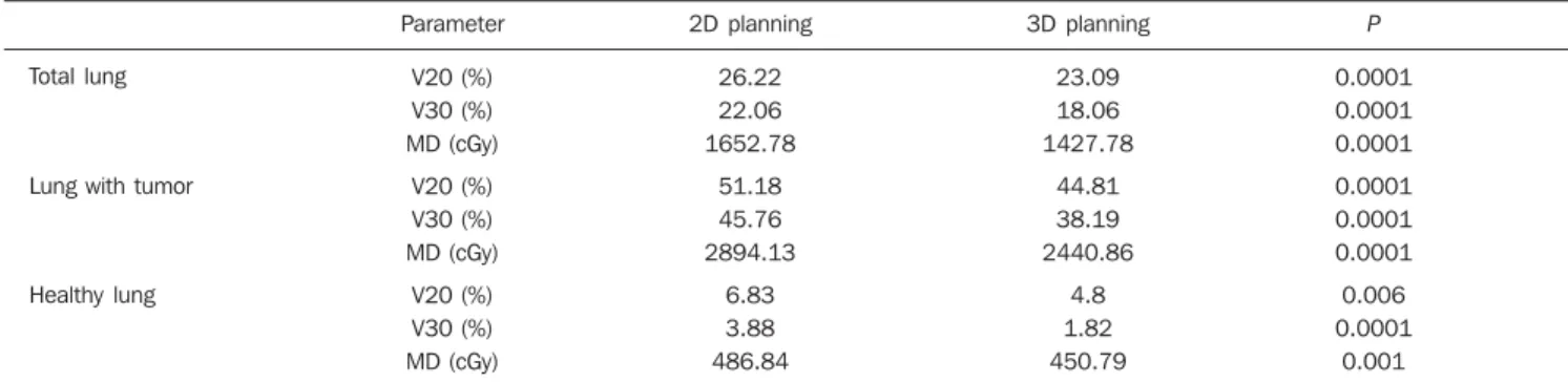
Tables 1 to 4 present the results from data regarding doses to lungs.
Both irradiated volumes and mean doses were significantly lower when 3DCRT was utilized, independently from the volume of the GTV (Tables 1 and 2). With the exception of V20 and the mean dose to the healthy lung for initial tumors, all the other evaluated parameters pre-sented significant absolute benefit in favor of 3DCRT. The observed benefit was even greater for larger volume tumors when compared with the initial tumors (Tables 3 and 4).
Figures 1 and 2 present the comparative dose-volume histograms of the studied pulmonary volumes, respectively for a tu-mor considered as a small one, and a large one.
DISCUSSION
Table 1 Comparison between the 2D and 3D plannings. The mean values of each variable are presented analyzing both lungs as a single organ (total lung) or separated (lung with tumor and without tumor respectively).
Total lung
Lung with tumor
Healthy lung
Parameter
V20 (%) V30 (%) MD (cGy)
V20 (%) V30 (%) MD (cGy)
V20 (%) V30 (%) MD (cGy)
2D planning
26.22 22.06 1652.78
51.18 45.76 2894.13
6.83 3.88 486.84
3D planning
23.09 18.06 1427.78
44.81 38.19 2440.86
4.8 1.82 450.79
P
0.0001 0.0001 0.0001
0.0001 0.0001 0.0001
0.006 0.0001
0.001
2D, conventional 2D radiotherapy; 3D, conformal 3D radiotherapy; V20, percentage of lung volume receiving 20 Gy; V30, percentage of lung volume receiving 30 Gy; MD, mean dose.
Table 2 Comparison between 2D and 3D plannings considering GTV.
Total lung
Lung with tumor
Healthy lung
Parameter
V20 (%) 2D 3D V30 (%)
2D 3D MD (cGy)
2D 3D
V20 (%) 2D 3D V30 (%)
2D 3D MD (cGy)
2D 3D
V20 (%) 2D 3D V30 (%)
2D 3D MD (cGy)
2D 3D
GTV ≤ 125 cm3
24.99 22.98
21.78 17.73
1486.78 1366.65
48.50 42.97
42.61 35.75
2613.38 2248.84
6.04 5.03
3.28 1.62
365.18 457.41
P
0.006
0.001
0.003
0.0001
0.001
0.0001
0.417
0.011
0.410
GTV > 125 cm3
27.06 23.18
22.31 18.37
1806.92 1484.53
53.67 46.52
48.69 40.46
3154.83 2619.16
7.57 4.58
4.44 2.00
599.81 444.65
P
0.0001
0.0001
0.0001
0.0001
0.0001
0.0001
0.001
0.015
0.002
GTV, gross tumor volume; 2D, conventional 2D radiotherapy; 3D, conformal 3D radiotherapy; V20, percentage of lung receiving 20 Gy; V30, percentage of lung volume receiving 30 Gy; MD, mean dose.
Table 3 Mean values of the absolute benefit obtained with the conformal 3D technique in relation to irradiated pulmonary volumes (mean GTV = 189.65cm3)
(P < 0.05).
Benefit with 3D
Both lungs
Lung with tumor
Healthy lung
V20
11.94%
12.45%
29.72%
V30
18.13%
16.54%
53.09%
MD
13.61%
15.66%
7.40%
normal structures to be visualized and iden-tified with higher accuracy in patients sub-mitted to radiotherapy. The possibility of greater technical variations such as the use of several angled fields, non-coplanar fields and mainly the quantification of dose
delivered to a given organ or tissue volume by means of the dose-volume histograms have consolidated the method that is cur-rently widely used. In Brazil, this technol-ogy is already available in many centers, including the public health services.
How-Table 4 Mean values of absolute benefit obtained with the conformal 3D technique in relation to irradiated pulmonary volumes according to GTV (≤ 125 cm3
or > 125 cm3).
Benefit with 3D
Mean GTV = 62.94 cm3
Both lungs Lung with tumor
Healthy lung
Mean GTV = 307.36 cm3
Both lungs Lung with tumor
Healthy lung
V20
8.04% 11.40%
16.72%*
14.34% 13.32%
39.50%
V30
18.59% 16.10%
50.61%
17.66% 16.90%
54.95%
MD
8.08% 13.95%
–25.26%*
17.84% 16.98%
25.87%
3D, conformal 3D radiotherapy; V20, percentage of lung receiving 20 Gy; V30, percentage of lung receiving 30 Gy; MD, mean dose.
* Non significant differences. In the case of mean dose in the normal lung, there was an increase of 25.26% between the means with 3D, however not significant.
ever, 3D planning is more time consuming for the radiation oncologist and the physi-cist, due to greater detailing in the delimi-tation of the target-lesion and structures at risk, and increased planning possibilities. In centers with a high demand, many times
Figure 1. Dose-volume histogram of the lungs for a 49 cm³ tumor, with the respective V20 and V30 marked. A: Total lung. B: Lung with tumor and healthy lung.
A B
Figura 2. Dose-volume histogram of the lungs for a 183 cm³ tumor, with the respective V20 and V30 marked. A: Total lung. B: Lung with tumor and healthy lung.
the benefit of a 3D planning may be ques-tioned, particularly in the case of patients with advanced tumors, or those that should only receive a palliative treatment.
Furthermore, the actual benefit of 3DCRT in relation to survival of patients with lung cancer is not yet well established. Its’ main advantage is the evaluation and possibility of decreasing or preventing the potential radiotherapy toxicity, on an indi-vidual basis(12). For this reason, only from the advent of this technology innumerable studies on dose escalation(13–17) are being developed, in association or not with more advanced techniques such as image-guided radiotherapy (IGRT)(18), respiratory gating or breath-holding radiotherapy(19), hypo-fractionated radiotherapy(20) or still, the association with functional diagnosis meth-ods such as positron emission tomography (PET), that allows a more accurate identi-fication of the target volume(21).
The present study did not intend to dis-cuss the lung tolerance doses, but to evalu-ate and quantify the benefit of 3DCRT for a group of patients with lung cancer under-going treatment at the institution.
In spite of maintaining the same pre-scription doses utilized in the 2D plannings, 3DCRT provided a greater sparing of the lungs in practically all situations, specially the healthy lungs. Such benefit may be even greater, considering that the 2D plan-ning was the best possible, as it was carried out in a 3D planning system, based on CT images, and not on plain simulation radio-graphs. However, even with a significant decrease of irradiated pulmonary volumes with 3DCRT (V20 and V30), the increase in the number of fields may lead to an in-crease of volumes receiving low doses, particularly for smaller tumors (Figure 1). Therefore, the evaluation of the mean dose is of great value in such situations, for ana-lyzing each case individually.
The possibility of reduction of field margins by itself, besides the construction of individualized shielding blocks, in-creases the protection to healthy tissues, with appropriate target coverage in the 3D planning. When the PTV is subtracted from the pulmonary volume, the results in rela-tion to the lungs sparing would be even better. Notwithstanding, the worst possible situation was analyzed, considering the
whole “useful” volume of the lung. In the simulations that were made, the field mar-gins were equal, and we have chosen to compare patients regarding only the GTV, since it is better defined in a 2D planning than the PTV, and can be easily estimated on a diagnostic CT. With this type of analy-sis it was possible to observe an absolute reduction of irradiated lung volumes of about 15%, independently of the tumor size (Table 3). In patients for whom the dose escalation becomes more complex due to irradiation of large volumes of healthy tis-sue, the decrease in toxicity is a key issue. This fact may be of particular advantage in cases of patients with impaired pulmonary function, where chemotherapy may be as-sociated.
The sparing of other organs at risk (esophagus, spinal cord and heart) was not evaluated in the present study, as it was possible to keep the doses for these organs below their tolerance limits, even on the 2D plannings. Additionally, both the esopha-gus and spinal cord are organs whose tol-erance doses depend very little on the re-spective irradiated volumes and the dose in the heart may vary significantly according to the lesion location. The lungs, however, object of the present study, present toler-ance doses much lower than those of the surrounding organs at risk(7,8).
Finally, specifically in Brazil, the num-ber of fields that were utilized does not in-validate the technique regarding its use in the public health services, as most of the times it falls within the limits established by the Brazilian public health system (“Sistema Único de Saúde”) (maximum of 90 fields)(22) for reimbursement of treat-ments.
CONCLUSIONS
3DCRT allowed the sparing of approxi-mately 15% of the irradiated pulmonary volumes, both in the cases of initial and advanced tumors.
The benefit was greater for the lung without tumor, which can be better spared by the appropriateness of the irradiation technique.
The possibility of greater sparing of pul-monary volumes at the observed levels, supports the conclusion that 3DCRT
should be utilized in patients with lung tumors, regardless of size.
REFERENCES
1. Shibuya K, Mathers CD, Boschi-Pinto C, et al. Correction: Global and regional estimates of can-cer mortality and incidence by site: II. results for the global burden of disease 2000. BMC Cancer. 2003;3:20.
2. Jemal A, Siegel R, Ward E, et al. Cancer statis-tics, 2008. CA Cancer J Clin. 2008;58:71–96. 3. Brasil. Ministério da Saúde. Instituto Nacional do
Câncer – INCA. Estimativa de incidência e mor-talidade por câncer. Rio de Janeiro: INCA; 2008. [cited 2009 May 25]. Available from: http://www. inca.gov.br/estimativa/2008
4. Bradley J, Govindan R, Komaki R. Lung. In: Perez CA, Brady LW, Halperin EC, et al., editors. Principles and practice of radiation oncology. 4th ed. Philadelphia: Lippincott Williams & Wilkins; 2004. p. 1201–43.
5. Robinson LA, Ruckdeschel JC, Wagner H Jr, et al. Treatment of non-small cell lung cancer-stage IIIA: ACCP evidence-based clinical practice guide-lines (2nd edition). Chest. 2007;132(3 Suppl): 243S–65S.
6. Jett JR, Schild SE, Keith RL, et al. Treatment of non-small cell lung cancer, stage IIIB: ACCP evidence-based clinical practice guidelines (2nd edition). Chest. 2007;132(3 Suppl):266S–76S. 7. International Commission on Radiation Units and
Measurements. Report 50 (ICRU 50). Prescrib-ing, recordPrescrib-ing, and reporting photon beam therapy. Bethesda: ICRU; 1993.
8. International Commission on Radiation Units and Measurements. Report 62 (ICRU 62). Prescrib-ing, recordPrescrib-ing, and reporting photon beam therapy (Supplement to ICRU Report 50). Bethesda: ICRU; 1999.
9. Emami B, Lyman J, Brown A, et al. Tolerance of normal tissue to therapeutic irradiation. Int J Radiat Oncol Biol Phys. 1991;21:109–22. 10. Milano MT, Constine LS, Okunieff P. Normal
tis-sue tolerance dose metrics for radiation therapy of major organs. Semin Radiat Oncol. 2007;17: 131–40.
11. Miller KL, Shafman TD, Marks LB. A practical approach to pulmonary risk assessment in the radiotherapy of lung cancer. Semin Radiat Oncol. 2004;14:298–307.
12. Armstrong J, McGibney C. The impact of three-dimensional radiation on the treatment of non-small cell lung cancer. Radiother Oncol. 2000;56: 157–67.
13. Arriagada R, Komaki R, Cox JD. Radiation dose escalation in non-small cell carcinoma of the lung. Semin Radiat Oncol. 2004;14:287–91. 14. Bradley J, Graham MV, Winter K, et al. Toxicity
and outcome results of RTOG 9311: a phase I-II dose-escalation study using three-dimensional conformal radiotherapy in patients with inoperable non-small-cell lung carcinoma. Int J Radiat Oncol Biol Phys. 2005;61:318–28.
15. Bradley J. A review of radiation dose escalation trials for non-small cell lung cancer within the Radiation Therapy Oncology Group. Semin Oncol. 2005;32(2 Suppl 3):S111–3.
Induc-tion and concurrent chemotherapy with high-dose thoracic conformal radiation therapy in unresect-able stage IIIA and IIIB non-small-cell lung can-cer: a dose-escalation phase I trial. J Clin Oncol. 2004;22:4341–50.
17. Lee CB, Stinchcombe TE, Rosenman JG, et al. Therapeutic advances in local-regional therapy for stage III non-small-cell lung cancer: evolving role of dose-escalated conformal (3-dimensional) radiation therapy. Clin Lung Cancer. 2006;8:195– 202.
18. Chang JY, Dong L, Liu H, et al. Image-guided
radiation therapy for non-small cell lung cancer. J Thorac Oncol. 2008;3:177–86.
19. Giraud P, Yorke E, Jiang S, et al. Reduction of organ motion effects in IMRT and conformal 3D radiation delivery by using gating and tracking techniques. Cancer Radiother. 2006;10:269–82. 20. Salazar OM, Sandhu TS, Lattin PB, et al. Once-weekly, high-dose stereotactic body radiotherapy for lung cancer: 6-year analysis of 60 early-stage, 42 locally advanced, and 7 metastatic lung can-cers. Int J Radiat Oncol Biol Phys. 2008;72:707– 15.
21. Mac Manus MP, Hicks RJ. Impact of PET on ra-diation therapy planning in lung cancer. Radiol Clin North Am. 2007;45:627–38, vi. 22. Ministério da Saúde, INCA, SAS, DAE, CGAC,