Abstract
Objectives: To identify and characterize respiratory viruses that infect children from daycare centers with symptoms of respiratory infection and to evaluate the association of clinical and epidemiological disease data with the identiied virus.
Methods: We conducted a study between 2003 and 2005 in 176 children with respiratory infection symptoms attending a municipal daycare center. Samples from nasopharyngeal secretion were tested by reverse transcriptase polymerase chain reaction and positive samples for picornavirus were sequenced.
Results: All 782 collected samples were analyzed and 31.8% were positive for at least one of the studied respiratory viruses. Respiratory infections were characterized by the presence of mild symptoms of the upper respiratory tract, the most common of which were runny nose and cough. In the 2 years of study, most cases of infection occurred in autumn and winter, but respiratory viruses were detected throughout all the study period.
Conclusions: Respiratory viruses and respiratory infections caused by them are part of the daily life of children attending daycare centers. Our results show the great impact that respiratory infections have on these children and suggest that more attention must be paid to viral pathogens.
J Pediatr (Rio J). 2011;87(5):439-44: Respiratory viruses, acute respiratory infection, daycare children.
ORiginAl ARtiCle
Copyright © 2011 by Sociedade Brasileira de Pediatria439
introduction
Acute respiratory infections (ARIs) are the major cause of morbidity in young children,1,2 particularly in developing countries.3 Children are more susceptible to
respiratory infections due to anatomical, physiological and immunological characteristics.4 These respiratory infections have a signiicant impact on health worldwide and are predominantly of viral origin.5
Viral pathogens account for 30-40% of ARI cases.6 The
viruses most frequently involved are human respiratory syncytial virus (HRSV), human metapneumovirus (hMPV), inluenza A and B (FluA and FluB), parainluenza virus (PIV) 1, 2, and 3, and rhinovirus.
Currently, daycare centers are increasingly present in the lives of children aged 0-72 months, and several studies
Frequent respiratory pathogens of respiratory tract infections
in children attending daycare centers
Caroline M. Bonim,1 Maurício L. Nogueira,2 Paulo Vítor M. Simas,1 Luis Gustavo A. Gardinassi,1 Edison L. Durigon,3 Paula Rahal,4 Fátima P. Souza5
1. Mestre em Microbiologia, Universidade Estadual Paulista (UNESP), São José do Rio Preto, SP, Brazil.
2. Livre docente, Faculdade de Medicina de São José do Rio Preto (Famerp), São José do Rio Preto, SP, Brazil. Chefe, Laboratório de Virologia (Famerp), São José do Rio Preto, SP, Brazil.
3. Doutor em Microbiologia, Universidade de São Paulo (USP), São Paulo, SP, Brazil. 4. Livre docente, UNESP, São José do Rio Preto, SP, Brazil.
5. Doutora em Biofísica, UNESP, São José do Rio Preto, SP, Brazil.
No conflicts of interest declared concerning the publication of this article.
Financial support: Coordenação de Aperfeiçoamento de Pessoal de Nível Superior (CAPES) and Fundação de Amparo à Pesquisa do Estado de São Paulo (FAPESP).
Suggested citation: Bonfim CM, Nogueira ML, Simas PV, Gardinassi LG, Durigon EL, Rahal P, et al. Frequent respiratory pathogens of respiratory tract infections in children attending daycare centers. J Pediatr (Rio J). 2011;87(5):439-44.
Manuscript submitted Mar 02 2011, accepted for publication Jun 27 2011.
suggest that attending a daycare center is an important risk factor for respiratory infections.7 Children in preschool
and daycare centers have a high frequency of respiratory infections due to contact with other children every day.8
Furthermore, infant morbidity associated with daycare centers has economic implications, since diseases result in the increased use of health care services and in increased family expenses on medical care and medications.9 However, studies on the viral etiology of ARIs in children attending daycare centers are relatively few in the literature.10
The purpose of this paper was to make a descriptive study reporting results on daycare-based ARI, carried out over a 24-month period to identify the frequency of viral agents in children with symptoms of respiratory infection.
Materials and methods
Study design and population
We conducted a longitudinal study of ARI cases in children from a daycare center in the city of São José do Rio Preto, SP, Brazil, from July 2003 to September 2005. All children attending the daycare aged 4-72 months and diagnosed by a pediatrician with typical symptoms of respiratory infection, such as sneezing, runny nose, fever, cough, and shortness of breath, were selected to participate in the study. Our study population consisted of 176 children, 100 boys and 76 girls. It is important to note that the daycare center was closed in the holiday period, which corresponded to the months of December, January, and February. This study was approved by the Research Ethics Committee of Universidade Estadual Paulista Júlio de Mesquita Filho (UNESP), Instituto de Biociências, Letras e Ciências Exatas (IBILCE), by opinion no. 062/2001 on June 11, 2001 in São José do Rio Preto, SP, Brazil. In addition, a consent form for collection of samples was obtained from parents or guardians of all children enrolled.
Sample collection
At the time of sample collection, to ensure that the genetic material found was characteristic of an ongoing respiratory infection and not residual genetic material from previous respiratory infections, a new episode of respiratory infection was considered only if there was an interval of 7 days or more between the end and restart of symptoms.
The collection of material from nasopharyngeal secretion was performed by a single nurse, using 0.5 mL of sterile 1X phosphate buffered saline (PBS) for streamlining the secretion and swabs and neonatal suction catheter to harvest nasal material. The material was stored in a vial containing 3 mL of 1X PBS and informing name and protocol number of the child. Once collected, the nasal aspirates were placed in an insulated cooler with ice and sent to the laboratory (Laboratory for Genomic Studies, Laboratório de Estudos
Genômicos, UNESP, São José do Rio Preto) for processing. The nasal aspirates were diluted in PBS pH 7.2. Samples were divided into aliquots of 250 µL and treated with 25 µL of streptomycin and 2.5 µL of antifungal agents to prevent contamination. After an incubation period of 1 hour, samples were added to 750 µL of Trizol-LS (Gibco) and the material was stored in a freezer at -80 ºC for subsequent RNA extraction.
Molecular viral detection
Aliquots (250 μL) of each sample were frozen at -70 ° C, each with 750 μL of Trizol-LS (Gibco). The extraction of total RNA / DNA followed the instructions of the manufacturer of Trizol. The nucleic acid extracts were diluted with 50 μL of MilliQ water treated with diethylpyrocarbonate (Sigma) and containing 0.5 μL of RNase OUT (Invitrogen) at a inal concentration of ~ 1 unit/μL. Extracts were immediately tested by reverse transcriptase polymerase chain reaction (RT-PCR). Reverse transcription was performed with High Capacity cDNA Archive kit (Applied Biosystems), according to manufacturer's instructions. For ampliication of the samples, we used the method of polymerase chain reaction (PCR) with speciic primers for detection of HRSV, hMPV, FluA, FluB, PIV-1, PIV-2, PIV-3, and picornavirus.11-14
Typing of picornaviruses to detect rhinovirus or enterovirus was performed by direct sequencing with the speciic primers OL26 (5’- GCA CTT CTG TTT CCC C -3’) and OL27 (5’- CGG ACA CCC AAA GTA -3’).15 The PCR products were puriied by column with the QIAquick PCR Puriication Kit (Qiagen) according to manufacturer’s instructions, and the BigDye® Terminator v 3.1 Cycle Sequencing Kit was used for luorescent reaction. Samples were precipitated and then sequenced on a 3130xl Genetic Analyzer sequencer (Applied Biosystems). All sequences of rhinovirus and enterovirus samples were analyzed for homology with the sequence corresponding to the 5’ untranslated region of the genomes of the respective virus using the Basic Local Alignment Search Tool (BLAST) program (Table 1)
.
Results
A total of 782 samples were collected throughout the 2 years of study. Of the 782 samples tested, 37.6% (294/782) were positive for at least one of the respiratory viruses analyzed. Among the positive samples, we found 20.7% (61/294) of HRSV, 5.8% (17/294) of hMPV, 6.5% (19/294) of FluA, 17.3% (51/294) of FluB, 3.1% (9/294) of PIV-1, 3.4% (10/294) of PIV-2 and PIV-3, 37.7% (111/294) of human rhinovirus (HRV), and 2% (6/294) of human enterovirus (HEV), as illustrated in Figure 1.
Figure 1 - Prevalence of respiratory viruses in samples of nasopharyngeal aspirates collected from children attending a daycare center
FluA and FluB = influenzatype A and B; HEV = human enterovirus; hMPV = human metapneumovirus; HRSV = human respiratory syncytial virus; HRV = human rhinovirus; 1, 2, and PIV-3 = parainfluenza virustype 1, 2, and 3.
Virus Primer Sequence BP Reference
HRSV sense (+) 5’- AAC AGT TTA ACA TTA CCA AGT GA -3’ 379 Mazzulli et al., 199911
antisense (-) 5’- TCA TTG ACT TGA GAT ATT GAT CG -3’
hMPV sense (+) 5’- GAG CCA ATT GAA AAT CCC AGA CA -3’ 343 Falsey et al., 200312
antisense (-) 5’- GAA AAC TGC CGC ACA ACA TTT AG -3
FluA sense (+) 5’- CTA AGG GCT TTC ACC GAA GA -3’ 191 Claas et al., 199213
antisense (-) 5’- CCC ATT CTC ATT ACT GCT TC -3’
FluB sense (+) 5’- ATG GCC ATC GGA TCC TCA AC -3’ 240 Claas et al., 199213
antisense (-) 5’- TGT CAG CTA TTA TGG AGC TC -3’
PIV-1 sense (+) 5’- CCG GTA ATT TCT CAT ACC TAT G -3’ 317 Echevarria et al., 199814
antisense (-) 5’- CCT TGC AGC GGA GTT GTT AAG -3’
PIV-2 sense (+) 5’- CCA TTT ACC TAA GTG ATG GAA T -3’ 204 Echevarria et al., 199814
antisense (-) 5’- GCC CTG TTG TAT TTG GAA GAG A -3’
PIV-3 sense (+) 5’- ACT CCC AAA GTT GAT GAA AGA T -3’ 102 Echevarria et al., 199814
antisense (-) 5’- TAA ATC TTG TTG TTG AGA TTG A -3’
HRV sense (+) 5’- GGC CCC TGA ATG YGG CTA A -3’ 114 Arruda & Hayden, 199315
antisense (-) 5’- GAA ACA CGG ACA CCC AAA GTA -3’ table 1 - Speciic primers used for virus detection
BP = base pair; FluA and FluB = influenza type A and B; hMPV = human metapneumovirus; HRSV = human respiratory syncytial virus; HRV = human rhinovirus; PIV-1, PIV-2, and PIV-3 = parainfluenza virus type 1, 2, and 3.
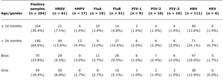
and HEV, which affected more children under 24 months. Not all children who had symptoms of respiratory infection were positive for a virus, and 124 of the 176 children studied (70.4%) were really affected by some viral respiratory infection. Boys were infected in 56.4% (70/124) of the cases and girls in 43.5% (54/124). There were cases in which the same child was affected by a virus more than once in different collections. The most prevalent virus in both genders was HRV (Table 2).
All episodes of respiratory infection were characterized by the absence of severe symptoms and were generally limited to upper airways, with no need of hospitalization during the respiratory infection. The most frequent symptom in the study population was runny nose (93.2%), followed by cough (58.2%), nasal obstruction (14.6%), wheezing (3.7%), and fever (2.4%). Some symptoms considered common in respiratory infections, such as shortness of breath and sibilance, were not observed in our study population.
Regarding the seasonal distribution of viral respiratory infections among the children from the daycare center, it was observed that rhinovirus was detected more frequently in autumn, but the virus occurred throughout all the study period, with low frequency in the summer months (December, January, and February) (1%, n = 1). In all years, we observed that, in the summer, the number of respiratory infections was reduced. When we analyzed outbreaks per year, we noted that, in 2003, the most prevalent virus was HRSV, in 2004 there was a decrease in infections by HRSV and an increase in HRV outbreaks, and in 2005 HRSV was the predominant virus once again (Figure 2).
Discussion
Positive
samples HRSV hMPV FluA FluB PIV-1 PIV-2 PIV-3 HRV HEV
Age/gender (n = 294) (n = 61) (n = 17) (n = 19) (n = 51) (n = 9) (n = 10) (n = 10) (n = 111) (n = 6)
≤ 24 months 104 21 4 10 14 3 4 4 40 4
(35.4%) (7.1%) (1.4%) (3.4%) (4.8%) (1.0%) (1.4%) (1.4%) (13.6%) (1.4%)
> 24 months 190 40 13 9 37 6 6 6 71 2
(64.6%) (13.6%) (4.4%) (3.0%) (12.6%) (2.0%) (2.0%) (2.0%) (24.1%) (0.7%)
Boys 70 28 9 11 28 6 7 6 47 5
(23.8%) (9.5%) (3.0%) (3.7%) (9.5%) (2.0%) (2.4%) (2.0%) (16.0%) (1.7%)
Girls 54 20 8 8 15 3 3 3 38 0
(18.4%) (6.8%) (2.7%) (2.7%) (5.1%) (1.0%) (1.0%) (1.0%) (12.9%) (0.0%)
table 2 - Distribution of each respiratory virus according to gender and age of the children
FluA and FluB = influenza type A and B; HEV = human enterovirus; hMPV = human metapneumovirus; HRSV = human respiratory syncytial virus; HRV = human rhinovirus; PIV-1, PIV-2 and PIV-3 = parainfluenza virus type 1, 2 and 3.
Figure 2 - Seasonal distribution of viral respiratory infections among daycare children between July 2003 and September 2005
FluA and FluB = influenzatype A and B; HEV = human enterovirus; hMPV = human metapneumovirus; HRSV = respiratory syncytial virus; HRV = human rhinovirus; PIV-1, PIV-2 and PIV-3 = parainfluenza virus type 1, 2, and 3.
37.6% (n = 294) were positive for a respiratory virus. These results are similar to those from the study conducted by Tsuchiya et al. in Curitiba, state of Paraná, Brazil,16 which identiied a viral agent in 30% of the nasopharyngeal aspirate or bronchoalveolar lavage samples tested using shell vial culture and immunoluorescence assay (IFA). Other study analyzed nasopharyngeal specimens of children under 5 years of age with acute respiratory disease, and the application of IFA and RT-PCR allowed to detect a viral agent in 75.5% of the samples.17 However, these studies were performed with hospitalized children. In Bahia, Brazil, one of the few studies conducted with daycare children found 43% positivity for any of the respiratory viruses.18
However, in this case, only children under 24 months were selected, and the literature reports a high percentage of viral infection in children from this age group. Therefore, the lower positivity rate of our work is probably because most children from the daycare center were older than 24 months.
In our study, the most frequent respiratory pathogen found was HRV (37.7%), followed by HRSV (20.7%) and FluB (17.3%). In a study conducted by Souza et al.18 with
children from daycare centers, of the 264 samples available for testing by RT-PCR and hybridization, 78% were rhinovirus positive. A study by van der Zalm et al.19 indicates that the occurrence of HRV infection is 13 times higher than that of HRSV infection, and also that the number of medical visits made during an episode of HRV infection is seven times higher than during HRSV infection. Overall, their study conirms the high occurrence of HRVs in respiratory tract infections. Despite the fact that rhinovirus presents high positivity, there are few data available in Brazil about the characteristics and
functions of this viral type in respiratory infections, which complicates the diagnosis and demonstrates the need for further studies to obtain new information.
HRSV was the second most prevalent virus, an unexpected high frequency, because our samples were from daycare children, and HRSV is mostly found in samples from children attending hospitals, who present more severe cases of infection.20,21
The study by van der Zalm et al.19 found higher rates of inluenza infection, with 83% of type A and 17% of type B. This difference can be explained by the fact that this study was performed with hospitalized children, who had more severe infections characteristic of FluA. The children in our study had mild symptoms of respiratory infection, which is a characteristic of FluB infection, justifying thus the higher incidence of this less aggressive virus.
Regarding the age of children with respiratory infections, although there were cases of respiratory infection in all age groups, we found that there were more infections in children between the ages of 13 and 48 months. According to Fiterman et al.,22 age is a risk factor for mortality from respiratory diseases. In a study conducted in Taiwan, of the 523 children infected by a virus, 32.5% were younger than 1 year old, 37.7% were aged between 1 and 3 years, and 17.2% between 3 and 6 years old.23 Children affected by HRV typically have higher ages than those affected by other viruses such as HRSV, for example, which is usually detected in children younger than 1 year.24,25 However, in our study, the majority of HRSV-positive children was older than 24 months. This result can be explained by the fact that most children in our study population were from this age group.
In our study, when we analyzed the viral positivity for any of the nine viruses in relation to gender, there was a greater percentage of infections in boys (56.4%) compared to girls (43.5%). However, the number of boys included in our study is higher than the number of girls. Several studies have reported the high susceptibility of boys to respiratory infections; among them, a study in the Amazon also observed that boys were more frequently hospitalized due to respiratory diseases and had a 1.5 times greater risk of hospitalization for respiratory diseases as compared to girls.26 Among other factors, anatomical differences between boys (for example, the smaller airway diameter) may be the cause of this preponderance of boys with regard to respiratory infections.27,28
The predominant clinical symptoms in our study were runny nose (93.2%) and cough (58.2%), which shows the absence of severe symptoms. These results are consistent with studies by Thomazelli et al.,29 in which one of the most common symptoms was cough, found in 86% of the cases of respiratory infection. Pecchini et al.30 also found cough (92.3%) and runny nose (64.7%) as the most common symptoms. However, in their study and in other conducted in India,31 different from our results, fever was also a common symptom, found in high percentages. Our study population showed only mild symptoms, and this may have occurred due to the fact that these children live daily in the daycare center and have close contact with other children, and are daily exposed to different viral types, which gives them immunity without presenting severe symptoms of respiratory infection.
Regarding the seasonal distribution of viral respiratory infections among the children in the daycare center, it was found that outbreaks of infection occurred throughout all the study period, especially in autumn and winter. We observed that, in the summer, the number of respiratory infections was greatly reduced. However, infections were reduced in December, January, and February (summer) because it was vacation time in the daycare centers of the city; consequently, there was a little contact between the children and possibly a reduction of transmission of pathogens, which could have inluenced the reduction of respiratory viruses in these 3 months. A study by Rosa et al.26 in the Amazon region showed that hospitalizations
due to respiratory diseases were lower in the months of December, January and February, which is consistent with our results. Furthermore, it is known that the circulation of respiratory viruses shows different patterns according to each region.32
We have also observed interference between the annual outbreaks of HRSV and HRV. Previous studies have ascribed speciic patterns to the seasonal and annual variations in respiratory virus epidemics and suggested that interference between viruses is the reason for peak luctuations.33,34
However, these studies report the interference between outbreaks of HRSV and inluenza infections. We suggest that, in addition to inluenza, HRSV may have a possible interference also with HRV. Thus, further research is needed to elucidate these hypotheses.
It is important to note some limitations linked to the results, since, although São José do Rio Preto has 43 daycare centers, only one was authorized by the City Department of Education to implement this study. Hence, we believe that undertaking the study in a single daycare is not enough sample to represent the distribution of respiratory viral infections and to assess how respiratory viruses circulate in the city. Moreover, the percentage of viruses detected may be due to the lack of homogeneity of population in terms of age, because there were variations in the number of children in each age group. As already said, the literature reports a high percentage of viral infection in children under the age of 24 months, and the majority of children in our study population were older than this age group.
Therefore, we highlight the importance of further studies with larger number of daycare centers and with homogenization of age groups, which would allow a comparison between the results and a delineation of the proile of the children affected by respiratory infections, as well as increased knowledge about the behavior of viral agents that circulate in the daycare centers of the city.
References
1. Bryce J, Boschi-Pinto C, Shibuya K, Black RE, WHO Child Health Epidemiology Reference Group. WHO estimates of the causes of death in children. Lancet. 2005;365:1147-52.
2. Cabello C, Manjarrez ME, Olvera R, Villalba J, Valle L, Paramo I. Frequency of viruses associated with acute respiratory infections in children younger than ive years of age at a locality of Mexico City. Mem Inst Oswaldo Cruz. 2006;101:21-4.
3. Rodríguez L, Cervantes E, Ortiz R. Malnutrition and gastrointestinal and respiratory infections in children: a public health problem.
Int J Environ Res Public Health. 2011;8:1174-205.
4. Lanata CF. Incidência e evolução de pneumonia em crianças a nível comunitário. In: Benguigui Y, Antuñano FJL, Schmunis G, Yunes J. Infecções respiratórias em crianças. Washington (D.C.): OPAS; 1998. p. 63-87.
5. Christ-Crain M, Müller B. Biomarkers in respiratory tract infections:
Diagnostic guides to antibiotic prescription prognostic markers and mediators. Eur Respir J. 2007;30:556-73.
6. Cilla G, Oñate E, Yarza EG, Montes M, Vicente D, Perez-Trallero E. Viruses in community-acquired pneumonia in children
aged less than 3 years old: high rate of viral coinfection. J Med Virol. 2008;80:1843-9.
7. Kamper-Jørgensen M, Wohlfahrt J, Simonsen J, Grønbaek M, Benn CS. Population-based study of the impact of childcare attendance
on hospitalizations for acute respiratory infections. Pediatrics.
2006;118:1439-46.
8. Lu N, Samuels ME, Shi L, Baker SL, Glover SH, Sanders JM. Child day care risks of common infectious diseases revisited. Child Care Health Dev. 2004;30:361-8.
9. Lambert SB, Allen KM, Carter RC, Nolan TM. The cost of community-managed viral respiratory illnesses in a cohort of healthy preschool-aged children. Respir Res. 2008;9:11.
10. Ruohola A, Waris M, Allander T, Ziegler T, Heikkinen T, Ruuskanen O. Viral etiology of common cold in children, Finland. Emerg Infect Dis. 2009;15:344-6.
11. Mazzulli T, Peret TC, McGeer A, Cann D, MacDonald KS, Chua R, et al. Molecular characterization of a nosocomial outbreak of human respiratory syncytial virus on an adult leukemia/lymphoma ward.
J Infect Dis. 1999;180:1686-9.
12. Falsey AR, Erdman D, Anderson LJ, Walsh EE. Human
metapneumovirus infections in young and elderly adults.J Infect Dis. 2003;187:785-90.
13. Claas EC, Sprenger MJ, Kleter GE, van Beek, R, Quint WG, Masurel N. Type-speciic identiication of inluenza viruses A, B and C by the polymerase chain reaction. J Virol Methods. 1992;39:1-13. 14. Echevarría JE, Erdman DD, Swierkosz EM, Holloway BP, Anderson LJ.
Simultaneous detection and identiication of human parainluenza viruses 1, 2, and 3 from clinical samples by multiplex PCR. J Clin Microbiol. 1998;36:1388-91.
15. Arruda E, Hayden FG. Detection of human rhinovirus RNA in nasal washings by PCR.Mol Cell Probes. 1993;7:373-9.
16. Tsuchiya LR, Costa LM, Raboni SM, Nogueira MB, Pereira LA, Rotta I, et al. Viral respiratory infection in Curitiba, Southern Brazil. J Infect. 2005;51:401-7.
17. Costa LF, Yokosawa J, Mantese OC, Oliveira TF, Silveira HL, Nepomuceno LL, et al. Respiratory viruses in children younger than ive years old with acute respiratory disease from 2001 to 2004 in Uberlândia, MG, Brazil. Mem Inst Oswaldo Cruz. 2006;101:301-6.
18. Souza LS, Ramos EA, Carvalho FM, Guedes VM, Souza LS, Rocha CM, et al. Viral respiratory infections in young children
attending day care in urban Northeast Brazil.Pediatric Pulmonol. 2003;35:184-91.
19. van der Zalm MM, Uiterwaal CS, Wilbrink B, de Jong BM, Verheij TJ, Kimpen JL, et al. Respiratory pathogens in respiratory tract illnesses during the irst year of life: a birth cohort study. Pediatr Infect Dis J. 2009;28:472-6.
20. Rodrigues JC, da Silva Filho LV, Bush A. Diagnóstico etiológico das pneumonias – uma visão crítica. J Pediatr (Rio J). 2002;78 Suppl 2:S129-40.
21. Lichenstein R, King JC Jr, Lovchik J, Keane V. Respiratory viral infections in hospitalized children: implications for infection control.
South Med J. 2002;95:1022-5.
22. Fiterman J, Chatkin JM, Chatkin M. Epidemiologia das infecções respiratórias agudas (IRAs). In: Silva LCC, Menezes AM, editores. Epidemiologia das doenças respiratórias. Rio de Janeiro: REVINTER; 2001. p. 90-103.
23. Tsai HP, Kuo PH, Liu CC, Wang JR. Respiratory viral infections
among pediatric inpatients and outpatients in Taiwan from 1997
to 1999.J Clin Microbiol. 2001;39:111-8.
24. Manoha C, Espinosa S, Aho SL, Huet F, Pothier P. Epidemiological and clinical features of hMPV, RSV and RVs infections in young
children. J Clin Virol. 2007;38:221-6
25. Fabbiani M, Terrosi C, Martorelli B, Valentini M, Bernini L, Cellesi C, et al. Epidemiological and clinical study of viral respiratory tract infections in children from Italy.J Med Virol. 2009;81:750-6. 26. Rosa AM, Ignotti E, Hacon S de S, Castro HA. Análise das internações
por doenças respiratórias em Tangará da Serra – Amazônia Brasileira. J Bras Pneumol. 2008;34:575-82.
27. Iwane MK, Edwards KM, Szilagyi PG, Walker FJ, Grifin MR, Weinberg GA, et al. Population-based surveillance for hospitalizations associated with respiratory syncytial virus, and parainluenza
viruses among young children. Pediatrics. 2004;113:1758-64. 28. Weigl JA, Puppe W, Belke O, Neusüß J, Bagci F, Schmitt HJ. The
descriptive epidemiology of severe lower respiratory tract infections
in children in Kiel, Germany. Klin Padiatr. 2005; 217:259-67. 29. Thomazelli LM, Vieira S, Leal AL, Souza TS, Oliveira DB, Golono
MA, et al. Vigilância de oito vírus respiratórios em amostras clínicas de pacientes pediátricos no sudeste do Brasil. J Pediatr (Rio J). 2007;83:422-8.
30. Pecchini R, Berezin EN, Felício MC, Passos SD, Souza MC, Lima LR, et al. Incidence and clinical characteristics of the infection by the respiratory syncytial virus in children admitted in Santa Casa de São Paulo Hospital. Braz J Infect Dis. 2008;12:476-9. 31. Yeolekar LR, Damle RG, Kamat AN, Khude MR, Simha V, Pandit
AN. Respiratory viruses in acute respiratory tract infections in western India. Indian J Pediatr. 2008;75:341-5.
32. Costa LF. Vírus respiratórios em crianças menores de cinco anos de idade, com doença respiratória aguda, em Uberlândia, MG, no período de 2001 a 2004 [dissertation]. Universidade Federal de Uberlândia; 2006.
33. Ånestad G. Interference between outbreaks of respiratory syncytial virus and inluenza virus infection. Lancet. 1982;1:502. 34. Greer RM, McErlean P, Arden KE, Faux CE, Nitche A, Lambert
SB, et al. Do rhinoviruses reduce the probability of viral co-detection during acute respiratory tract infections? J Clin Virol.
2009;45:10-5.
Correspondence: Fátima Pereira de Souza
Universidade Estadual Paulista Júlio de Mesquita Filho
Instituto de Biociências, Letras e Ciências Exatas de São José do Rio Preto - Departamento de Física
Rua Cristóvão Colombo, 2265 - Jardim Nazareth CEP 15054-000 - São José do Rio Preto, SP - Brazil Tel.: +55 (17) 3221.2463
Fax: +55 (17) 3221.2247 E-mail: fatyssouza@yahoo.com.br the clinical and epidemiological importance of respiratory
tract infections caused by respiratory viruses in children. Furthermore, our results reinforce the importance to be given to rhinovirus.
Acknowledgements