Abstract
Objectives: To estimate survival and evaluate prognostic factors of pediatric patients with central nervous system (CNS) tumors treated in a single center.
Methods: Retrospective analysis of survival of 103 children with primary brain tumors diagnosed consecutively from January 2000 to December 2006. Cox regression was used for multivariate analysis of factors that affect overall survival to deine possible prognostic factors.
Results: Median and mean ages were 7.2 and 7.6 years. There was a male predominance (1.22:1). Most patients had medulloblastomas or primitive neuroectodermal tumors (PNET, 38%), or low-grade astrocytomas (18%). The anatomic site of most tumors was the cerebellum (49%) and the brain stem (21%). Five-year survival after diagnosis was 84% for low-grade astrocytomas and 51% for medulloblastomas and PNET. Prognostic factors for overall survival were histopathological type (high-grade astrocytomas and ependymomas; hazard ratio = 3.7 to 3.9), surgery (hazard ratio of 0.5 for completely resected tumors) and radiotherapy (hazard ratio of 0.5 for patients who underwent radiotherapy).
Conclusions: Overall survival of pediatric patients with brain tumors in this study was similar to that found in populations of the United States and Europe. The prognostic factors deined for overall survival are also similar to those published in previous studies.
J Pediatr (Rio J). 2011;87(5):425-32: Brain neoplasms, survival analysis, prognostic.
O
RiginAlA
RtiCle Copyright © 2011 by Sociedade Brasileira de Pediatria425
introduction
Central nervous system (CNS) tumors are the second most common cancer among children and the main solid tumor in childhood in the United States. It affects about 21.3% of all children with malignant diseases,1 and its annual incidence is 2.5 cases per 100,000.2 In the whole world, about 8 to 15% of the pediatric tumors are estimated to be in this group, and it is the most frequent pediatric solid tumor.2,3 In developing countries, CNS cancers have the
third highest incidence rate among children.3 In the city of Fortaleza, Brazil, age-adjusted incidence from 1998 to 2002 was 1.3 cases per 100,000 children younger than 18 years, which corresponds to an annual incidence of 0.26 cases per 100,000 children. It accounts for 11% of all pediatric cancer diagnoses and is the third most frequent type of childhood cancer, after only leukemia (30%) and lymphoma (15%).4
Analysis of survival and prognostic factors
of pediatric patients with brain tumor
Orlandira l. de Araujo,1 Karine M. da trindade,2 nadia M. trompieri,3
Juvenia B. Fontenele,4 Francisco H. C. Felix3
1. Pediatra cancerologista, Hospital Infantil Albert Sabin, Fortaleza, CE, Brazil. 2. Médica, Faculdade de Medicina de Juazeiro do Norte, Juazeiro do Norte, CE, Brazil.
3. Mestre em Farmacologia, Universidade Federal do Ceará, Fortaleza, CE, Brazil. Pediatra cancerologista, Hospital Infantil Albert Sabin, Fortaleza, CE, Brazil. 4. Professora, Disciplina de Farmácia, Faculdade de Farmácia, Odontologia e Enfermagem, Universidade Federal do Ceará, Fortaleza, CE, Brazil.
No conflicts of interest declared concerning the publication of this article.
Suggested citation: de Araujo OL, da Trindade KM, Trompieri NM, Fontenele JB, Felix FH. Analysis of survival and prognostic factors of pediatric patients with brain tumor. J Pediatr (Rio J). 2011;87(5):425-32.
Manuscript submitted Jan 06, 2011, accepted for publication Jun 27, 2011.
One third of CNS tumors are diagnosed before 3 years of age. More boys than girls are affected depending on tumor type and patient age.5 The incidence of this type of tumor has been growing progressively, and survival has improved less than for other cancers.5 Although CNS tumors are the second most common childhood cancer, they are the most common cause (30%) of death due to cancer in adolescence, and one of the most common causes of death of children after the irst year of life, second only to accidents.5 There was a 1.1% decrease in annual mortality associated with CNS tumors from 1975 to 1995 in the US.2 Brazilian authors have found no reduction in mortality among children with a diagnosis of brain tumor from 1980 to 1998.6 In Fortaleza, Brazil, there was a slight reduction in the number of deaths due to brain tumors among children younger than 15 years from 1.3 in 1980-1982 to 1.1 per 100,000 inhabitants in 1995-1997.6
Tumors have three main treatment modalities: surgery, radiotherapy and chemotherapy. Surgery (complete resection) is the main treatment for CNS tumors; it is the only treatment necessary for many patients with low-grade astrocytomas and has the greatest impact on patient survival. Radiotherapy is necessary for patients for whom surgery does not control the disease or for patients that cannot undergo surgery, such as those with medulloblastoma or iniltrative tumors of the pons. Radiotherapy, however, is not free of short- and long-term side effects, and may especially affect cognition and growth depending on the dose used and the area exposed. Moreover, it is not a routine procedure for children younger than 3 years.7 Up to the 1990s, the use of chemotherapy for brain tumors was controversial, but a growing number of patients has beneited from this treatment. Currently, chemotherapy is well established for pediatric patients with medulloblastoma and low-grade astrocytomas.7
Our hospital is a reference center in our state because it is the only one to receive, treat and follow up pediatric patients with brain tumors. As recent studies conirmed the effectiveness of adjuvant radiotherapy and chemotherapy, the progression, prognosis and survival of these patients should be evaluated.
Materials and Methods
Study design
This retrospective cross-sectional study used the databases of the Pediatric Oncology and Hematology Service and of the Medical Record Service of Hospital Infantil Albert Sabin. Data were collected for patients diagnosed from January 2000 to December 2006.
Population and sample
Patients aged 0 to 18 years were included in the study if they had primary brain tumors diagnosed in
our center during the study time according to the World Health Organization (WHO)8 classiication and selected according to the International Classiication of Childhood Cancer (ICCC group III).9 Neuroblastoma and similar tumors (ICCC group IV) were included because they are classiied as primitive neuroectodermal tumors (PNET). Exclusion criteria were: germ-cell tumors (ICCC group X) and secondary CNS tumors or other metastases. ICCC classiies ependymomas and astrocytomas together as gliomas.
Deinition of study variables
A standardized study form was illed out with data from patient records. Survival was calculated electronically as months (primary outcome) from diagnosis date to death due to disease or censoring on October 10, 2010, or before that date when speciied. Death due to disease progression was classiied as failure. Deaths of patients in complete remission due to treatment complications or non-associated causes were censored. Data for patients lost to follow-up were censored.10 The secondary variables under analysis were: sex; age at diagnosis (absolute age, and younger or older than 3 years); origin (Fortaleza and metropolitan area or any other place in the state of Ceará); histological type (low-grade astrocytomas, high-grade astrocytomas, medulloblastoma and PNET, ependymoma, no biopsy); anatomic site (cerebellum, brain stem, diencephalon, other supratentorial sites); treatment (complete resection, radiotherapy, chemotherapy) and recurrence or tumor progression.
Analysis and data interpretation
Results were computed and described as absolute values and percentages and summarized as measures of central tendency (median and mean) and dispersion (standard deviation of the mean, 95% conidence interval) and compared with current indings in the literature.
Univariate analysis compared the overall curves obtained using the Kaplan-Meyer product limit estimates and log-rank test. Cox regression was used for the multivariate analysis of factors affecting primary outcome. The covariance matrix was examined to determine independence between variables. The proportional hazards test was used to test weighted residuals.14 The results of multivariate analysis were described as statistical chances of type I error (maximum likelihood estimate) and hazard ratio estimates (HR) using a Wald test. Only the factors that had a statistically signiicant association with the primary outcome (prognostic factors) are reported.
Calculations and analysis were performed electronically using BrOfice.org 3.X (Sun Microsystems, 2000-2009) and R 2.X (R Development Core Team, 2009).15
Ethical issues
This study followed the recommendations of Resolution no. 196/96 of the National Committee on Ethics in Research of the Brazilian Health Council. It was registered in the Brazilian National Information System on Ethics in Research with Human Beings and approved by the Committee of Ethics in Research of the institution where it was conducted.
Results
Epidemiological proile
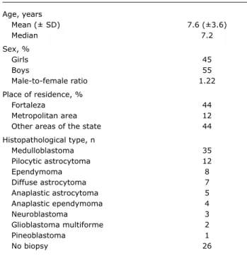
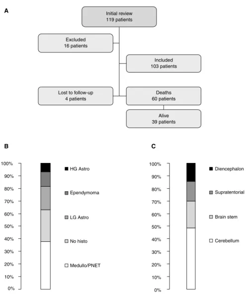
Of the 103 patients included in the study, 90 underwent chemotherapy in our center. There were complete records for 99 patients; 4 were lost to follow up. Four patients in full remission died due to treatment complications or non-associated causes. Mean age at diagnosis was 7.6 (±3.6), median age was 7.2, and prevalence was greater among boys (1.22:1 male-to-female ratio). A little more than half of the patients came from the metropolitan area (56%). Tumor histopathological types are summarized in Table 1. Medulloblastomas and PNET were the most prevalent tumors (38%), followed by low-grade, pilocytic and diffuse astrocytomas (18% altogether), adding up to 56% of the total. The percentage of tumors that were not biopsied was signiicant (25%), and most affected the brain stem. The cerebellum was the most frequent anatomic site (49%), followed by brain stem tumors (21%). Therefore, 70% of the patients had infratentorial tumors (Figure 1).
Figure 1A describes the review of our service database, which yielded data for 119 patients diagnosed with central
nervous system tumors from January 2000 to December 2006, as mentioned above. Of these patients, 16 were excluded because they did not meet inclusion criteria. The other patients (103) were included in the survival analysis. At the end of the evaluation, 4 patients had been lost to follow-up, 60 had died and 39 were alive. Figure 1B shows indings about histopathological type: medulloblastoma/ PNET (38%), no histological indings (25%), low-grade astrocytoma (18%), ependymoma (12%) and high-grade astrocytoma (7%). Figure 1C shows the rates of primary tumor anatomic sites: cerebellum (49%), brain stem (21%), other supratentorial tumors (15%) and diencephalon (15%).
Age, years
Mean (± SD) 7.6 (±3.6)
Median 7.2
Sex, %
Girls 45
Boys 55
Male-to-female ratio 1.22
Place of residence, %
Fortaleza 44
Metropolitan area 12
Other areas of the state 44
Histopathological type, n
Medulloblastoma 35
Pilocytic astrocytoma 12
Ependymoma 8
Diffuse astrocytoma 7
Anaplastic astrocytoma 5
Anaplastic ependymoma 4
Neuroblastoma 3
Glioblastoma multiforme 2
Pineoblastoma 1
No biopsy 26
table 1 - Epidemiological evaluation of patients with central nervous system tumors treated in the Service of Pediatric Oncology and Hematology of Hospital Infantil Alberto Sabin from January 2000 to December 2006
SD = standard deviation.
Survival analysis
HG Astro = high-grade astrocytoma; LG Astro = low-grade astrocytoma; PNET = primitive neuroectodermal tumors.
Figure 1 - (A) Flowchart of patient inclusion in the study. (B) Tumor distribution according to histopathological indings. (C) Tumor distribution according to primary anatomic site
tumors, in contrast, had a survival rate of about 51% at 5 years.
Prognostic factors of primary outcome for the whole cohort
Diagnosis of high-grade astrocytoma (HR = 3.7; 95%CI, 1.04-13; p = 0.04), ependymoma (HR = 3.9; 95%CI, 1.1-14; p = 0.04) and high grade according to WHO classiication (grade III-IV; HR = 2.4; 95%CI, 1.1-5.5; p < 0.01) were independent factors for poor prognosis. Complete surgical resection, in contrast, was associated with a better prognosis than incomplete resection (Figure 2) (HR – 0.5; 95%CI,
0.2 – 0.9; p = 0.05). In the same way, radiotherapy was a factor of better prognosis (HR = 0.5; 95%CI, 0.3-0.9; p < 0.05) (Figure 2).
Prognostic factors for the primary outcome only in the subgroup of gliomas (astrocytomas and ependymomas)
Figure 2 - (A) Survival curves according to histopathological type. (B) Survival curves according to tumor anatomic site. (C) Survival curves according to surgical resection (HR = 0.5; 95%CI 0.2-0.9; p = 0.05). (D) Survival curves according to radiotherapy (RT) (HR = 0.5; 95%CI 0.2-0.9; p = 0.05)
HG = high-grade; LG = low-grade; RT = radiotherapy.
Prognostic factors for primary outcome only in the subgroup of medulloblastomas and PNET
The only independent prognostic factor in this group was treatment with radiotherapy, which was associated with better survival rates (HR = 0.3; 95%CI, 0.1-0.8; p = 0.05). Patients with medulloblastoma or PNET had a better prognosis when compared with patients with high-grade astrocytomas, adjusted for the other variables, except treatment (HR – 0.4; 95%CI, 0.1-0.9; p < 0.01).
Prognostic factors for primary outcome only in the subgroup of cases with no biopsy
Age below 3 years, which correlated with better survival (HR = 0.2; 95%CI, 0.1-0.6; p = 0.05), radiotherapy (HR = 0.3; 95%CI, 0.1-0.6; p < 0.001) and recurrence (HR = 2.7; 95%CI, 1.2-5.9; p < 0.05), was an independent prognostic factor in this subgroup.
Survival
Median survival 12 months 24 months 60 months
All 28 months 67% 52% 45%
Histopathological type
Gliomas – 76% 76% 59%
Low-grade astrocytoma – 84% 84% 84%
High-grade astrocytoma 24 months 57% 57% 0%
Ependymoma 24 months 67% 33% 33%
Medulloblastoma/PNET 76 months 94% 57% 51%
Anatomic site
Other Supratentorial tumors 24 months 79% 47% 31%
Diencephalon – 65% 59% 59%
Cerebellum 76 months 79% 60% 51%
Brain stem 8 months 33% 29% 19%
table 2 - Evaluation of median survival at 12, 24 and 60 months of patients with central nervous system tumors according to histopathological type and anatomic site from diagnosis to date of death or censoring on October 10, 2010
PNET = primitive neuroectodermal tumors.
difference in survival according to site, and patients with diencephalic tumors had a 59% survival at 60 months, whereas patients with brain stem tumors had a survival of only 19% in the same period (Figure 2).
Discussion
The main prognostic factors for the primary outcome found for the whole group were histopathological type (high-grade astrocytoma and ependymoma), complete surgical resection and radiotherapy. Age, sex and the other variables did not have any statistically signiicant effect on survival of the whole cohort. The patients that underwent partial resection or biopsy and the inoperable patients were included in the same group because their survival curves were similar. There was a signiicant difference between survival curves for patients with completely and incompletely resected tumors, in agreement with other reports in the literature.16,17
Tumor site and histopathological type did not have an important correlation with surgical resection, showing that the effect did not depend on patient selection. Radiotherapy also seemed to have an association with better survival in this series. The analysis of subgroups showed that radiotherapy affected the survival of patients with medulloblastomas and PNET or with brain stem tumors, but not those with low-grade astrocytomas. This inding has already been described in the literature, but there are no studies with children only.18 The indication of radiotherapy in cases of low-grade astrocytoma is usually limited to patients that did
not undergo complete resection or had recurrent disease, which limits the evaluation of the role of radiotherapy. Because of the small number of patients that did not undergo chemotherapy in our series, its impact on survival could not be statistically evaluated. Despite advances in speciic pathologies, the role of chemotherapy remains to be better deined.19
Patients with medulloblastomas or PNET had a better prognosis if they underwent radiotherapy. Those that did not receive radiotherapy were children younger than 3 years, the age group with the worst results and lower survival rates. Postoperative radiotherapy is the main adjuvant treatment for medulloblastoma and PNET.17 Prognostic factors for overall survival of patients with gliomas in childhood are not found in the literature, except in a recent SEER retrospective analysis.20 SEER found that age below 3 years correlated with better prognosis of brain stem tumors, whereas incomplete resection and high-grade histology were factors of poorer prognosis in the group of patients under study. Our data are in agreement with those indings, and age and WHO high grade were risk factors for patients with gliomas in our study. Moreover, patients with an undeined histopathological type, most with brain stem tumors, had a better prognosis associated with age below 3 years. This is the irst report of prognostic factors among patients with gliomas in the Latin American literature and one of the few in the world.
References
1. American Cancer Society. Cancer Facts & Figures 2010. Atlanta: American Cancer Society, 2010.
2. Ries LAG, Smith MA, Gurney JG, Linet M, Tamra T, Young JL, Bunin GR, editors. Cancer incidence and survival among children and adolescents: United States SEER Program 1975-1995. Bethesda: National Cancer Institute, SEER Program; 1999.
3. Little J. Introduction. In: Little J. Epidemiology of childhood cancer. Lyon: International Agency for Research on Cancer: World Health Organization; 1999.
4. Brasil. Instituto Nacional de Câncer. Câncer da criança e adolescente no Brasil: dados dos registros de base populacional e de mortalidade. Rio de Janeiro: INCA; 2008.
5. Gurney JG, Smith MA, Bunin GR. CNS and miscellaneous intracranial and intraspinal neoplasms. In: Ries LAG, Smith MA, Gurney JG, Linet M, Tamra T, Young JL, Bunin GR, editors. Cancer incidence and survival among children and adolescents: United States SEER Program 1975-1995. Bethesda: National Cancer Institute, SEER Program; 1999.
6. Monteiro GTR, Koifman S. Mortalidade por tumores de cérebro no Brasil, 1980-1998. Cad Saúde Pública. 2003;19:1139-51. 7. Blaney SM, Kun LE, Hunter J, Rorke-Adams LB, Lau C, Strother
D, et al. Tumors of the Central Nervous System. In: Pizzo PA, Poplack DG, editors. Principles & Practice of Pediatric Oncology, 5th edition. Philadelphia: Lippincot Williams & Wilkins; 2006. 8. Louis DN, Ohgaki H, Wiestler OD, Canenee WK, editors. WHO
classiication of tumors of the central nervous system. Lyon: International Agency for Research on Cancer; 2007.
9. Steliarova-Foucher E, Stiller C, Lacour B, Kaastch P. International Classification of Childhood Cancer, Third Edition. Cancer. 2005;103:1457-67.
10. Kleinbaum DG, Klein M. Survival Analysis. A Self-Learning Text, 2nd edition, New York: Springer; 2005.
11. Adamo MB, Johnson CH, Ruhl JL, Dickie LA, editors. 2010 SEER Program Coding and Staging Manual. [Internet]. Bethesda: National Cancer Institute; 2010 [cited 2011 Jul 20]. Available from: http://seer.cancer.gov/manuals/2010/SPCSM_2010_maindoc. pdf.
12. Tamburrini G, D’Ercole M, Pettorini BL, Caldarelli, Massimi L, Di Rocco M. Survival following treatment for intracranial ependymoma: a review. Childs Nerv Syst. 2009;25:1303-12. tumor adjusted for age was 1.5 to 12 times greater than
that reported for Fortaleza from 1998 to 2002.4,6 This might probably be assigned to substantial under-referral. Although our medical service was the only center to receive pediatric patients with brain tumors for oncologic treatment during the study time, a signiicant number of patients might not have been referred to our center. Starting on December 2006, a restructuring of local health care services led to improvements in the referrals to our center. From 2007 to 2009, there were 96 new diagnoses of pediatric brain tumors (data not published or reported in this study), which would correspond to an annual incidence of 1.0 to 1.2 cases per 100,000 inhabitants, closer to the rates reported for Brazil and South America (from 0.9 to 3.7).
We compared our indings with data published by SEER.5 Astrocytomas were found at a smaller percentage (65% in SEER versus 25% in our series); medulloblastomas and PNET occurred in greater proportions (20% in SEER versus 30% in our series); and we had no other gliomas or spinal tumors in our series. The fact that most brain stem and diencephalic tumors were not biopsied in our series may be a source of the differences in astrocytoma frequencies. Another cause may be the fact that patients with completely resected low-grade cerebellar astrocytomas were not referred to our service and are, therefore, underrepresented in our center’s sample. In our series, brain stem and cerebellar tumors occurred at a greater proportion, in contrast with supratentorial tumors, including diencephalic tumors. Mean age of our patients conirms the predominance of cerebellar and brain stem tumors over telencephalic tumors.5
Overall 5-year survival among pediatric patients with brain tumors was similar to the SEER rates for medulloblastomas and PNET (55% in SEER versus 51% in our series). It was somewhat lower than rates reported in the literature for astrocytomas (73% in SEER versus 59% in our series) and much lower for ependymomas (56% in SEER versus 33%). We believe that such differences may relect a reduced number, probably due to under-referral, of low-grade astrocytomas (only 18% of the total number of tumors in our series). In a epidemiological series of 1195 pediatric patients with brain tumors operated on in Hospital de Clínicas of São Paulo from 1974 to 2003, 24.4% of the diagnoses were low-grade astrocytomas.21 The results for ependymomas are similar to the SEER indings up to 1984 (39% versus 33%). A comparison between SEER and European Population-based Data (EUROCARE) show that 5-year survival among children with ependymomas in Europe was closer to our series, and the same was seen in the comparison with a population series in England and Wales (43% versus 33% in our series).22,23 However, the small number of ependymomas in our study (n = 12) does not warrant deinitive conclusions.
The analysis of survival of pediatric patients with brain tumors in Latin America remains limited. A series of 39
patients with astrocytomas diagnosed from 1989 to 1995 in Mexico24 showed that overall survival was 52% ive years after diagnosis. Another report found an overall 5-year survival of 58% among 101 pediatric patients with medulloblastoma treated in the Brazilian National Institute of Cancer (INCA) from 1983 to 2001.25 An evaluation of 64 pediatric patients with brain stem tumors, including tumors of the pons, found a median survival of 8.8 to 14.6 months and prolonged survival of 6%.26 We found no publications of Brazilian studies that evaluated prognostic factors in series of pediatric patients with CNS tumors in journals indexed in SciELO, LILACS and PubMed.
13. Wilne S, Collier J, Kennedy C, Koller K, Grundy R, Walker D. Presentation of childhood CNS tumors: a systematic review and meta-analysis. Lancet Oncol. 2007;8:685-95.
14. Grambsch P, Therneau T. Proportional Hazards tests and diagnostics based on weighted residuals. Biometrika. 1994;81:515-26. 15. R Development Core Team. R: A language and environment
for statistical computing [Internet]. Vienna: R Foundation for Statistical Computing; 2009 [cited 2011 Jul 20]. Available from: http://www.R-project.org.
16. Wisoff JH, Boyett JM, Berger MS, Brant C, Li H, Yates AJ, et al. Current neurosurgical management and the impact of the extent of resection in the treatment of malignant gliomas of childhood: a report of the Children’s Cancer Group Trial No. CCG-945. J Neurosurg. 1998;89:52-9.
17. Packer RJ, Vezina G. Management of and prognosis with medulloblastoma: therapy at a crossroads. Arch Neurol. 2008;65:1419-14.
18. Berg G, Blomquist E, Cavallin-Stahl E. A systematic overview of radiation therapy effects in brain tumors. Acta Oncol. 2003;42:582-8.
19. Bouffet E, Tabori U, Huang A, Bartels U. Possibilities of new therapeutic strategies in brain tumors. Cancer Treat Rev. 2010;36:335-41.
20. Qaddoumi I, Sultan I, Gajjar A. Outcome and prognostic features in pediatric gliomas: a review of 6212 cases from the Surveillance, Epidemiology, and End Results database. Cancer. 2009;115:5761-70.
21. Rosemberg S, Fujiwara D. Epidemiology of pediatric tumors of the
central nervous system according to the WHO 2000 classiication:
a report of 1,195 cases from a single institution. Childs Nerv Syst. 2005;21:940-4.
Correspondence: Francisco H. C. Felix Hospital Infantil Albert Sabin Rua Tertuliano Sales, 544
CEP 60410-790 - Fortaleza, CE - Brazil Tel.: +55 (85) 3257.9613
E-mail: [email protected]
22. Gatta G, Capocaccia R, Coleman MP, Ries LAG, Berrino F. Childhood cancer survival in Europe and the United States. Cancer. 2002;95:1767-72.
23. Tseng JH, Tseng MY. Survival analysis of children with primary malignant brain tumors in Englang and Wales: a population-based study. Pediatric Neurosurg. 2006;42:67-73.
24. Lopez-Aguillar E, Cerecedo-Diaz F, Sepulveda-Vidosola AC, Rivera-Marquez H, Castellanos-Toledo A, Arias-Gomez J, et al. Astrocitomas en pediatria. Factores pronósticos y sobrevida. Gac Med Mex. 1997;133:231-5.
25. Santos MA, Viegas CMP, Servidoni RA, Barros MHM, Pinel MI, Araujo CMM. Timing of radiation in children with medulloblastoma/PNET. Pediatr Blood Cancer. 2007;48:416-22.