Vascular injuries of the upper extremity
Lesões vasculares de membros superiores
Raafat Shalabi1, Yoysifh Al Amri2, Elham khoujah3
271
Abstract
Objective: T his study analyzes the causes of injuries, presentations, surgical approaches, outcome and complications of vascular trauma of the upper limbs, in spite of limited hospital resources.
Methods: A 5-year retrospective analysis. From 01/01/2001 to 31/12/2005, 165 patients were operated for vascular injuries at King Fahd Hospital, Medina, Saudi Arabia. O f all peripheral vascular trauma patients (115), upper limb trauma was present in 58. Diagnosis was made by physical examination and hand-held D oppler alone or in combination with Doppler scan/angiography. Primary vascular repair was performed whenever possible; otherwise, the interposition vein graft was used. Fasciotomy was considered when required. Patients with unsalvageable lower extremity injury requiring primary amputation were excluded from the study.
Results: Fifty patients were male (86%) and eight were female (14%), aged between 2.5-55 years (mean 23 years). Mean duration of presentation was 8 h after the injury. T he most common etiological factor was road traffic accidents, accounting for 50.5% in the blunt trauma group and 33% among all penetrating and stab wound injuries. Incidence of concomitant orthopedic injuries was very high in our study (51% ). T he brachial artery was the most affected (51% ). Interposition vein grafts were used in 53% of the cases. Limb salvage rate was 100%.
Conclusion: Patients who suffer vascular injuries of the upper extremities should be transferred to vascular surgery centers as soon as possible. Decisive management of peripheral vascular trauma will maximize patient survival and limb salvage. Priorities must be established in the management of associated injuries, and delay must be avoided when ischemic changes are present.
Keywords: Vascular trauma, upper extremity, vein interposition.
Resumo
Objetivo: Este estudo analisa as causas de lesões, apresentação, abordagens cirúrgicas, desfechos e complicações do trauma vascular de membros superiores, apesar de recursos hospitalares limitados.
Métodos: Análise retrospectiva de 5 anos. De 01/01/2001 a 31/ 12/2005, 165 pacientes foram operados devido a lesões vasculares no King Fahd Hospital, Medina, Arábia Saudita. De todos os pacientes com trauma vascular periférico (115), trauma de membros superiores esteve presente em 58. O diagnóstico foi realizado por exame físico e Doppler manual isoladamente ou associado com ultra-som Doppler/ angiografia. A restauração vascular primária foi realizada sempre que possível; do contrário, utilizou-se a interposição de veia. A fasciotomia foi considerada quando necessário. Pacientes com lesão de membro inferior não resgatável necessitando de amputação foram excluídos do estudo.
Resultados: Cinqüenta pacientes eram homens (86%) e oito eram mulheres (14%), com idade entre 2,5 e 55 anos (média de 23 anos). A duração média de apresentação foi 8 h pós-lesão. O fator etiológico mais comum foi acidente em estradas, sendo responsável por 50,5% no grupo de trauma contuso e 33% entre as lesões penetrantes e por arma branca. A incidência de lesões ortopédicas concomitantes foi muito alta em nosso estudo (51%). A artéria braquial foi a mais afeta-da (51%). A interposição de veias foi utilizaafeta-da em 53% dos casos. A taxa de preservação de membros foi de 100%.
Conclusão: Pacientes que sofrem lesões vasculares de membros superiores devem ser transferidos para centros de cirurgia vascular o mais rápido possível. O tratamento imediato do trauma vascular peri-férico aumentará a sobrevida dos pacientes e a preservação dos mem-bros. Devem-se estabelecer prioridades no tratamento de lesões asso-ciadas e evitar o atraso quando alterações isquêmicas estiverem pre-sentes.
Palavras-chave: Trauma vascular, membro superior, interposição de veia.
1. MD. Consultant in Vascular Surgery, King Fhad Hospital, Medina Mou-nwara, Saudi Arabia.
2. MD. Consultant in Causality and Orthopedic Surgery, King Fhad Hospi-tal, Medina Mounwara, Saudi Arabia.
3. MD, Consultant in Vascular Surgery, King Fhad Hospital, Medina Mou-nwara, Saudi Arabia.
Artigo submetido em 13.11.06, aceito em 27.12.06. J Vasc Bras 2006;5(4):271-6.
Copyright © 2006 by Sociedade Brasileira de Angiologia e de Cirurgia Vascular.
Introduction
Vascular injury is a major complication of military and civilian trauma. Major developments in this field have been related to military conflicts during the past 100 years.1
the majority of upper extremity vascular injuries are due to penetrating trauma, with progressively increasing numbers attributable to iatrogenic causes. Blunt injuries account for 6-10% of upper extremity vascular trauma and are often associated with musculoskeletal injuries and neural injuries.3
However, the mechanism of injury seems to differ between different parts of the world.4,5 While successful treatment of major arterial injuries may be life-saving as well as allowing limb salvage and restoration of function,6 return of function is often related to the presence of concomitant injury to peripheral nerves.3
Upper limb vascular trauma can therefore be associated with major morbidity and mortality, but little is known about its incidence or nature in Medina, Saudi Arabia. A retrospective study of 58 patients requiring operative intervention for upper limb vascular trauma over a 5-year period between 01/01/2001 and 31/12/2005 was performed. In this report we present the different mechanisms of trauma, arteries involved, associated orthopedic or nerve injuries and types of vascular repairs employed. T o the best of our knowledge, this is the first report on upper limb vascular trauma from this region of Saudi Arabia, which is highly respectable to all Muslims worldwide.
Patients and methods
During the 5-year period, 58 patients presented with upper extremity vascular injuries at King Fahd Hospital, Medina, Saudi Arabia. All patients underwent full physical examination and resuscitation according to the principles of the advanced trauma and life support (AT LS) guidelines. T he patients were either first assessed by emergency room residents or were referred from the orthopedic surgeon after finding absent distal pulses in a patient with fracture or fracture/ dislocation of the extremity (Figures 1 and 2).
T he diagnosis of an upper extremity vascular injury is initially made by physical examination as part of the full trauma assessment. T he classic five P’s – pain, pulselessness, pallor, paresthesias, and paralysis – may be partially present or may be absent in many patients. Some patients with axillary and proximal brachial artery injuries may have palpable pulses at the wrist. T he injury type and location are noted, and the axillary, brachial, radial, and ulnar arteries are palpated for pulsations. D epending on the mode of presentation, most patients were taken immediately
to the operating room for vascular or orthopedic/ vascular management. In others with soft signs or doubtful vascular injury (especially with hematoma, compartmented limb or for medico-legal reasons) and when patients were stable, preoperative duplex ultrasonography/angiography were performed (Figure 3).
All patients with associated orthopedic injury underwent reduction of joint dislocation or bone fracture and immobilization by internal or external fixation (Figure 4).
It always preceded vascular repair unless the extremity was threatened and required immediate revascularization. Endoluminal shunts were not used in any patient. Patients with more severe soft tissue
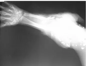
Figure 1 - X-ray of massive bone fracture (gun missiles)
Figure 4 - Pre-revascularization bone fixation
Figure 3 - Preoperative angiography, supracondylar fracture and muscle injuries were treated with thorough debridement of all grossly nonviable tissue, with removal of foreign bodies and copious irrigation with isotonic saline performed by us and the orthopedic surgeons (Figure 5). Suitable covers for the defect were developed by plastic surgeons with split-skin grafting or with the application of flap techniques.
Repaired vessels, especially at the anastomotic suture lines and graft location, were compulsory covered with muscles and soft tissue to prevent desiccation and disruption. I n all patients, management of vascular injuries was performed in the operating room under general anesthesia using standard vascular techniques. D epending on the condition of the limb after revascularization, open full fasciotomy was carried out liberally to either relieve existing compression or to avoid one from occurring in the postoperative period (Figure 6).
Fasciotomy wounds were usually covered later by a delayed primary, split-thickness skin graft (Figure 7).
An intraoperative angiogram was performed in few cases, whenever distal pulses were absent after the revascularization or assessing injury site or spasm.
Figure 7 - Delayed primary, split-thickness skin graft
Figure 6 - Fasciotomy after brachial artery repair
Successful repair was assessed by the return of distal pulses at the end of the operation. Although associated nerve injuries were not usually repaired at the time of vascular repair, major associated venous injuries were repaired whenever possible, in an attempt to prevent postoperative venous hypertension and to minimize development of compartment syndrome. Patients with unrepaired nerve injuries were postoperatively followed by our colleagues in neurosurgery. Four weeks after hospital discharge, patients were routinely examined in the outpatient department (OPD) to assess functional status of the limb. T hereafter, they were followed at longer periods of time. All patients received intravenous preoperative prophylactic antibiotics, which were continued postoperatively for 5 to 7 days, unless prolonged use was dictated by the presence of contamination or infection, or else advised by the attending orthopedic/plastic surgeons. All patients also received intravenous heparin for a period of 5-7 days postoperatively and were discharged home on oral aspirin 100 mg tablet/day for a period of 12 weeks.
Patients with isolated venous trauma and patients with unsalvageable lower extremity injury requiring primary amputation were excluded from the study.
Results
Successful outcome in vascular trauma depends on early diagnosis and referral to specialists. In our series, most patients presented what is considered as the “golden period.” T he time interval between the beginning of the trauma and arrival to our center was 8 h in average.
T he patients consisted of 50 males (86% ) and eight females (14% ) with mean age of 23 years (range 2.5-55 years). T he left and right upper limbs were equally involved in 3 9 patients (5 0 % ). T he mechanism of trauma was blunt in 39 patients (67% ) and penetrating in the remaining 19 patients (33% ). However, road traffic accident (RT A) was the most common cause of upper limb vascular injury in this group of patients, occurring in 35 patients (52.5% ). Stab injury was the most frequent form of penetrating trauma (11 out of 19). O ther forms of trauma in a descending order of frequency were cut wrist in four patients (7% ), industrial or machinery such as electric saw or hand drilling machine in two patients (3.5% ), gunshot in three patients (5% ) and fall to the ground in six patients (10% ).
T hirty patients (51%) presented with ischemia, 20 patients with bleeding (34%) and eight with hematoma (13.5%).
T he brachial artery was the most frequently affected (30 patients, 51%), followed by the ulnar artery alone in three patients (5%), both the ulnar and radial arteries in 12 patients (20.5%) and the radial artery alone in seven patients (12%). T he axillary artery was involved in five patients (8.5%), and the subclavian arteries in one patient (1.7%).
T he vascular injury was more often associated with orthopedic injuries (30 patients, 51%). Orthopedic injuries were in the form of supracondylar fracture in 13 patients (21%), fracture/dislocation in seven patients (12%) and dislocation alone in seven patients (12%).
Concomitant vein or nerve injury also occurred in 35 patients (59.5%). Associated nerve injury occurred in 13 patients (22%), vein injury in 22 patients (37.5%) and both occurred in 15 patients (22.5%).
In 50 patients (86%) the diagnosis of arterial injury was based on clinical and hand-held D oppler examination. Preoperative angiography was used in two patients (3.5%). Duplex scan was used in only 10 patients (17%).
Arterial repair performed by interposition vein graft in 31 patients (53% ) was the most frequently used single technique of arterial repair. O ther techniques used were ligation in 10 patients (17% ); primary anastomosis by end-to-end anastomosis in 17 patients (29% ). Venous bypass grafting was used in one patient (1.7% ). Repair of major venous injuries was performed in two patients (3.5% ). T herapeutic or prophylactic fasciotomy was performed in nine patients (15% ).
No patient underwent amputation and no one died as a consequence of upper limb vascular injury. A limb salvage rate of 100% was therefore achieved.
Discussion
Specific surgical techniques8 must be mastered if successful vascular repair is to be achieved. T hese include proximal and distal exposure for control with vascular clamps and loops; dissection and isolation of injured vessels including veins; local and/or systemic heparinization; use of vascular sutures; magnification loops; assessment of injury: debridement, contusion, intimal flap and distal dissection and thrombosis; selective use of shunting; anatomic repairs, with vein patch, end/end anastomosis without tension and reversed autologous vein graft for larger defects; technical details of spatulated ends, running vs. interrupted sutures; distal thrombectomy;9 completion arteriography;10 fasciotomy and soft tissue coverage. Proper handling of the autogenous vein graft is important.11
T he timing of the vascular repair in relation to fracture management has long been a source of controversy. T he standard recommendation is for vascular repair to precede orthopedic management. Prevention of prolonged tissue ischemia is the objective. While there are no prospective studies, McHenry,12 in a retrospective study, suggested an increased need for fasciotomy when fractures are stabilized before revascularization. No cases of disruption of vascular repair occurred in 22 cases of subsequent fracture stabilization. Most fractures can be adequately stabilized with traction or posterior plaster splinting, but external fixation may be necessary in some cases. Volgas13 gives a good review of ballistic injury management.
Imaging, in particular contrast arteriography, has played an important role in the development of vascular surgery. While it is clear that advanced imaging techniques are important in the management of zones 1 and 3 neck injuries14 and thoracic aortic disruption15, for most extremity vascular injuries preoperative arteriography is not necessary. However, on-table operative angiograms are easily carried out with a minimum amount of equipment and provide important information about the extent of injury and the adequacy of repair.10 In suspected vascular injury exclusion, arteriography has been shown to be cost-effective.16 T he literature is full of epidemiological studies describing the features of vascular trauma in various countries.17-25 T here is wide variation in the incidence, cause and mechanism of injury, depending on the local conditions. In a civilian population in Australia21,25, vascular injuries represent 1-2% of total trauma patients. However, they account for 20% of all trauma-related death.21 Deaths from vascular injury vary considerably with anatomical
location and mechanism of injury. T horacic and abdominal injuries routinely have death rates between 30-50%; vascular injuries to extremities are significantly lower in the range of 5%. In an unparalleled large study in Vietnam, Rich26 reported a total death of only 1.7% for all vascular injuries. It may be that life-threatening vascular injuries were preselected by their failure to survive transportation. In the current warfare conditions of the American intervention in Iraq and Afghanistan27, vascular trauma represents 7% of total battle injuries, 88% of these were extremity injuries. T he amputation rate was only 8% after vascular repair.
In North India,19 with a low risk of personal violence, blunt injuries, mostly motor vehicle accidents, account for 84% of vascular injuries. Whereas in Medellin, Colombia,22 93% of vascular injuries are penetrating and in Georgia18 they represent 85% of the total. Surprisingly, in the European experience,24 up to 40% of vascular injuries are iatrogenic, as a result of vascular and other surgical interventions. Kuwait17 strikes a middle ground with 41% penetrating, 23% a result of RT A and 22% iatrogenic. In Malaysia,20 over 50% of vascular injuries occur as a result of RT A.
As far as anatomic site of injury is concerned, variability is less. In Australia21,25, injuries are split almost equally between thorax, abdomen and upper and lower extremities, with cervical injuries being less common. In Latin America,23 extremity injuries are twice as common as thoracic and abdominal, although these later result in higher mortality. As far as extremities are concerned, upper and lower injuries occur with similar frequency and the brachial, femoral and popliteal arteries are the most commonly injured vessels. With special relevance to conditions in Africa, in the Latin American survey,23 68% of cases were managed on a clinical basis alone, i.e., without arteriography, and 78% were managed within 6 h of injury.
Among other findings, this study showed that the main victims of upper limb vascular injury in this region are males, with female patients forming 14% of the total.
T he brachial artery was the most frequently affected artery in our patients at a rate of 51%, which is in agreement with most previous reports of between 37-66%. T he most frequent type of vascular repair was interposition vein graft,using the thigh long saphenous vein, at a rate of 53%. Intraluminal shunts were not used in any of our patients without adverse effect, this agreed with W ally. Fasciotomy is especially recommended in cases of established ischemia, as previously pointed out by Fletcher & Little. No amputation performed in this series compared with reports from Wally (4%), Brown et al. (6%) and Kruse-Andersen et al. (28%).2
T he functional outcome depended on the associated nerve injuries as previously pointed out by different authors.2 No mortality was reported in our patients.
Conclusion
Prompt vascular repair of the upper extremity and attention to associated injuries result in a minimum morbidity and zero mortality.In our study, we dealt with vascular injuries and associated problems in one session and these patients performed very well in the follow-up.
Increasing number of religious tourists, especially in our region (Medina Mounwara), has resulted in increasing number of RT A, higher than stab or penetrating trauma.
Correspondence: Raafat Shalabi
King Fhad Hospital, Medina Mounwara, Saudi Arabia Phone: +9664846150s0 / 3219, +966506319117 Fax: +96648461190
E-mail: [email protected]
References
1. Hood DB, Yellin AE, Weaver F. Vascular trauma. In: Dean RH, Yao JST , Brewster DC, editors. Current diagnosis & T reatment in Vascular surgery. Connecticut: Lange; 2000. p. 405-28. 2. Wali MA. Upper limb vascular trauma in the Asir region of
Saudi Arabia. Ann T horac Cardiovasc Surg. 2002;8:298-301. 3. Hunt CA, Kingsley JR. Vascular injuries of the upper extremity.
South Med J. 2000;93:466-8.
4. Creagh T A, Broe PJ, Grace PA, Bouchier-Hayes DJ. Blunt trauma-induced upper extremity vascular injuries. J R Coll Surg Edinb. 1991;36:158-60.
5. Shaw BA, Kasser JR, Emans JB, Rand FF. Management of
vascular injuries in displaced supracondylar humerus fractures without arteriography. J Orthop T rauma. 1990;4:25-9. 6. Kruse-Andersen S, Lorentzen JE, Rohr N. Arterial injuries of
the upper extremities. Acta Chir Scand. 1983;149:473-7. 7. Iriz E, Kolbakir F, Sarac A, Akar H, Keceligil HT , Demirag MK.
Retrospective assessment of vascular injuries: 23 years of experience. Ann T horac Cardiovasc Surg. 2004;10:373-8. 8. Rutherford RB. Basic vascular surgical techniques. In: Rutherford
RB, editor. Vascular surgery. Philadelphia: WB Saunders; 2004. p. 395-404.
9. Fogarty T J. Fogarty catheter thrombectomy. In: Rutherford RB, editor. Vascular surgery. Philadelphia: WB Saunders; 2004. p. 410-4.
10. Subber SW. Contrast arteriography (excerpt). In: Rutherford RB, editor. Vascular surgery. Philadelphia: WB Saunders; 2004. p. 195-202.
11. T owne JG. T he autogenous vein. In: Rutherford RB, editor. Vascular surgery. Philadelphia: WB Saunders; 2004. p. 482-91. 12. McHenry T P, Holcomb JB, Aoki N, Lindsey RW. Fractures with major vascular injuries from gunshot wounds: implications of surgical sequence. J T rauma. 2002;53:717-21.
13. Volgas DA, Stannard JP, Alonso JE. Current orthopaedic treatment of ballistic injuries. Injury. 2005;36:380-6. 14. Ferguson E, Dennis JW, Vu JH, Frykberg ER. Redefining the
role of arterial imaging in the management of penetrating zone 3 neck injuries. Vascular. 2005;13:158-63.
15. Blackmore CC, Zweibel A, Mann FA. Determining risk of traumatic aortic injury: how to optimize imaging strategy. AJR Am J Roentgenol. 2000;174:343-7.
16. Keen JD , Keen RR. T he cost-effectiveness of exclusion arteriography in extremity trauma. Cardiovasc Surg. 2001;9: 441-7.
17. Asfar S, Al-Ali J, Safar H, et al. 155 vascular injuries: a retrospective study in Kuwait, 1992-2000. Eur J Surg. 2002;168:626-30. 18. Razmadze A. Vascular injuries of the limbs: a fifteen-year
Georgian experience. Eur J Vasc Endovasc Surg. 1999;18: 235-9.
19. Menakuru SR, Behera A, Jindal R, Kaman L, Doley R, Venkatesan R. Extremity vascular trauma in civilian population: a seven-year review from North India. Injury. 2005;36:400-6. 20. Lakhwani MN, Gooi BH, Barras CD. Vascular trauma in Penang and Kuala Lumpur Hospitals. Med J Malaysia. 2002;57:426-32.
21. Sugrue M, Caldwell EM, Damours SK, Crozier JA, Deane SA. Vascular injury in Australia. Surg Clin North Am. 2002;82: 211-9.
22. Morales-Uribe CH, Sanabria-Quiroga AE, Sierra-Jones JM. Vascular trauma in Colombia: experience of a level I trauma center in Medellin. Surg Clin North Am. 2002;82:195-210. 23. Sonneborn R, Andrade R, Bello F, et al. Vascular trauma in
Latin America: a regional survey. Surg Clin North Am. 2002;82: 189-94.
24. Fingerhut A, Leppaniemi AK, Androulakis GA, et al. T he European experience with vascular injuries. Surg Clin North Am. 2002;82:175-88.
25. Gupta R, Rao S, Sieunarine K. An epidemiological view of vascular trauma in Western Australia: a 5-year study. ANZ J Surg. 2001;71:461-6.
26. Rich NM. Complications of vascular injury management. Surg Clin North Am. 2002;82:143-74.