Licenciado sob uma Licença Creative Commons
[T]
Determination of muscle fatigue index for strength
training in patients with Duchenne dystrophy
[I]
Determinação do índice de fadiga muscular para treinamento de força
em pacientes com distroia de Duchenne
[A]
Adriano Rodrigues Oliveira[a], Fernanda Ishida Corrêa[b], Márcia Moreira Valim[c],
Cláudia Santos Oliveira[d], João Carlos Ferrari Corrêa[e]
[a] Fisioterapeuta, Mestrando em Ciências da Reabilitação, Universidade Nove de Julho (UNINOVE), São Paulo, SP - Brasil, e-mail:
[b] Fisioterapeuta, Doutora em Engenharia Biomédica, Universidade Nove de Julho (UNINOVE), São Paulo, SP - Brasil, e-mail:
[c] Fisioterapeuta, Mestranda em Ciências da Reabilitação, Universidade Nove de Julho (UNINOVE), São Paulo, SP - Brasil, e-mail:
[d] Fisioterapeuta, Doutora em Ciências da Saúde, Universidade Nove de Julho (UNINOVE), São Paulo, SP - Brasil, e-mail:
[e] Fisioterapeuta, Doutor em Morfologia, Universidade Nove de Julho (UNINOVE), São Paulo, SP - Brasil, e-mail: [email protected]
[R]
Abstract
by Pearson’s test on the median frequency of each strength level. Conclusion: Electromyographic mea-surements of the strength index for muscle training proved to be a simple accessible assessment method, as well as an extremely valuable tool, allowing the design of a muscle strength training program with an individualized load threshold.
[P]
Keywords: Duchenne. Fatigue. EMG. [B]
Resumo
Introdução: Sabe-se que a fraqueza muscular é o prejuízo mais proeminente na distroia muscular de Duchenne (DMD), envolvendo frequentemente perda da habilidade funcional e outras restrições relativas à vida diária. Pensando nisso, existe a necessidade de se manter a força muscular de grandes grupos musculares, tais como o quadríceps femoral, responsável por diversas habilidades funcionais; porém, a carga e a periodização de treina-mento para tal reabilitação tem se mostrado uma grande incógnita, principalmente pelo aparecitreina-mento indesejado da fadiga muscular, fator severo para o acometimento lesional das ibras musculares. Objetivos: Este estudo objetivou determinar, por meio da eletromiograia de superfície (EMG), um índice de fadiga que sirva de parame-trização para treinamento isioterapêutico de fortalecimento muscular. Metodologia: Participaram deste estudo transversal (série de casos) quatro pacientes com DMD. Três pares de eletrodos de superfície foram colocados sobre o ponto motor dos músculos reto femoral, vasto lateral e vasto medial do membro dominante, mantendo o joelho em lexão de 60º, sendo instruídos a realizar o movimento de extensão desta articulação em quatro níveis de força (100%, 80%, 60% e 40% da contração voluntária máxima isométrica). Resultados: Para determinar o índice de fadiga, utilizou-se o ângulo de inclinação para a linha de regressão linear, realizada pelo teste de Pearson da frequência mediana de cada nível de força coletado. Conclusão: Medidas eletromiográicas do índice de força para treinamento muscular mostraram-se um método simples e acessível de avaliação, além de se apresentar como uma ferramenta extremamente valiosa, permitindo traçar um programa de treinamento de força muscular com limiar de carga especíico individualmente.
[K]
Palavras-chave: Duchenne. Fadiga. EMG.
Introduction
Duchenne muscular dystrophy (DMD) is characterized by a recessive genetic disorder with a high mutation rate of a gene in the short arm of the X chromosome in a region denominated Xp21 (1). Unless an older brother had previously developed the condition or there is a signiicant family history of the disease, most infants are not monitored for symptoms. However, in children in whom there has been an early identi-ication because of a family history, the Gowers’ sign (compensatory maneuver when standing up from the ground, in which the patient uses his/her hands to climb up the lower limbs due to a lack of muscle strength in the thighs and hips) may be evident by 15 months of age.
Most children with DMD are not identiied until reaching three to ive years of age. Beginning at this age, there is notable muscle weakness that selectively affects the proximal muscles before the distal muscles and the lower limbs before the upper limbs (2, 3). When the child begins to stand or walk, he/she tends to fall and the gait is called “anserine”. With the progression of the disease, there is increasing dificulty in walking, which generally becomes impossible between 8 and 12 years of age (4, 5).
the principal muscles makes the musculature work at a greater percentage of maximal capacity, recruiting less eficient secondary muscles, which increases energy expenditure and leads to fatigue (6-9).
Muscle weakness is the most prominent feature of muscular dystrophy and impedes mobility. This frequently involves a loss of the ability to walk and leads to other limitations to daily living. The usefulness of resistance strength training for such patients has been discussed (10-12). Progressive resistance strength training increases muscle size, strength and endurance in healthy individuals. In muscular dystrophy, how-ever, the situation is not as clear (13). The load and training period for such rehabilitation has proven to be a great unknown thus far, principally due to the appearance of fatigue, which is a severe factor for the injury of muscle ibers in this population. Due to this constant weakness, there is a need to maintain the muscle strength of the large muscle groups, such as the femoral quadriceps muscle, which is responsible for different functional abilities such as walking, climbing up and down stairs and getting up from a chair or bed (14, 15).
Considering the importance of these muscles in activities that affect the independence of patients with DMD, the aim of the present study was to determine a fatigue index by means of surface electromyog-raphy (EMG) that can serve as parameterization for physiotherapy training.
Methodology
A cross-sectional case series study was carried out at the Physiotherapy Clinic of the Universidade Nove de Julho (São Paulo, Brazil), involving four patients with DMD. Orientation was given to the volunteers regarding the physical activities to be executed during the data collection and the subjects were informed as to the objectives of the study. The volunteers were told that they could remove themselves from the study at any time with no negative repercussions. The subjects read and signed terms of informed consent agreeing to participate. The study received approval from the Ethics Committee of the institution (process n. 133.260/2007).
The EMG signal acquisition system consisted of a load cell (SV-100 model) with a nominal capac-ity of 100 kg, made with anodized aluminum, with a sensitivcapac-ity of ± 10% and three pairs of active, bipolar, differential electrodes for capturing the electrical activity of the muscles. The signal was pre-ampliied in the differential electrode with a ten-fold gain, with a common rejection of 80 dB and sampling frequency of 2.000 Hz. The components of the signal acquisition system were connected to a signal conditioner module, in which the analogue signals were iltered in a 10 Hz to 1.000 Hz band-pass ilter and ampliied again with a 100-fold gain, achieving a inal gain of 1000. The three pairs of surface electrodes were placed on the motor point of the Rectus femoris (RF), Vastus lateralis (VL) and Vastus medialis (VM), muscles of the dominant limb following the longitudinal direction of the muscle ibers (ixed inwardly to the skin by double-faced adhesive tape, with another piece of adhesive tape used outwardly, thereby providing better ixation of the electrodes) (16, 17).
The patients were positioned sitting in a chair, maintaining the knee joint a 60º of lexion and were instructed to perform extension of this joint at four strength levels (100%, 80%, 60% and 40% of maximal voluntary isometric contraction – MVIC) sustained for thirty seconds, with a three-minute rest period between each strength level. The same procedure was repeated twice, for a total of three series (18).
Results
An important limitation of our study was the sample number. The initial sample had 13 patients, from which nine were excluded, resulting in just four patients. Four patients were excluded for presenting severe postural deviations associated with DMD; one patient was excluded for presenting hearing alteration; and four patients were excluded for their absence in the scheduled dates for data collection and evaluation. Another limitation found wasn’t using a control group, that because the musculoskeletal and functional char-acteristics can not be compared.
In order to exemplify and simplify the visualization of the results obtained in the present study, only the data on the Rectus femoris muscle of volunteer 1 are used, which is suficient for the understanding of how these data were analyzed. Another point that should be stressed is that the entire analysis of the data was performed as intra-subject analysis, since biological individuality – which is the primary principle of physical training – does not permit inter-subject analysis.
Fatigue and Range of the Myoelectrical Signal
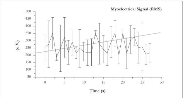
Figure 1 displays the EMG signal of the Rectus femoris (mean curve with respective standard deviation representative of three contractions performed by volunteer 1), given as RMS. Based on the linear regression line, there was an initial increase in values, thereby demonstrating the need of the Rectus femoris to recruit a greater number of motor units in order to maintain the strength level stipulated for the data collection (80% of MVIC).
Figure 1 - Alterations in myoelectrical activity (RMS) over time, resisted at 80% of MVIC (Rectus
femoris of Volunteer 1)
Fatigue and Fmed
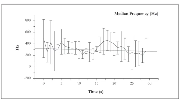
Figure 2 presents the mean curve and standard deviation of the representative Fmed of three con-tractions of the Rectus femoris performed by volunteer 1 and shows the linear regression line determined by Pearson’s test in the time domain. The initial Fmed values underwent a decline during the sustaining of muscle contraction, which demonstrates the appearance of muscle fatigue.
500
450
400
350
300
250
200
150
100
50
0 5 10
Time (s)
Myoelectrical Signal (RMS)
(u.V
.)
Muscle fatigue index
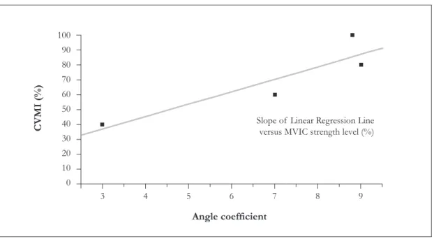
The coeficients of the angle of the slope of the linear regression line (determined by Pearson’s test) for the median frequency of the four strength levels (100%, 80%, 60% and 40% of MVIC) of the RF, VL and VM muscles in volunteer 1 are displayed in Figures 3, 4 and 5, respectively.
Figure 2 - Alterations in Fmed over time, resisted at 80% of MVIC (Rectus femoris of Volunteer 1)
Figure 3 - Slope of linear regression line (determined by Pearson’s test) for median frequency of each strength level measured for the Rectus femoris of Volunteer 1 (abscissa axis) crossed with respective strength levels (ordinate axis). The linear regression line obtained by the slope of each strength level (100, 80, 60, 40% of MVIC) establishes a new linear regres-sion line, which crosses the ordinate axis (percentage of strength level), demonstrating the fatigue index for the muscle analyzed
-200 0 200 400 600 800
0 5 10 15 20 25 30
Time (s)
Median Frequency (Hz)
Hz
0 10 20 30 40 50 60 70 80 90 100
4 5 6 7 8 9 10
Slope of Linear Regression Line versus MVIC strength level (%)
Angle coefficient
Figure 4 - Slope of linear regression line (determined by Pearson’s test) for median frequency of each strength level measured for the Vastus lateralis of Volunteer 1 (abscissa axis) crossed with respective strength levels (ordinate axis). The linear regression line obtained by the slope of each strength level (100, 80, 60, 40% of MVIC) establishes a new linear regres-sion line, which crosses the ordinate axis (percentage of strength level), demonstrating the fatigue index for the muscle analyzed
Figure 5 - Slope of linear regression line (determined by Pearson’s test) for median frequency of each strength level measured for the Vastus medialis of Volunteer 1 (abscissa axis) crossed with respective strength levels (ordinate axis). The linear regression line obtained by the slope of each strength level (100, 80, 60, 40% of MVIC) establishes a new linear regres-sion line, which crosses the ordinate axis (percentage of strength level), demonstrating the fatigue index for the muscle analyzed
0 10 20 30 40 50 60 70 80 90 100
3 4 5 6 7 8 9
Slope of Linear Regression Line versus MVIC strength level (%)
Angle coefficient
CVMI (%)
0 10 20 30 40 50 60 70 80 90 100
3 4 5 6 7 8 9
Slope of Linear Regression Line versus MVIC strength level (%)
Angle coefficient
The muscle fatigue values obtained from the new regression line were 35% of MVIC for the Rectus femoris, 30% of MVIC for the Vastus lateralis and 32% of MVIC for the Vastus medialis. Thus, the muscle fatigue index for the femoral quadriceps muscle of volunteer 1 was 32.33 ± 2.51 of MVIC. For the other volunteers analyzed in the present study, the muscle fatigue index for the femoral quadriceps muscle was 43.54±3.25 of MVIC in volunteer 2, 25.12±7.45 of MVIC in volunteer 3 and 56.87±5.42 of MVIC in volunteer 4.
Discussion
The present study sought to demonstrate the muscle fatigue index through the Fmed and amplitude of the electromyographic signal (given in RMS) during MVIC of the Rectus femoris, Vastus medialis and Vastus lateralis of patients with DMD. This proposal came about from the need to understand the fatigue process in individuals with DMD in order to obtain more reliable orientation regarding muscle strengthening work without the occurrence of fatigue and consequent reversion of rehabilitation.
In a study carried involving 77 patients with muscular dystrophy, Natterlund (19) reports that muscle weakness is the main dificulty stemming from the disease, as it impedes mobility, thereby leading to a consequent loss in the ability to walk and perform activities of daily living. Sveen et al. (20) carried out endurance training on 11 patients with Becker’s muscular dystrophy for 12 weeks, involving muscle biopsies, echocardiography (ECG) and the determination of creatine kinase (CK) enzyme levels; they authors report that training may be harmful to muscle integrity, even though they found an approximately 11% improvement in the ECG signal, no increase in the CK enzyme and a 13 to 40% increase in strength. Thus, the indings of the present study represent a very useful tool for combating fatigue in patients with muscular dystrophy, as it is possible to quantify the load that leads to weakness processes (muscle fatigue index) and design a bet-ter muscle strengthening training program, thereby providing patients with a betbet-ter quality of life through enhanced independence.
Lindeman (21) also agrees that the reduction in strength is the most prominent impairment in muscular dystrophy and stresses that the literature offers no clear, speciic answers regarding the eficacy of strength training. According to Ansved (22), progressive-resistance strength training increases muscle size and strength in healthy individuals. Regarding muscular dystrophy, there are reports of a moderate increase in strength without additional apparent morphological harm. Vignos (23) and Ansved (22) have the same opinion that the increase in strength in limited in individuals with muscular dystrophy, but is possible when carefully supervised. With the present study, we offer a new option for muscle strength training by means of a quantitative assessment.
It is well known that strengthening is indicated in cases of muscle weakness. With muscular dystrophy, however, one factor cannot be forgotten, namely, the fatigue imposed by muscle strengthening programs. Thus, training is an unknown, as an overestimated strengthening program could lead to fatigue and consequent irreversible muscle injury in patients with DMD. On the other hand, an under-estimated training protocol would merely lead to a limited increase in strength after several weeks of training.
to each patient, with no parameters for assessing the ideal load and we do not know whether the increase in strength was due to the load or electrostimulation (29, 30).
Mccartney (13) published a study questioning the eficacy of muscle training in patients with mus-cular dystrophy. The author reports that results regarding increases in muscle strength in neuromusmus-cular diseases are subjective and limited improvement potential is expected in investigations involving patients with DMD. We disagree with this, for we believe that gains in strength have been limited because the evaluation has not been quantitative, but rather merely subjective, thereby underestimating an adequate training load. Limitations are expected due to the disease, but the increase in strength could be optimized if the assessment were more objective and precise. Therefore, the assessment protocol described in the present study proved to be an extremely valuable tool, allowing the design of a more objective strength-ening program for patients with DMD after determining the adequate load to be applied without harm to the patient.
Conclusion
The electromyographic measurement of the muscle fatigue index for muscle strength training is a simple, accessible assessment method. Based on the results demonstrated in the present study, the method proved extremely reliable and effective in determining the muscle fatigue index. This assessment protocol constitutes an extremely valuable tool, allowing the design of an individualized muscle strength training pro-gram based on the speciic load threshold of each patient.
However, more studies are needed to verify the reliability within and between examiners, as well as the applicability of the development of an individualized program of muscular strength training based on the threshold of speciic load of each patient.
References
1. Caromano FA, Kuga LS, Passarela J, Sá CSC. Efeitos isiológicos da sessão de hidroterapia em crianças portadoras de Distroia Muscular de Duchenne. Fisioter Pesq. 1998;5(1):49-55.
2. Ratel S, Duché P, Williams CA. Muscle fatigue during high intensity exercise in children. Sports Med. 2006;36(12):1031-65. 3. Heydemann A, McNally E. No more muscle fatigue. J Clin Invest. 2009;119(3):448-50.
4. Emery AEH. Duchenne muscular dystrophy. London: Oxford; 1993.
5. Leitão RA, Leitão AVN, Lancellotti CLP. Distroias musculares. In: Lianza S. Medicina de reabilitação. 3a ed. Rio de Janeiro: Guanabara Koogan, 2001. p. 381-93.
6. Moura RCF, Cunha MCB, Monteiro AP. Orientações isioterapêuticas motoras para pacientes portadores de dis-troia muscular de Duchenne, na fase I. Fisioterapia Brasil. 2002;3(1):46-52.
7. Kilmer DD, Aitkens S. Doenças neuromusculares. In: Frontera WR, Dawson DM, Slovik DM. Exercício físico e reabilitação. Porto Alegre: Artmed; 2001. p. 235-44.
8. Ratel S, Duché P, Williams CA. Nitric oxide deiciency determines global chromatin changes in Duchenne muscular dystrophy. Sports Med. 2006;36(12):1031-65.
9. Caromano FA. Características do portador de Distroia Muscular de Duchenne. Arq Ciências Saúde UNIPAR. 1999;3(3):211-18.
11. Bobo JK, Kenneson A, Kolor K, Brown MA. Adherence to american academy of pediatrics recommen-dations for cardiac care among female carriers of duchenne and becker muscular dystrophy. Pediatrics. 2009;123(3):e471-5.
12. Lindeman E, Spaans F, Reulen J, Leffers P, Drukker J. Surface EMG of proximal leg muscles in neuro-muscular patients and in healthy controls. Relations to force and fatigue. J Electromyogr Kinesiol. 1999;9(5):299-307.
13. MacCartney N, Moroz D, Garner SH, McComas AJ. The effects of strength training in patients with select neu-romuscular disorders. Med Scienc Sports Exerc. 1988;20(4):362-8.
14. Weidner NJ. Developing an interdisciplinary palliative care plan for the patient with muscular dystrophy. Pediatr Ann. 2005;34(7):546-52.
15. Lindeman E, Spaans F, Reulen J, Leffers P, Drukker J. Quadriceps strenght and timed motor performances in myotonic dystrophy, Charcot-Marie-Tooth disease, and healthy subjects. Clin Rehabil. 1998;12(2): 127-35.
16. Corrêa FI, Tessarolo A, Melo AS, Corrêa JCF, Sampaio LMM, Costa MS, et al. Avaliação do ácido lático em indi-víduos portadores de hemiparesia em decorrência de AVE após utilização de FES para fortalecimento muscular. Rev Fisioter Pesq. 2009;16:122-6.
17. Corrêa JCF, Rocco CCM, Andrade D, Oliveira CS, Corrêa FI. Functional implication of gait after left or right-sided stroke. Electromyog Clin Neurophysiol. 2008;48:323-7.
18. Corrêa JCF, Rocco CCM, Andrade D, Corrêa FI. Electromyograhic and neuromuscular analysis in patients with post-polio syndrome. Electromyog Clin Neurophysiol. 2008;48:329-33.
19. Natterlund B, Ahlstrom G. Activities of daily living and quality of life in persons with dystrophy muscular. J Rehab Med. 2001;33(5):206-11.
20. Sveen ML, Jeppesen TD, Hauerslev S, Køber L, Krag TO, Vissing J. Endurance training improves itness and strength in patients with Becker muscular dystrophy. Brain. 2008;131(11):2824-31.
21. Lindeman E, Leffers P, Spaans F, Drukker J, Reulen J, Kerckhoffs M et al. Strenght training in patients with Myotonic dystrophy and hereditary motor and sensory neuropathy: a randomized clinical trial. Arch Phys Med Rehabil. 1995;76(7):612-20.
22. Ansved T. Muscle training in muscular dystrophies. Acta Physiol Scand. 2001;171(3):359-66.
23. Vignos PJ. Physical models of rehabilitation in neuromuscular disease. Muscle Nerve. 1983;6(5): 323-38.
24. Milner-Brow HS, Miller GR. Muscle strengthening through high-resistance weight training in patients with neuromuscular disorders. Arch Phys Med Rehabil. 1988;69(1):14-9.
25. Milner-Brow HS, Miller GR. Muscle strengthening through electric stimulation combined with low-resistance weights in patients with neuromuscular disorders. Arch Phys Med Rehabil. 1988; 69(1):20-4.
26. Allen DG. Skeletal muscle function: role of ionic changes in fatigue, damage and disease. Clin Exp Pharmacol Physiol. 2004;31(8):485-93.
27. Rose KJ, Burns J, Wheeler DM. Interventions for increasing ankle range of motion in patients with neuromuscular disease. Cochrane Database Syst Rev. 2010;17(2):CD006973.
29. Okafor UA, Akinbo SR, Sokunbi OG. Comparison of electrical stimulation and conventional physiotherapy in functional rehabilitation in Erb's palsy. Nig Q J Hosp Med. 2008;18(4):202-5.
30. Dolhem R. The history of electrostimulation in rehabilitation medicine. Ann Readapt Med Phys. 2008;51(6):427-31.
Received: 08/05/2009
Recebido: 05/08/2009 Approved: 04/14/2010
Aprovado: 14/04/2010
Reviewed: 06/22/2010