INSTITUTO SUPERIOR DE CIÊNCIAS DA SAÚDE EGAS MONIZ
ERASMUS MUNDUS MASTERS IN FORENSIC SCIENCE
ASSESSMENT OF NEURONAL CYTOTOXICITY OF JWH-073 AND JWH-250
Work submitted by
Carlos Victor Montefusco Pereira
for the obtaining of the Master's Degree in Forensic Science
Work supervised by
Dr. Alexandre Quintas M.Sc. Joana Couceiro
Acknowledgments
Thank you God for being by my side always and everywhere.
Thank you mom and dad for supporting my idea of leaving the perfect place I had with you.
Thank you sis, Naísa Karla, and bro, Renanzinho, for never stop loving me. Thank you to my great grandmothers Nair and Luzia.
Thank you to my uncle Afonso and my grandfathers João and Afonso (in memoriam).
Thank you to all the members and ‘aggregates’ (Meire Motta too) of the Montefusco Family. Thank you to the Silva Family (Regina, Deolinda [Carlos e Guilerme], Gerson [Andreza, Bia, Rapha], Ricardo [Maricélia, Arthur e Tiago], Sérgio e Ellen Cristina), the Novo Family (Valéria, Luiz Alberto, Glaúria, Geísa, Neto, Júnior), the Dantas Family at Ribeirão Preto (Mariza, Graça e Lu), the Naveca Lima Family (Clotilde, Valéria, Sandra and family).
Thank you to all my Brazilian friends that has not abandoned me yet, Adriano, Patrícia, Débora, Misael, Samuel, Jonas, Rodrigo, Edival, Aline Scalia, Júlia Calderoni, to all my former work colleagues from the Manaus Health City Office.
Thank to another Brazilian, Dr. Howard Lopes Ribeiro Júnior, you give me hope on the Brazilian scientists evoling in a daily basis, together with a sincere and loving friendship. We are far, but never apart!
Thank you Dr. Alessandro Cerri for the patience and for seeing hope in me.
Thank you for the funding from the European Commission through the Education, Audiovisual and Culture Executive Agency (EACEA).
Thank you Laboratory of Molecular Pathology, to Dr. Alex, Joana, Rita, Carla, Carlos and Suzana (Lab 304.).
Thank you for the amazing classmates of this crazy degree, to the girls, Inês, Rocío, Teresa, Megan, Heather, Thana, Mukhil, Chantal, Hannah and Gillian; to the boys Alex, Bobmanuel, Carlos, Emmanuel and Kunal. Special thanks to Rocío on the huge help on statistical treatment.
“O Homem que deseja ser Cientista e à Ciência dedicar seu tempo e amor
tem ao menos três certezas: a de que morrerá um dia (como todo mundo), a de que não ficará rico (como quase todo mundo) e a de que se divertirá muito (como pouca gente).
Certificate of originality
This is to certify that I am responsible for the work submitted in this thesis and that the work is original and not copied or plagiarised from any other source, except as specified in the acknowledgements and in references. Neither the thesis nor the original work contained therein has been previously submitted to any institution for a degree.
Signature:
Abstract
Synthetic cannabinoids from marijuana herbal blends like ‘Spice’ and ‘K2’ are drawing
the attention of drug of abuse organizations, including the UNODC1, the EMCCDA2 and emergency hospital all over the world. This concern rises from clinical episodes of psychotropic effects that go beyond the regular range of marijuana and THC – namely, panic attacks, psychosis, catatonia, addiction and withdrawal symptoms. Our study addressed two emergent synthetic cannabinoids (napthtoylindoles) denominated JWH-073 and JWH-250 that are currently detected on ‘Spice’-like products, in order to observe their cell toxicity profile on neuronal cells in vitro model (SH-SY5Y). Using 0.2% DMSO as negative control, MTT and LDH results revealed that within concentrations of 1, 5, 10, 25, 37.5 and 50 µM, JWH-250 is identified as ‘toxic’ in a statistically significant manner at higher concentrations. This work did not detect any statistically significant toxicity from JWH-073. This data suggests to extend these studies on new synthetic cannabinoids to neuronal cells with increased concentrations, as well as the application of assays assessing apoptosis (conditions and signalling), neuronal function and activity (as cell membrane potential assay) within differentiated cells as neurons and glia. At the same time, the evaluation of herbal mixtures of more than one cannabinoids and plant types is advisable in order to understand synergic effects.
Keywords: Synthetic cannabinoids. Forensic toxicology. Illicit drugs. Napthtoylindoles. Phenylacetylindoles. Marijuana.
List of figures
Figure 1. Pharmacological activities of non-psychotropic cannabinoids (and its suggested mechanisms of action). (Izzo et al., 2009) ... 15
Figure 2. Types of cannabinoids. (Greydanus et al., 2013) ... 16
Figure 3. Structure classes of cannabinoids (Console-Bram et al., 2012) ... 20
Figure 4. Structures of representative synthetic cannabinoids categories, and
representative compound, commonly found in “Spice/K2” products (taken from Fattore and Fratta, 2011). ... 21
Figure 5. NPS notification from member countries of the EU Early Warning system (EMCDDA, 2014) ... 22
Figure 6. Structural comparison for JWH073 (1-Butyl-1H-indol-3-yl)(1-naphthyl)methanone, C23H21NO) and JWH018 (1-Naphthyl(1-pentyl-1H-indol-3-yl)methanone, C24H23NO) (Adapted from JWH-018 and JWH-073, ChemSpider, 2014) .. 25
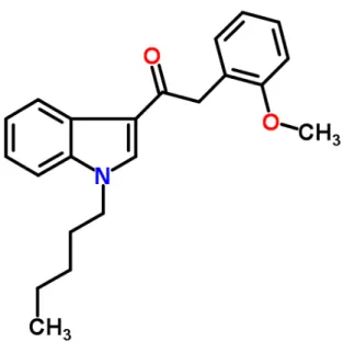
Figure 7. Structural molecule for JWH250 2-(2-Methoxyphenyl)-1-(1-pentyl-1H-indol-3-yl)ethanone, C22H25NO2 (Adapted from JWH-250, ChemSpider, 2014) ... 27
Figure 8. Optimization for MTT assay. Without drug exposure and yielding an optical density at 595nm of 1.2, the concentration of 3x10⁴ cells per well (1,5x10⁵ cells per ml) was selected for cytotoxicity assays. ... 34
Figure 9. MTT results of SH-SY5Y exposure to JWH-073 (95% confidence interval, Kruskal-Wallis, n=3 independent assays of 6 replicates each). ... 35
Figure 10. MTT results of SH-SY5Y exposure to JWH-250 (p < 0.05, Kruskal-Wallis, n=3 independent assays of 6 replicates each). ... 36
Figure 11. LDH results of SH-SY5Y exposure to JWH-073 (p < 0.02, Student’s T test, n=1)…… ... 37
Figure 12. LDH results of SH-SY5Y exposure to JWH-250 (p < 0.0006, Student’s T test, n=1). ... 38
Figure 13. Assay comparison and observation of tendencies for 250 and JWH-073……. ... 38
Figure 14. Graphic view of the Neubauer chamber. Counting was performed at quadrants 1, 5 and 4, then average from it divided by 3, multiplied by 5 (as the dilution factor) and 104 (possible amount on cells in 1cm² of the quadrants). ... 59
Figure 15 (compilation). Outlier detection tests for JWH-073 MTT data... 66
Figure 17 (compilation). Outlier detection tests for JWH-250 MTT data... 73
List of tables
Table 1. Scheme for cell MTT/LDH assay on a 96-wells microplate. ... 58
Table 2. Scheme for cell optimization on a 96-wells microplate. ... 59
Table 3. Scheme for obtaining required cell density for cell optimization on a 96-wells microplate. ... 60
Table 4. Absorbance data minus blank from MTT results of JWH-073. ... 61
Table 5. Means of each independent assay, final mean equals the calculation of % MTT and standard deviation (SD) to JWH-073. ... 61
Table 6. Absorbance data minus blank from MTT results of JWH-250. ... 70
Table 7. Means of each independent assay, final mean equals the calculation of % MTT and standard deviation (SD) to JWH-250. ... 70
Table 8. Raw data from LDH results of JWH-073 ... 77
Table 9 (Compilation). F-tests for LDH results of JWH-073 ... 79
Table 10 (Compilation).Student’s T-tests for LDH results of JWH-073 ... 80
Table 11. Raw data from LDH results of JWH-250 ... 81
Table 12 (Compilation). F-tests for LDH results of JWH-250. ... 82
List of abbreviations
Δ9THC - delta-9-tetrahydrocannabinol µM - micromolar
ATCC - American Type Culture Collection ANOVA – Analysis Of Variance
AEA - N-arachidonylethanolamide; anandamide cAMP - Cyclic adenosine monophosphate CB - cannabinoid receptors
CDC – Center for Diseases Control CNS - Central Nervous System CP - cyclohexylphenol
DEA - Drug Enforcement Administration DMEM - Dulbecco's Modified Eagle's Medium DMSO – dimethylsulfoxide
DNA - Deoxyribonucleic acid
EMCDDA - European Monitoring Centre for Drugs and Drug Addiction et al. - et alii
EU – European Union
GABA - Gamma-AminoButyric Acid
GC-MS - gas chromatography – mass spectrometry GHB - gamma hydroxy butyrate
GPCR - G protein-coupled receptors g/mol – grams per mol
GRK – G protein-coupled receptors kinases h – hours
H+ - hydrogen ion
HEK-293 - Human Embryonic Kidney 293 cells HU210 - Hebrew University 210
IC50 - inhibitory concentration 50%
LC-MS/TOF - liquid chromatography – mass spectrometry/time of flight LDH – Lactate desidrogenase
MTT – 3-(4,5-Dimethylthiazol-2-yl)-2,5-Diphenyltetrazolium Bromide mTOR - mammalian target of rapamycin
mg/kg – milligrams per quilo mg/g – milligrams per gram mL – millilitre
NAD+ - Nicotinamide adenine dinucleotide
NADHH+ - Nicotinamide Adenine Dinucleotide - Hydrogen (reduced) ng/mL – nanograms per millilitre
nM - nanomolar
NPS - novel psychoactive substances OD – optical density
pen strep – penicillin streptomycin pg/mg – pictograms per milligrams PKB - protein kinase B
PPAR - peroxisome proliferator-activated receptor S.D. – standard deviation
SC - synthetic cannabinoid TB – Trypan blue
THC – Tetrahydrocannabinol
TRP - transient receptor potential cation channel US – United States
UHPLC-MS/MS - ultra high performance liquid chromatography-tandem mass spectrometry UV – ultraviolet
UK – United Kingdom
Content
Abstract…… ... 6
List of figures ... 7
List of tables ……….9
List of abbreviations ... 10
Introduction.. ... 14
1.1. Background ... 14
1.2. Cannabinoid system, receptors and types of chemicals ... 18
1.3. Synthetic cannabinoids precedent ... 20
1.4. JWH-073 (1-butyl-1H-indole-3-yl)-1-naphthalenyl-methanone) ... 24
1.5. JWH-250 (2-(2-Methoxyphenyl)-1-(1-pentyl-1H-indol-3-yl)ethanone) ... 26
1.6. Study rationale ... 28
1.7. Research questions, Aim and Objectives ... 29
1.7.1. Key research questions/hypotheses ... 29
1.7.2. Aim… ... 29
1.7.3. Objectives ... 29
2. Experimental ... 30
2.1. Reagents and Equipment ... 30
2.2. Cell Culture ... 30
2.3. Trypan blue (TB) counting ... 31
2.4. MTT (3-(4,5-dimethylthiazol-2-yl)-2,5-diphenyltetrazolium bromide) assay: optimization and cytotoxicity assay ... 32
2.5. Lactate Desidrogenase (LDH) assay ... 33
2.7. Statistical Analysis ... 33
3. Results….. ... 34
3.1. MTT Optimization ... 34
3.2. JWH 073 MTT Assay ... 35
3.4. JWH 250 MTT Assay ... 36
3.5. JWH 073 LDH Assay ... 36
4.1. JWH-073 results ... 41
4.2. JWH-250 results ... 43
5. Conclusions ... 44
6. Recommendations for further work ... 45
7. References ... 48
1. Introduction
1.1. Background
In 2013, a 31 year old female was brought to the Emergency Department, following a three stories fall from her apartment’s fire escape. She was unresponsive and, at the scene, was diagnosed as a 3 on the Glasgow Coma Scale3: she could still open her eyes in response, but she uttered words and had abnormal reactions to pain stimulus. The woman was intubated with no need to use general anaesthesia. Her radiographic data showed a large subdural hematoma, facial fractures, pelvic fractures, liver laceration and elbow fracture. On day 10 of hospitalisation, the patient died from her traumatic injuries.
According to her husband, on that evening, while rehearsing for a play, she ingested a
“pot brownie”. Shortly after, she began to feel “weird” and called her husband at work for
help; when he got home she expressed her intention to jump off the balcony. He gave her water and induced vomiting and physically restrained her to the bed twice afterwards. She however broke free and jumped.
The medical doctors reasoned that marijuana does not normally cause such symptoms and while the patient’s urine routine toxicology was negative for cannabinoids, further testing was performed with gas chromatography – mass spectrometry (GC-MS), searching for delta-9-tetrahydrocannabinol (Δ9THC) and other products of THC or marijuana metabolism. Those were all negative, however, the patient’s ante mortem serum was tested with liquid chromatography – mass spectrometry/time of flight (LC-MS/TOF). A new compound never detected in patients before was found in her serum; it was JWH-175, one of the many emerging members of the synthetic cannabinoid (SC) family. This case was reported by Dr. Armenian, a medical doctor from the Emergency Department of the Hospital-University of California, at San Francisco (Armenian, 2014).
phytocannabinoids: as cannabinol, cannabidiol, delta-9-tetrahydrocannabivarin, cannabichromene, cannabigerol, delta-9-tetrahydrocannabinolic acid and cannabidiolic acid.
Figure 1. Pharmacological activities of non-psychotropic cannabinoids (and its suggested mechanisms of action). (Izzo et al., 2009)
Figure 2. Types of cannabinoids. (Greydanus et al., 2013)
The cannabinoid system consists of Central Nervous System (CNS) cannabinoid receptors and their endogenous ligands or triggering molecules that bind to a target protein site. Tetrahydrocannabinol (THC) is the main active principle in marijuana. In the 1980s, researchers began to synthesise THC analogues and look for marijuana metabolites (Greydanus et al., 2013). These studies led to the development of new classes of bicyclic cannabinoids (as CP55,940), the aminoalkylindoles (as WIN55,212-2), among others; the present study is focusing on members of the naphthoylindoles class.
The JWH4 type of synthetic cannabinoids belongs to naphthoylindole class. The physical and psychological effects of JWH cannabinoids are similar – but stronger – to those of Δ9THC. In fact, it has been stated that JWH binds 4x more than THC to the CB1 and 10x more to the CB2 receptor (Wintermeyer et al. 2010).
Dr. Huffman intended to explore the analgesic potential properties of CB1 agonists, but by describing and publishing JWH synthesis he de facto primed the production of this synthetic drug family.
brand names: K2, Spice Gold or Silver, Yucatan Fire, and several others. After 2008, various cannabinoids were detected in marijuana blends.
The use by humans of novel psychoactive substances (NPS), such as synthetic cannabinoids, instead of traditional illegal substances – (e.g. cannabis) is a fairly recent practice (UNODC, 2013). However, the majority of European countries list prohibited/controlled synthetic cannabinoids and their commercialisation is being scrutinised by the authorities. These drugs are hard to deal with from the point of view of their legality. They are labeled as `legal highs`, an umbrella term impliying that they have not yet been
“reviewed” by legislators, therefore being indirectly legal. This is then an area characterised by limited data with unknown risks. (EMCDDA, 2009)
Legislators are delayed on evaluating the danger of NPS, but emergency rooms see an increased number of cases. Izzo et al. (2009) showed a summary of the first agonists of cannabinoid system and the way they could modify our physiology, when in fact, these have the characteristic of being non-psychoactive drugs. This has suggestive implications on the effect of psychotropic, psychostimulant agonists of CB1 and 2 such as synthetic cannabinoid compounds.
About 20 case studies with full medical and toxicological assessment are published in the literature and two of them are summarized here. In 2012, a 16-yr old was submitted at the emergency room with altered mental status and was hospitalised overnight in catatonic state
after smoking “K2”; a 18-yr old was described as agitated, aggressive and had profuse sweating (Cohen et al., 2012) after using “Spice”. In 2013, a fatal case was described in which postmortem blood showed the presence of JWH-018 (0.1 ng/mL) and JWH-122 (0.3 ng/mL) together with amphetamine. The conclusion of this report alerted that synthetic cannabinoids are more potent and effective than THC, potentially leading to life-threatening situations (Schaefer et al., 2013).
bicyclic classes of synthetic cannabinoids tested, were cytotoxic in a concentration-dependent manner. They further found that cell death was mediated by the CB1 receptors and not the CB 2 receptors (Tomiyama & Funada, 2011). Another study demonstrated that all 3 types of cannabinoids (endo, phyto and synthetic) can induce apoptosis in cells, even in the absence of CB receptors (Athanasiou et al., 2007).
There is an emerging body of evidence that these compounds are being pursued with the original intention of developing new therapeutics. This establish another field of research on synthetic cannabinoids. There are numerous pieces of evidence of non-toxic potential use of SC on the recovery of myelination in multiple sclerosis neuronal models (Arévalo-Martín, 2003; Downer, 2011) and still onto the analgesic effects for cancer patients (Reynolds, 2013). The paradox is what some SC do possess therapeutical use but the abuse of other SC in spiked marijuana can cause human intoxication. Therefore, toxicological assays are paramount to know how harmful these new psychoactive substances can be.
With the spreading use of marijuana nowadays (plant consumption; pharmaceuticals for cancer-related symptoms – Bar-Sela et al., 2013; epilepsy – Devinsky et al., 2014; obesity
– Le Foll et al., 2013; schizophrenia – Robson et al., 2014; improved appetite and sense of taste – Brisbois et al., 2011) and the near future possibility of marijuana legalization worldwide, there is a chance of illegal markets investing in synthetic cannabinoids for their potency and addictive potential. Toxicological assays are vital to assess the harm potential of these novel psychoactive substances.
1.2. Cannabinoid system, receptors and types of chemicals
Cell biologists found that CB1 can stimulate the initiation of mitogen activated protein kinases signalling, (Daigle et al., 2008) influencing cell movement, growth and diversification (Derkinderen et al., 2003). With continued stimulation, the CB1 receptor is regulated by desensitisation and internalisation of receptors (Hsieh et al., 1999). In fact, desensitisation is thought to be regulated by kinases of GPCR (so called GRK2 and GRK3) and β-arrestin (Premont & Gainetdinov, 2007). While other molecular pathways like protein kinase B (PKB or Akt) and mammalian target of rapamicin (Manning and Cantley, 2007) are induced by CB1 receptor, CB1 agonists, like WIN55212-2 (one of the first synthetic cannabinoids) was shown to be involved in the rodent and human brain of cocaine addiction (Alvaro-Bartolome et al., 2011). The CB1 receptor was also found intracellularly in mitochondrial membranes (Benard et al., 2012), while on transfected HEK-293 cells CB1 is inside intracellular vesicles and plasma membrane (Leterrier, et al., 2004).
The CB1 receptor has also been characterised on neuronal cells, including its involvement on the inhibition of voltage gated Ca+ channels and activation of K+ channels (Mackie et al., 1995). It has been found to reduce cellular excitability and the chances of neurotransmitter release (Shen et al., 1996), as well as detaining inhibitory and excitatory synapses, as found with glutamatergic and GABA terminals (Chevaleyre et al., 2006).
Overall, CB1 activity affects neurotransmission, but mostly for its predominance in crucial locations in the human body. Mackie (2005) described the expression of CB1 in the central nervous system, particularly in axon terminal and primarily at the cortex (anterior cingulate, limbic area), cerebellum, hippocampus, amygdala, basal ganglia, hypothalamus and not often in the brain stem – helping cannabinoids to have low toxicity if accidental (Herkenham et al., 1990). Since the CB2 receptor is predominantly located on immune cells (Galiegue et al., 1995), but also in the CNS as in microglia, spinal cord and brainstem (Van Sickle, et al., 2005), it was suggested as a novel target for pain and inflammation management (Onaivi et al., 2012).
1.3. Synthetic cannabinoids precedent
Endocannabinoids are part of marijuana-related research, whilst AEA (arachidonic acid moiety, N-arachidonylethanolamide), was entitled “anandamide” - according to Sanscrit
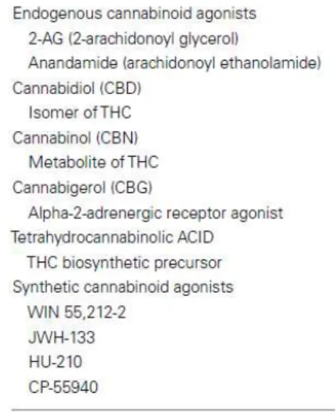
for “bliss” -, a range of pharmacological tools and new synthetic cannabinoids have been established. The classification of cannabinoid compounds is settled with five categories, including the classical (for instance, Δ9THC and HU210 – developed by Dr. Mechoulam at Hebrew University), non-classical (as CP-55,940), indoles (WIN 55,212), eicosanoids (mostly endogenous as AEA and 2-arachidonylglycerol) and pharmacological antagonists (AM251 and AM630, developed by Pfizer) (Devane et al., 1992). And its structures are presented below:
Figure 3. Structure classes of cannabinoids (Console-Bram et al., 2012)
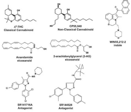
Figure 4. Structures of representative synthetic cannabinoids categories, and representative compound, commonly found in “Spice/K2” products (taken from Fattore and Fratta, 2011).
Gaoni and Mechoulam (1964) first exposed Δ9THC as the utmost psychoeffective constituent in marijuana and from that point, numerous additional typical and non-typical cannabinoids have been examined for medicine usage. However, structure-activity relationship study of Δ9THC shows cannabinoid pharmacological properties for any chemical with affinity for the cannabinoid receptor. This presents a challenge in producing a single effect selectivity of a cannabinoid drug. (Compton et al., 1993)
Currently, we are not aware of the entire chemical content of these new drugs, their acute or chronic toxicity, what can be translated into a worrisome obstacle to public health. For instance, in the United Kingdom, some first ‘legal highs’ (to be noted: piperazines,
“spice” and mephedrone) were already deliberated by the Misuse of Drugs Act of 1971, nonetheless, the introduction of legal boundaries over these drugs presented little changes on the drug scenario and, at least online, the banned drugs continue to be sold as new brands. These new brands are commercialized as greater products, and, as licit options to the banned drugs (Ramsey et al., 2010). We are not aware of the new brands’ content, if it comprises of new synthetic and legal compounds or if it possesses the amount of illegal compounds that do explain the link of a number of related deaths (Baron et al., 2011; EMCCDA, 2011). In Europe, the EU Early Warning System has received 81 new psychoactive substance in 2013, which 29 were synthetic cannabinoids, as presented on Figure 5, that shows constancy of NPS apprehensions all over Europe.
There is then a confusing perception of safety to some of the users (Sheridan and Butler, 2010), when actually there is no information on the psychology or behaviour consequences from the use in humans, especially for drugs like the synthetic cannabinoids (EMCCDA, 2009). In fact, products, when labelled, not always show its real content (or more worrisome, the manufactures could not even tell the content), even for products with the same name and brand (Davies et al., 2010). In the end, users are having their health comprimised when exposed to unknown drugs of unidentified concentrations and even frequent users of the same product could be purchasing a different drug or a more potent one. This possibility can be extended to the concern of drugs interaction and the effects of metabolites in human physiology. Although, some research is being done to unravel toxicity cases (Salmner et al., 2010), the current challenge puts the non-identification or association of adverse effects of these drugs by clinicians at emergency rooms (Smith et al., 2011), together with the difficulty in identifying the drugs, the new ones and unique compounds (Houston, 2011).
Meanwhile, a minor quantity is necessary in order to provoke a result and the minimum amount that can be bought is of one gram. Consumers will still go to hospital departments across the world, and medical doctors will need prompt information on these new drugs, their consequences and risks. Daily, the professionals, who are in contact with NPS cases, suffers with the scarcity of scientific and medical data. Investigation in this field is not all consolidated, medical clinical cases do not contemplate the reality and amount of episodes at the various hospitals and the health system of countries have not yet develop a central database that combines toxicology and forensics (Boyce, 2011).
Contributing to the risk scenario, the absence of safety guidance (how to use, overdosage, adverse effects) on the online market of NPS drugs is justified by putting labels
Even though synthetic cannabinoids were designed to provide insights into the cannabinoid organization (Huffman et al., 1994), numerous synthetic cannabinoids turned out as drugs of abuse, apparently as these could present advantages resembling marijuana; to mention, the non-appreciation and awareness of drug legislation or detectability on conventional drug-urine exams (Seely et al., 2012). From that, studies endorse comparable physiological reactions from ‘Spice’ and cannabis use (Zimmermann et al., 2009, this paper describes a patient case that was undertaking cannabis-like withdrawal and tolerance
symptoms subsequently the stop of ‘Spice’ use). Disturbingly, the usage of K2 (one of the synthetic cannabinoids commercial product) has a significant occurrence of serious adverse effects, that are not usually described with marijuana (tachycardia with hypertension, anxiety attacks, seizures, psychosis and hallucinations (Harris & Brown, 2013).
With the occurrence of synthetic cannabinoids in commercial herbal blends from 2008, various research cores have use liquid or gas chromatography with mass spectrometry (LCMS; GC-MS) in order to point out its presence on the content of ‘K2/Spice’ (Lindigkeit et al., 2009). Remarkably, Δ9THC was not found in the analyses of ‘Spice’ samples, proposing that the physiological impact of these substances were happening because of the synthetic cannabinoid ‘cutting agents’ (Auwarter et al., 2009). The investigating group that used LC/GCMS and identified synthetic cannabinoids did find ones as JWH-018, JWH073 and CP-47,497 in a concurrent presence as components of the marijuana products. The prediction
nowadays is that more new compounds will be encountered to be sprayed on ‘Spice’ and it is already true to findings of JWH398 and JWH250 in Germany and the UK (Vardakou et al., 2010).
1.4. JWH-073 (1-butyl-1H-indole-3-yl)-1-naphthalenyl-methanone)
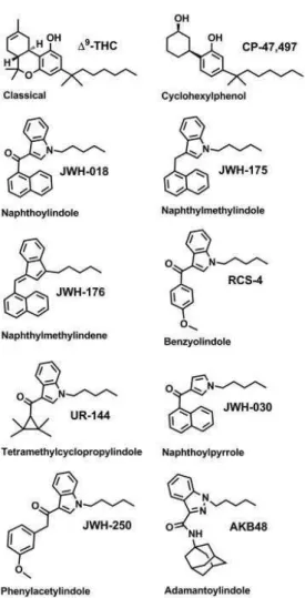
Dresen et al. (2010) mention the initial cannabimimetics had succumbed to new, yet similar, synthesised ones that represents an escape of the European legislation assessing and
Figure 6. Structural comparison for JWH073 (1-Butyl-1H-indol-3-yl)(1-naphthyl)methanone, C23H21NO) and
JWH018 (1-Naphthyl(1-pentyl-1H-indol-3-yl)methanone, C24H23NO) (Adapted from JWH-018 and JWH-073,
ChemSpider, 2014)
November 24th of 2010 was the date of ban of JWH-073 and other four synthetic cannabinoids, by including these compounds as Schedule I (meaning, high potential for abuse, no accepted medical use, lack of accepted safety) of the Substance Act from the DEA (Drug Enforcement Administration) in the United States (Young et al, 2012). This action is already a consequence from intoxication cases, although it pushes the DEA to figure out toxicology in order to describe and explain why the ban has happened. JWH-073 in high concentration on synthetic marijuana is considered unlikely, or more as an additive or impurity of producing JWH-018, but also, there is a chance for JWH-073 detection as consequence of metabolism of AM-2201, a newer synthetic cannabinoid, or from the decarboxylation of JWH-018 (Hutter, 2013).
JWH-073 is described to possess increased efficacy, nearly, 5-fold greater than
Δ9THC (Brents et al., 2012) and, although, JWH-018 and JWH-073 are scheduled as narcotics in Germany, this does not mean these compounds have been banned from the commercial products. In fact, a study using new method of LC-MS (Kneisel and Auwater, 2012) identified JWH-073 in serum of patients from emergency rooms and cases of criminal investigation on levels of 7.1 ng/mL, in an average of 0.85 ng/mL. As far as cases of intoxication are concerned, JWH-073 has been described to be involved on Cannabinoid hyperemesis syndrome (Hopkins and Gilchrist, 2013) or cardiotoxicity (Young et al., 2012).
cannabinoids and the main assessment is through receptor sensitisation and internalisation; in which, JWH-073 is considering for Atwood et al. (2011) to produce slower internalisation (about 105 minutes) than JWH-018 (37 minutes). However, Wu et al. (2008) describes faster desensitisation for JWH-073, instead of JWH-018. In the end, the studies provide inferences that needed to be assessed in vivo, but it can be strong evidence on clinical reports of withdrawal and tolerance cases (as described on Zimmermann et al., 2009).
In a structure-affinity study of indoles synthetic cannabinoids (Aung et al., 2000), there was the observation of influence of changes in the N-1 alkyl length of the chain on CB1/CB2 binding. For JWH-018, the kinetic values are CB1: 9.00 ± 5.00 and CB2: 2.94 ± 2.65 nM, while for JWH-073, the values for CB1: 8.90 ± 1.80 and CB2: 38.0 ± 24.0 nM
(while Δ9THC had values of CB1: 40.7 ± 1.7 and CB2: 36.4 ± 10 nM). These values and the increase of the alkyl chain shows a decrease of binding the bulkier the compound is, but interestingly presents JWH-073 low binding, gathering important data for the reflect on human physiology. That can be evidence to explain results as JWH-073 having the same discriminatory effects as Δ9THC in rhesus monkeys (Ginsburg et al., 2012), hypo motion, stillness, anti-nociception, and hypothermia in mice (Hruba et al., 2012).
Concerning cytotoxicity, JWH-073 has been evaluated in a range of 75-100 µM, to be toxic to cell lines from hepatoma, buccal epithelium and mammary tissue, using lactate desidrogenase assay (only at 100 µM), but this same drug was not considered toxic with XTT assay that assess mitochondrial function. There was evaluation of genotoxic effects with positive toxic results of JWH-073 (also at 100 µM) in buccal cells (remembering that JWH are smoked and buccal cells are one of the first ones affected) and hepatoma cells. (Koller et al., 2013)
1.5. JWH-250 (2-(2-Methoxyphenyl)-1-(1-pentyl-1H-indol-3-yl)ethanone)
In fact, JWH-250 is mentioned to be first detected in Germany, on 2009, and it is classified as a phenylacetylindole (see Fig. 7) with receptor CB1 affinity of Ki = 11nM; KI= 33nM for CB2 (Huffman et al., 2005; Dargan et al., 2011), and, according to the UNODC (2011), it is considered controlled substance in Austria (Oct 2010), Denmark (Mar 2010), Germany (2011), Japan (Sep 2010), Lithuania (May 2009), Romania (Feb 2010), Switzerland (Dec 2010) and the USA (Jul 2012, DEA 2013).
Figure 7. Structural molecule for JWH250 2-(2-Methoxyphenyl)-1-(1-pentyl-1H-indol-3-yl)ethanone, C22H25NO2 (Adapted from JWH-250, ChemSpider, 2014)
For this person, several methodologies of JWH-250 detection have been developed, even with the difficulties in detecting new synthetic cannabinoids, new ways of screening have been researched, including hair detection by ultra high performance liquid chromatography-tandem mass spectrometry (UHPLC-MS/MS) that showed real cases enclosing 4.8-83.4 pg/mg (Salomone et al., 2014). While at the emergency departments, cases of intoxication in which JWH-250 is present (in a mix of other cannabinoids) shows symptoms as:
- In 38% of the cases, symptoms as nervousness, impatience, acute psychosis, and hallucinations, and, also, light and external stimulus hypersensitivity and panic reactions (Hermanns-Clausen et al., 2013)
However these symptoms are from episodes in which JWH-250 was not the only synthetic cannabinoid involved and the detected levels of this drug on human serum was of 0,1-0.4 ng/mL, providing intoxication signs in 6 to 24h (WHO, 2014). In JWH-250 smoking self-studies (product ‘8-Ball’, a mix of JWH-250, JWH-019, JWH-081 and RCS-4 in approximately 10 mg/g), levels of this drug reached the blood as 10 ng/ml (after 20 minutes) and 10 ng/ml in oral fluid, while reached a peak of 140 ng/ml of its 4-hydroxylated metabolite in urine 1 hour after smoking (Adams and Logan, 2011). A review from the Expert Committee on Drug Dependence (WHO, 2014) points out that there is no study on
dependence data or abuse potential and “No pre-clinical safety data are available about the toxicity, reproductive impact and mutagenic/carcinogenic potential of JWH-250”.
1.6. Study rationale
This study intends to assess the cellular toxicity of JWH-073 and JWH-250, synthetic cannabinoids identified as drugs of abuse. The cell lineage model is SH-SY5Y, a human-derived neuroblastoma-glioma line. This cell line shows adherent behaviour and possesses the ability to differentiate along the neuronal type of cells (La Quaglia and Manchester, 1996).
Cell toxicity is initially evaluated with the metabolic reduction of 3-[4,5-dimethyl-thiazol-2-yl]-2,5-diphenyltetrazolium bromide (MTT). Via endocytosis or protein facilitation, MTT enter cells and is reduced, by mitochondrial enzymes, to produce a purple compound (formazan). This compound is generally not permeable to membranes, therefore accumulating inside live cells. Dissolution of formazan crystals in the cells releases a purple product that is identified using a spectrophotometer. The cells’ capability of reducing MTT signals mitochondrial physiology and therefore an indication of cell viability. (Maioli et al, 2009)
1.7. Research questions, Aim and Objectives
1.7.1. Key research questions/hypotheses
- Are JWH-073 and JWH-250 cytotoxic to neuronal cells? To what extent of dose dependency?
- If cytotoxic, do these chemicals act on cell metabolism and cytoplasmatic leakage? - Can CB1 and CB2 receptors facilitate synthetic cannabinoids binding and possibly
affect their toxicity?
1.7.2. Aim
To test neuronal toxicity of JWH-073 and JWH-250 on SH-SY5Y cell lineage.
1.7.3. Objectives
- Test neuronal toxicity focusing on mitochondrial damage with the MTT assay
- Test neuronal toxicity focusing on the release of cytoplasmatic content with the LDH assay
2. Experimental
2.1. Reagents and Equipment
All JWH were acquired from Lipomed, Switzerland. Stock solutions of JWH 018 (molecular mass = 341.5 g/mol), JWH 073 (molecular mass = 327.4 g/mol) and JWH 250 (molecular mass = 335.2 g/mol) were prepared to the final volume of 1ml and concentration of 25000 µM in pure sterile DMSO. The groups of samples applied were one blank (no cells, all other MTT components), one negative control (cells, 0.2% DMSO, no JWH)5, one positive control (cells, 1% Triton X-100, no JWH), 6 concentrations of each JWH (1, 5, 10, 25, 37.5, 50 µM)
MTT (Thiazolyl Blue Tetrazolium Bromide; Sigma-Aldrich, Portugal; 0.5 mg/ml) solution was prepared extemporanely in each time to read a new plate. For that, knowing the amount of wells to be used on the assay, we weighted in a semi analytical scale (Sartorius, Germany) of pure MTT to be dissolved on sterile conditions inside the vertical laminar flow.
LDH (Lactate desidrogenase Kit, Clontech, USA) was prepared according to
manufacturer’s instructions for 100 tests.
For cell culture and assays, we used a laminal flow hood (Scanlaf, Mars Safety Class 2), T25 culture plastic flasks (25 cm², volume of 60ml, VWR, Belgium), Dulbecco's Modified Eagle's Medium (DMEM, Gibco, Portugal) supplemented with 10% fetal bovine serum and 1% pen strep + glutamine (both from Gibco, Portugal), a cell culture incubator (Water jacket incubator, ShelLab, USA), an inverted light microscope (Zeiss, Germany), Phosphate Buffered Saline (Gibco, Portugal), TrypLE™ Express (1X; Life Technologies, Portugal), a Neubauer hemocytometer chamber (Hirschmann EM Techcolor, 0.0025 mm2, depth 0.1 mm) centrifuge machine (Sigma 3-16PK, Portugal), Trypan Blue dye (Sigma, Portugal), 96-well microplates (Corning Inc., USA).
2.2. Cell Culture
were sprayed with 70% ethanol, then the same procedure for every materials that need to be inside the hood and other safety protocols were followed.
SH-SY5Y cell line (human neuroblastoma-glioma) was obtained as a gift from Dr Tiago Outeiro, from the Cell and Molecular Neuroscience Unit of the Institute of Molecular from Lisbon, Portugal. The cell lines were stored in vials at -80°C and after cultivation were used for the experiments. This cell line was cultivated in T25 culture plastic flasks (25 cm², volume of 60ml, VWR, Belgium) in Dulbecco's Modified Eagle's Medium, supplemented with 10% fetal bovine serum and 1% pen strep + glutamine, incubated under standard conditions (37 °C, humidified atmosphere, 5% CO2).
Passages with changing of media occurred every 3–4 days; whilst proliferation was observed every 24h using an inverted light microscope. When the cultures reached confluence of 70-80%, cells were washed with Phosphate Buffered Saline, detached with TrypLE™ Express – acting for one minute under standard incubation conditions -, centrifuged, the supernatant was discard, the cells were re-suspended in 5 mL of DMEM and subcultured or proceed for counting. This counting was determined by exclusion of Trypan Blue dye before drug exposure growth and cell viability was of 90 % in the untreated cells.
For drug exposure, cells were seeded into 96-well microplates. Drug exposures were started after 24 h of each subculture. JWH 073 and 250 (2.6 µL from stock solutions) were dissolved in 100 % DMSO, as vehicle, then diluted with supplemented DMEM (final concentration of 0.2% of DMSO) and added to cells to a final concentration of 1, 2, 5, 10, 25, 37.5 and 50 µM (as presented on Table 1 – Appendices).
2.3. Trypan blue (TB) counting
TB counting measurement was described before (Reeb, 1992). Forty microliters of TB solution and 10 μL of cell suspension were used. The suspension was loaded into a Neubauer hemocytometer chamber with cover slip and scored with a light microscope at 40×, using quadrants 1, 5 and 4 (vide Figure 15). Cells that stained blue were scored as nonviable. Measurements were considered as follow:
No. of viable cells = [(Viable cells from Q1 + Viable cells from Q4 + Viable cells from Q5)/3] x 5 (Dilution factor) x 104(one well’s size on the Neubauer chamber)
Viability (%) = (No. of viable cells/ No. of viable cells + No. of NON-viable cells) x 100
2.4. MTT (3-(4,5-dimethylthiazol-2-yl)-2,5-diphenyltetrazolium bromide) assay: optimization and cytotoxicity assay
According to ATCC (2011), in order to define the ideal number of cells per well of the 96 well plate, trials must be done to measure the number of cells to be used. Cell suspension was obtained from T25 flasks and re-suspended at 6.2x10⁵ cells per ml. From this starting concentration, serial dilutions of cells in culture medium were seeded in a 96-well microplate (vide Figure x – Appendix), within a range of 1,5x10⁴ to 5x10⁵ cells per ml (1,5x10⁴, 2x10⁴, 2.5x10⁴, 3x10⁴, 3.5x10⁴, 4x10⁴, 4.5x10⁴, 5x10⁴, 1x10⁵, 1.5x10⁵, 2x10⁵, 2.5x10⁵, 3x10⁵, 3.5x10⁵, 4x10⁵, 4.5x10⁵, 5x10⁵ cells per ml). The selection of the cell density range for MTT was based on the reports about its IC50 in the literature (3x10⁴ - Cernaiani, 2008; 4x10⁴ - Kim, 2010; 1x10⁴ - Wang, 2011; 1x10⁵ - Tai, 2011) and our preliminary experiments.
The dilutions were done using the last concentration (5x10⁵) calculated to obtain 700 µl (triplicates; each well fits 200 µl) of each concentration (vide Table 2 and 3 for microplate scheme and summary of cell density obtention). On triplicates, 200 μL of the dilution samples were added into wells of a 96-well plate, including control wells of medium alone to provide the blanks for absorbance readings, then proceed to incubation under appropriate conditions for the cell line for 24 hours. From this step, proceed to MTT assay normally (to be described below), then after the formation of crystals, read absorbance in an UV spectrophotometer (Biorad, USA). Blanks will show values of zero (± 0.1). Defining the mean values from triplicate readings, we could subtract the mean value for the blank and use the MTT reduction formula (to be presented below).
MTT assay (described by Maioli, 2009) itself starts after cell trypsinization, when cells were again counted with Trypan Blue method to achieve a concentration of 3×10⁴ cells/well (1x10⁵ cells per ml defined from the cell optimization assay; area of each well was 0.32 cm2), into to a sterile 96-well microplate. As each well contains 200 µl, it was calculated the amount
of volume to apply 1x10⁵ cells/ml, plus the difference was complete with supplemented DMEM medium. After 24 hours of microplate culture, the medium was removed and
perform LDH assay. New 100 µl of DMEM was applied to wash drug-exposed cells on the microplate, then removed before MTT adding. Previously MTT solution (200μl; 0.5mg/ml)
was added to each well, and the plates were incubated in the dark for 2,5 h at 37°C/5% CO2 (this incubation will allow formazan crystals formation). Dissolve crystals with 200µl of pure
DMSO and carefully homogenising the liquid and perform absorbance readings at 595nm, calculate mean, standard deviation and error (%) of the samples and plot as dose range. This experiment was repeated in independent sample at least three times. For calculations, use the equation:
% reduced MTT = 100 x (mean of the sample / mean of the negative control)
2.5. Lactate Desidrogenase (LDH) assay
With the same microplate cell culture from MTT assay (already exposed to JWH and controls), we collect 150 µL of media samples of each well carefully, without disturbing the microplate bottom, into an eppendorf. We performed centrifugation of the tubes at 1000g/3min, and, without disturbing the cell pellet, transferred 100 µl of supernatant into a new 96-well plate. After, add 100 μl of the Reaction Mixture (freshly prepared; LDH kit from Clontech, USA) to each well, we incubated the plate at room temperature for 30 min, protecting from light, to allow the enzymatic reaction to take place. Then, absorbance was read at 490 nm and cytotoxicity was measured with the formula below. The experiment used six replicates and it was performed three times independently.
Cytotoxicity (%) = 100 x [(Triplicate Absorbance – Negative control) / (Positive Triton-X Control - Negative control)]
2.7. Statistical Analysis
Data is presented as the mean of the % MTT or % LDH ± S.D. for, at least, 3 independent experiments for MTT and one experiment for LDH assay. MTT results compiled from all independent experiments and evaluated for outlier detection (Grubbs, Sigma rule, Inner and Outer Fence Rule). As data was considered non-parametric, we used Kruskal-Wallis 1-way ANOVA test. LDH results were evaluated by F-test to find out variances
3. Results
3.1. MTT Optimization
Since Mossmann (1983) introduced the MTT assay, adjustments were done to suit different laboratories, different analysts and other variable as time of incubation. It is indeed important to adapt the assay to the laboratory, cell lineage as well as to the analyst methodologies. This will provide the best results in the given set-up but will necessarily need to be reflected in the results interpretation. In addition, these factors do influence on results’ interpretation. Before proceeding with the assay, preliminary laboratory work was performed in order to identify the cell concentration most suitable to study proliferation and death as a function of drug exposure. Appendices tables reports the detail of this preparatory phase. The exponential growth phase was defined from a search on the literature that provide a range of 3x10³ to 1x10⁵ cells per well. Assays here were done in three replicates of each cell density.
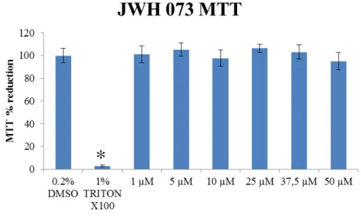
3.2. JWH 073 MTT Assay
The MTT method was applied to SH-SY5Y neuroblastoma cell line to JWH-073 (range of 1-50 µM), in parallel to a positive control of 1% Triton X-100 and a negative control of 0.2% DMSO. Drug exposure was performed after 24h of culture growth on a 96-wells microplate and then again incubated for more 24h. The fractional reduction in the SH-SY5Y cell culture was determined through optical density measurements, as detailed in the experimental section 2.4.
Fig. 9 shows the results for control samples as well as the exposure of the culture to a range of JWH-073 concentrations. Triton X-100 effectively worked as a positive control on cell non-viability (97 % ± 0.8 of MTT toxicity resembling negative control, p < 0.05, Kruskal-Wallis), mainly due to its characteristics as detergent and protein extractor (Stowe et al, 1995). Applying the same statistical treatment (see Appendices), none of the concentration applied of JWH-073 affected the colture significantly with respect to the negative control.
Figure 9. MTT results of SH-SY5Y exposure to JWH-073 (95% confidence interval, Kruskal-Wallis, n=3 independent assays of 6 replicates each).
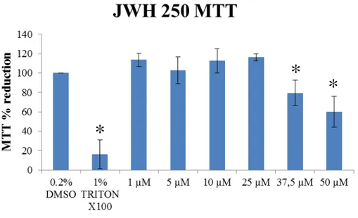
3.4. JWH 250 MTT Assay
MTT was also performed exposing SH-SY5Y neuroblastoma cell line to JWH-250 in the same manner as mentioned before for JWH-073.
Fig. 10 show results for a range of JWH-250 concentrations and control samples. Tritox X-100 effectively worked as a positive control on cell non-viability (84% ± 14.8 of MTT toxicity resembling negative control, p < 0.05, Kruskal-Wallis). Applying statistical treatment (see Appendices), there was statistical significance for the concentration of 50 µM applied of JWH-250 showing a MTT reduction of 40.1% ± 15.8 and the concentration of 37.5 showing MTT reduction of 20.4 % ± 13 µM with respect to the negative control (0.2% DMSO).
Figure 10. MTT results of SH-SY5Y exposure to JWH-250 (p < 0.05, Kruskal-Wallis, n=3 independent assays of 6 replicates each).
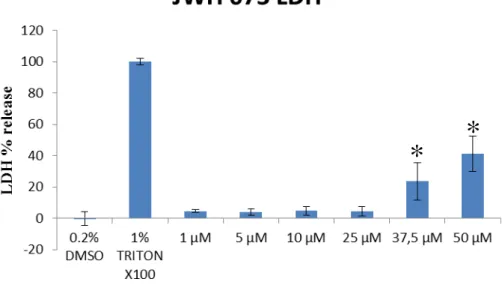
3.5. JWH 073 LDH Assay
In the range of in vitro assays that use enzymes as cellular markers of death, the choice in literature is usually between glucose-6-phosphate dehydrogenase, adenylate kinase and lactate dehydrogenase (LDH). Commercial kits to detect these enzymes are developed and available; however the stability of enzymes in between and within the assays is an issue. LDH
*
*
Samples for the LDH assay were collected from the MTT assay tray before applying the MTT test. JWH-073 (range of 1-50 µM), positive control of 1% Triton x100 and negative control of 0.2% DMSO were used. Drug exposure was performed after 24h of culture growth on a 96-wells microplate. On the determination of chemicals’ effect in SH-SY5Y culture, the optical density was obtained in which a calculation (see Experimental section 2.5) was done to obtain the percentage of LDH leakage detection.
Fig. 11 show results to JWH-073 range and controls. DMSO (0.2%) effectively worked as a negative control on the LDH assay (6% ± 4.3). Applying statistical treatment (see Appendices for data with mean and standard deviation – Table 6, F-test – Table 7 - and T-test for equal variances – Table 8), the concentrations of 37.5 and 50 µM were significantly different than the negative control and were considered ‘inducers of cell death’ with LDH leakage of 29 ± 12 % and 45 ± 11 %, respectively (while positive control, 1% Tritox X-100, showed 93.6 ± 1.9 %, compared to negative control).
Figure 11. LDH results of SH-SY5Y exposure to JWH-073 (p < 0.02, Student’s T test, n=1).
3.6. JWH 250 LDH Assay
LDH was also performed exposing SH-SY5Y neuroblastoma cell line to JWH-250 in the same manner as mentioned before for JWH-073. Fig. 12 show results to JWH-250 range and controls. DMSO (0.2%) effectively worked as a negative control on the LDH assay (-2 ± 2.8 %). Applying statistical treatment (see Appendices for data with mean and standard
deviation – Table 9, F-test – Table 10 - and T-test for equal variances), the concentrations of 25 and 50 µM were significantly different than the negative control and were considered
‘inducers of cell death’ with LDH leakage of 9 ± 3.1 % and 9 ± 3.4 %, respectively (while positive control, 1% Triton X-100, showed 93.1 ± 11.6 %, compared to negative control).
Figure 12. LDH results of SH-SY5Y exposure to JWH-250 (p < 0.0006, Student’s T test, n=1).
3.8. JWH comparison
In order to observe tendencies for both JWH synthetic cannabinoids, both MTT and LDH were assayed and JWH-073 and JWH-250 were studied as a function of drug concentration. Figure 15 reports the four line plots, comparing the reduction/release fractions as a function of concentration. It shows a correlation of MTT and LDH results in order to
support each other. Both JWH have similar MTT ‘behavior’, although LDH demonstrates the
statistical significance of JWH-073 toxicity on concentration of 37.5 and 50 µM. Nonetheless, futher characterization could be interesting, together with standard cannabinoids as THC, JWH-018 or WIN 55,212-2.
4. Discussion
Synthetic cannabinoids present as potent drugs, as far as 0.5 to 5 mg is considered a dose that can lead to psychoactive effects (Tuv et al., 2014), what presents as a challenges to forensic scientists throughout the world to provide insight into new methods of detection in biological matrices, while THC has been described as a strong impairment agent on cognition and motor skills, as driving (Ramaekers et al, 2000). In fact, an Australian report described driving accidents to have a prevalence of 10.8% of Cannabis use and 13,5% on fatal cases (Longo et al., 2000; Drummer et al., 2003). Concerning synthetic cannabinoids, these are considered stronger drugs then THC (Griffiths et al., 2010) and clinicians are step by step
describing the symptoms of a synthetic cannabinoids’ intoxication that includes euphoria, panic attacks, restlessness and anxiety (Bebarta et al., 2012).
The epidemiology of synthetic cannabinoids’ use is yet to be clarified and
organizations as the EMCDDA and the UNODC have provided efforts on it, however, local scenarios of epidemiologic research are needed. Together with clinical reports, that can
provide material for biological analysis, there have been large surveys on drug users’
behaviour, preferences and reactions. One example is the group of Winstock et al. (2011) assessing the British dance scene population, with a survey resulting on a prevalence of 13%
of ‘Spice’ users. A second example is a survey describing use of ‘Spice’ among US American
college students with a rate of 9% (Hu et al., 2011), while athletes are another segment investigated, Heltsley et al. (2012) performed 5956 urine screening and found synthetic cannabinoids in 4.5%.
The amount of side effects from the use of synthetics it is translated on the chemical
analysed of the herbal blends of ‘Spice’-products. Zuba et al. (2011) mentions that most frequently the blends do contain more than on synthetic cannabinoid and analysis from blood-work cases reveal intoxication mediated by more several synthetic cannabinoids; what is confirmed as 50% of the episodes from a series of toxicological cases performed by Yeakel and Logan (2013). Besides, the blends not only contain synthetic cannabinoids, but some do enclose known drugs and it can be detected as methamphetamine, benzodiazepines and THC itself – with other cases including GHB and codeine (Tuv et al., 2014). This entire scenario contributes to intoxication of multiple symptoms.
Toxicology. Remembering that ‘Spice’-products do vary intra and inter batches and it generates fractions of more than one JWH or other synthetic cannabinoid. The comprehension of polysubstances can lead to misunderstanding on inferences of toxicological effects. It is interesting to investigate these new drugs on its own specificity of influence on human physiology, but also to consider that the heterogeneity of compounds in one package of
‘Spice’ and its overall contribution to adverse neurological effects.
That is one of the reasons why this study here chose SH-SY5Y cell lineages to be applied. In addition, clinical reports are finally arising, as the one from Bernson-Leung et al. (2014), in which it identifies two cases of stroke symptoms on healthy individuals after experiencing synthetic cannabis for the first time.
SH-SY5Y as an option of in vitro model of toxicity focused on Δ9-THC been demonstrated as an inhibitor of the production of nucleic acids and proteins on neuronal cell lines, from human and mouse, but, also to influence on the cell membrane and cell growth
(Lew, 1996). In fact, Δ9THC has an emphasis on nerve cells, which type can be originated from neuroblastoma cultures (La Quaglia and Manchester, 1996). The SH-SY5Y has the most utilized cell line human-derived on toxicity studies, with more than 4900 hits on PubMed and it has been used for in vitro toxicology focusing on neurodegenerative disease, as Alzheimer’s (Harvey et al., 2012), as Parkinson’s (Choong and Say, 2011) and, for oxidative stress mediated by drugs (Halliwell, 2006), including on the test of cannabinoids toxicity or potential for therapy. These cells are considered easy to cultivate (Cheung et al., 2009) and are applied on the study or neuronal development (Radio et al., 2008). In addition, SH-SY5Y has been profiled to express enzymes as tyrosine- (Chen et al., 2013) and dopamine hydrolase (Ou et al., 1998). To complete information, the study of Sanfeliu et al. (1999) compared SH-SY5Y with primary culture of neuronal cells and found similar behaviour when exposed to toxicants, what brings advantage on the use of readily available immortal culture of SH-SY5Y, instead of having access primary human tissue sources. Altogether, it presents a good tool for in vitro investigation and that is why it was a chosen cell line to be used in the
European Union project for acute systemic toxicity “Acutox” that is reliably correlated to in vivo assays (Gustafsson et al., 2010).
4.1. JWH-073 results
MTT do rely on reducing a colouring reagent via a dehydrogenase enzyme to be functional in a live cell; this can be a measurement of cell viability. This test is also considered easy-going, safe and to possess good reproducibility, what makes it useful for either test of viability and cytotoxicity. These properties make it a common initial step in the toxicological assessment. MTT is frequently used as one of the method measuring the activity of mitochondria in live cells, while mitochondrial NADH converts MTT to formazan and this later as water-insoluble, it forms crystal needles of purple that can be readily dissolved by an organic solvent (in our case, DMSO). Altogether, MTT depends highly on mitochondrial state and not entirely on the cell itself.
LDH, as a marker of cell membrane damage, provided results that increased our suspicious to re-investigate concentrations of 50 µM and higher. Our MTT results do not show statistically significant evidence of toxicity for JWH-073 at the concentrations considered, however some caveats need to be taken into account. First, a total number of 18 replicates (6 on 3 independent assays) increased the uncertainty of the measurement and lead to interpretation difficulty, mainly on the last concentration (50 µM) of this drug. Cells acquiring genetic changes at each subculture – that can lead to differential growth or adaptation to the culture -, the need to use cell clones from different batches of culture flasks and the analyst handling of pippeting procedures are matters to be considered when reflecting on uncertainty.
Secondly, JWH-073 differs only by one methyl group from JWH-018 (the SC with greater binding energy to CB1, what can reflect its toxicity) and this similarity can be useful in comparing degrees of toxicity. However, the issue here lies on the expectative of obtaining comparable results to the chemical match of these two synthetic cannabinoids.
showed toxicity of 10-30 µM of drug. Again, here it can be discussed that forebrain culture are way more sensitive than immortalized cell lines as SH-SY5Y, yielding higher toxicities.. In fact, this same study puts in test other synthetic cannabinoids (HU-210, AM-2201, and MAM-2201), belonging to the differenct chemical classes rather than the JWH category. The Tomiyama and Funada study found all these cannabinoids to be cytotoxic, suggesting that chemical differences of cannabinoids do not matter as all these are considered toxic on these specific cells.
Following this lead,, Atwood et al. (2011) tested JWH-073 on primary culture of hippocampal cells and analyse the influence on neurotransmitter release. They do not find this drug to be as effective as JWH-018. This shows the sensitivity of primary culture to synthetic cannabinoids, is functional rather than toxicity-driven: besides cellular death, functional impairment needs to be assessed as another dimension.
When investigating complex systems rather than cells, Ginsburg et al. (2012) find that, JWH073 effectively mimics THC psychotropic effects in rhesus monkey in a concentration of 0.058 mg/kg in only 1 hour of exposure with intravenous administration, while JWH-018 was found to be more effective (0.013 mg/kg) in 2 hours. Although this study is important, we must consider realistic routes of administration when discussing drug dose effect. JWH is is usually self-administered through smoking. Marshell et al. (2014) compared both the smoking and intravenous routes, in which at 100 mg / 30L air of JWH-073 mimicked 50% of THC psychotropic effect, while JWH-018 induced 80% of effect at the same concentration. Also Poklis et al. (2012) exposed mice to ‘Magic Gold’ marijuana smoke (containing a mix of JWH-018, JWH-073 and JWH-398) and found JWH-073 levels in blood, after 20 minutes, were of 67-244 ng/ mL. However, in brain tissue the detected levels of JWH-073 were 412-873 ng/g. This data puts the brain as an important distribution site or point of accumulation of JWH-073: the brain is in fact fat-rich and is capable of biotransforming xenobiotics.
one synthetic cannabinoid (Lindigkeit et al., 2009). This shows the necessity of assessing synergy of effects (toxic or not) of synthetic marijuana combinations.
4.2. JWH-250 results
The Critical Review Report on JWH-250 from the World Health Organization (2014)
mentions “No pre-clinical safety data are available about the toxicity, reproductive impact and mutagenic/carcinogenic potential of JWH-250”. In this workhere we do provide initial toxicological assessment of this drug. The use of a neuronal cell line is instrumental to clarify the role of JWH-250 in psychopathological symptoms and in episodes of intoxication including confusion, hallucinations and convulsions (Papanti et al., 2013; Lonati et al., 2012).
JWH-250 show in this study at concentrations of 37.5 and 50 µM for MTT and of 25 and 50 µM for LDH. There are so far few reported cases of contamination of marijuana products with JWH-250, involving serum levels of 0.1-1.1 ng/mL, with effects in 6-24 hours (Kneisel et al., 2012). Our study shows a toxic effect of 50 µM of JWH-073 on SH-SY5Y neuronal cell line of 40.1% ± 15 (MTT) and 9 ± 3.4 % (LDH), in an exposure of 24 hours. This relatively small effect is however statistically significant and must be considered in perspective of potentially higher concentrations and longer exposures. Further studies are needed to understand its effect on neuronal function.
The study of Hermann-Clausen et al. (2013) presents JWH-250 as part of marijuana combination that provided acute intoxications, mainly together with JWH-081, JWH-018, JWH-122, THC and benzodiazepines. This variety of co-participants in the marijuana herbal blend suggests the need further investigations on the basal toxicity of JWH-250. Like in the JWH-073 case, synergy may play an important qualitative and quantitative role in adverse effects.
Mitochondrial effects of these drugs - as evaluated through MTT may be an important focal point: Athasaniou et al. (2007) conjecture a direct action of synthetic cannabinoid on mitochondria, which have a role in:
- the metabolism of brain aging (Chakrabarti et al., 2011) - the release of synaptic vesicles (Ivannikov et al., 2013)
- working memory of monkeys (which Hara et al., 2013, correlate with mitochondria morphology)
- the dynamics of DNA mutations and their interplay with energetic fluctutations (Picard and McEwen, 2014)
Results coming from experiments with SH-SY5Y allow to expand the approach to differentiated cells (recalling that SH-SY5Y neuroblastoma cells can be differentiated using all-trans-retinoid acid into neurons), including glial (astrocytes, microglia, oligodendrocytes), neuronal (neurons from different regions of the brain and that release different types of neurotransmitters) and nerve cells. These studies can be complemented by the use of primary cultures from brain segments (motor cortex, hippocampus, limbic system involved with addiction, etc.) and nerves from the peripheral system.
In fact, the use of MTT and LDH techniques would allow inferences on viability and death (or any type of cell), and could be expanded to assess energetic metabolism, uptake of glucose, oxidative stress, homeostasis of calcium. Emphasis can be put on measurements of neuronal type of cells as electrical activity, release of neurotransmitters, migration of axons, activation of specific receptors and channels, the chance of excitoxicity, the influence on the interaction between neurons and glial cells. Moreover, it can be expanded to the development of the nervous system in activities that include cell proliferation, movement, apoptosis signalling, commitment with neural cell type, and also, on the influence on progenitor cells, the growth of neurites, activation of glia (for instance, brain microglial cells on inflammatory states), the impact on myelin production and electrophysiology. (Suñol et al., 2008)
5. Conclusions
and the release of cytoplasmatic content on the results of LDH assay. Altogether, this confirms that phenylacetylindoles can have toxic effect , reflecting its binding strength to the CB1 receptor. SH-SY5Y cells possess CB1 receptors, but do not show clear evidence of heightened syntetic cannabinoid uptake in our preliminary study. Our results based on the SH-SY5Y cell line are definitely encouraging. These cells have proven to be a good model for cell toxicity in the nervous system. Their use in further characterizing the cannabinoids considered in this study - as well as other psychotropic susbtances - is definitely a promising line of research on the impact of a continuously evolving synthetic drug market.
6. Recommendations for further work
This dissertation is one of the first results in our laboratory on the use of neuronal culture model and the first characterization of JWH-250 on cell toxicity. The results presented here are an initial assessment of toxicological properties of these synthetic cannabinoids. These results clearly indicate the need to expand these drugs’ in vitro evaluation dalso independently from the cell model used. Our findings stress the importance of furthering the understanding of the influence of synthetic cannabinoids on apoptosis and its signalling mechanisms as caspases, Bcl/Bax, Akt or mTOR pathway. The drugs’ biotransformation needs to be addressed too, with special focus on mono-hydroxylated metabolites of other cannabinoids with stronger activity.
Our study will benefit from a study in a broader range of damages such as the effect on the genetic and hormonal levels (potentially linking the action of cannabinoids to the activation of CB1 and CB2 receptors). From the point of view of cannabinoid receptors, it is misleading to consider them the only mediators for psychotropic effects of cannabinoids: new receptors are in need to be discovered and classified, and other already known receptors and channels could be potentially involved with cannabinoid drugs. In addition, the concerning aspect of the abuse and dependence degrees of synthetic cannabinoids calls for further investigation of receptors desensitization and regulation.
7. References
Adams WR, Logan BK. (2011) Missouri K2 Administration Study, raw data. Available from http://www.nmslabs.com/uploads/PDF/Pharm%20of%20Synthetic%20Cann%20021712.pdf [Accessed 01 July 2014].
Alvaro-Bartolome M, La Harpe R, Callado LF, Meana JJ, Garcıa-Sevilla JA. (2011) Molecular adaptations of apoptotic pathways and signalling partners in the cerebral cortex of human cocaine addicts and cocaine-treated rats. Neuroscience, 196 1–15.
American Type Culture Collection – ATCC. (2011) MTT Cell Proliferation Assay -
Instruction Guide. Available from
http://www.atcc.org/~/media/DA5285A1F52C414E864C966FD78C9A79.ashx [Accessed 4 jun 2014].
Armenian, P. (2014) Week 4 (Stimulants) - Part 6 - Synthetic Cannabinoids with Dr. Roy
Gerona. Slides presentation from the course “Poisonings in the Home and Community:
Assessment and Emergency Response”. [online] San Francisco: University of California, San
Francisco. Available from https://www.coursera.org/course/poisonings [Accessed 27 May 2014].
Arévalo-Martín A, Vela JM, Molina-Holgado E, Borrell J, Guaza C. (2003) Therapeutic action of cannabinoids in a murine model of multiple sclerosis. Journal of Neurosciences, 23(7) 2511-6.
Athanasiou, A., Clarke, A.B., Turner, A.E., Kumaran, N.M., Vakilpour, S. (2007) Cannabinoid receptor agonists are mitochondrial inhibitors: A unified hypothesis of how cannabinoids modulate mitochondrial function and induce cell death. Biochemical and Biophysical Research Communications, 364 (1) 131-137.
Atwood BK, Lee D, Straiker A, Widlanski TS, Mackie K. (2011) CP47,497-C8 and JWH073,
commonly found in ‘Spice’ herbal blends, are potent and efficacious CB1 cannabinoid receptor agonists. European Journal of Pharmacology, 659 (2-3) 139-45.
Aung, M.M., Griffin, G., Huffman, J.W.,Wu, M., Keel, C., Yang, B., Showalter, V.M., Abood,M. E.,Martin, B.R. (2000) Influence of the N-1 alkyl chain length of cannabimimetic indoles upon CB(1) and CB(2) receptor binding. Drug Alcohol Dependence, 60 (1) 133–140. Auwarter V, Dresen S, Weinmann W, Muller M, Putz M, Ferreiros N (2009). ‘Spice’ and other herbal blends: harmless incense or cannabinoid designer drugs? Journal of Mass Spectrometry, 44 (1) 832–837.
Baron M, Eile M, Eile L. (2011) Analysis of legal highs do they contain what it says on the tin? Drug Test Analysis, 3 (1) 576-81.
Bar-Sela G, Vorobeichik M, Drawsheh S, Omer A, Goldberg V, Muller E. (2013) The medical necessity for medicinal cannabis: prospective, observational study evaluating the treatment in cancer patients on supportive or palliative care. Evidence-based Complementary and Alternative Medicine. 2013 (1) 510392.
Bebarta VS, Ramirez S, Varney SM. (2012) Complication of spice use in a deployedcombat setting-seizure while on duty. The American Journal on Addictions, 21 (5) 496–497.