In vitro
cyto to xicity o f the
LD E:dauno rubicin co m ple x in
acute m ye lo ge no us le uke m ia
blast ce lls
1Departamento de Hematologia e Hemoterapia, Faculdade de Medicina,
Universidade de São Paulo, São Paulo, SP, Brasil
2Fundação Pró-Sangue, Hemocentro de São Paulo, Universidade de São Paulo,
São Paulo, SP, Brasil
3Instituto do Coração, Universidade de São Paulo, São Paulo, SP, Brasil P.E. Dorlhiac-Llacer1,2,
M.V. Marquezini2, O . Toffoletto2, R.C.G. Carneiro2, R.C. Maranhão3 and D.A.F. Chamone1,2,3
Abstract
Acute myelogenous leukemia (AML) blast cells show high-affinity degradation of low-density lipoprotein (LDL), suggesting an increased expression of cellular LDL receptors. LDE is a lipid microemulsion easily synthesized in vitro which is known to mimic the metabolic pathway of LDL. We used LDE as a carrier for daunorubicin and assayed the cytotoxicity of the complex using AML blast cells since RT-PCR analysis showed that AML cells express LDL receptor mRNA. The LDE:daunorubicin complex killed 46.7% of blast cells and 20.2% of normal bone marrow cells (P<0.001; Student t-test). Moreover, this complex destroyed AML blast cells as efficiently as free daunorubicin. Thus, LDE might be a suitable carrier of chemo-therapeutic agents targeting these drugs to neoplastic cells and protect-ing normal tissues.
Co rre spo nde nce
P.E. Dorlhiac-Llacer Rua dos Escultores, 571 05469-010 São Paulo, SP Brasil
E-mail: llacer.ops@ zaz.com.br
P.E. Dorlhiac-Llacer and
M.V. Marquezini were supported by Fundação Pró-Sangue (Hemocentro de São Paulo). O . Toffoletto, R.C.G. Carneiro and R.C. Maranhão were supported by CNPq. Publication supported by FAPESP.
The present address of O . Toffoletto is Hospital do Rim e Hipertensão, Fundação O swaldo Ramos, Rua Borges Lagoa, 960, 04038-002 São Paulo, SP, Brasil.
Received January 19, 2001 Accepted July 12, 2001
Ke y wo rds
·Cytotoxicity ·Acute myelogenous
leukemia ·Daunorubicin
·Low-density lipoprotein ·LDE complex
·RT-PCR
Intro ductio n
Low-density lipoprotein (LDL) is the lipo-protein that carries most of the cholesterol contained in the plasma compartment and is removed from the circulation into cells by specific receptors on the cell surface (1). The receptors recognize apolipoprotein (apo) B100, which is the only protein moiety of LDL particles, and apolipoprotein E (apo E) (2). LDL degradation is up-regulated in sev-eral cancer cell lines (3), probably because of the great demand of lipids for synthesis of new membranes required by cell prolifera-tion in neoplasia. As originally described by Ho et al. (4) in 1978, LDL degradation reaches
up to one hundred-fold that of normal cells in acute myelogenous leukemia (AML) blast cells. It was then demonstrated that LDL could be used as a drug carrier to target neoplastic cells, while avoiding normal tis-sues and organs (5). Internalization of LDL-containing chemotherapeutic agents into neo-plastic cells resulting in accumulation of the drug in the cytoplasm was in fact
demon-strated in several in vitro studies (6-9).
can be safely given to cancer patients, the lipoprotein is still difficult to obtain and handle, so that its introduction in clinical practice is rather problematic. Moreover, the use of a human blood product carries the risk of transmission of infectious diseases.
In 1982, Ginsburg et al. (11) synthesized in vitro a microemulsion resembling the
li-pid portion of LDL. This emulsion (LDE) is composed of phospholipids and cholesteryl esters. When the microemulsion is incubated with high-density lipoproteins or injected into the bloodstream, several exchangeable apolipoproteins shift from the native lipo-proteins to the artificial emulsion (12) in-cluding apo E. Apo E may permit LDE bind-ing to the LDL receptor (rLDL) (13) and/or rLDL-related protein (14). Several experi-mental observations on humans and animals have indicated that LDE may be taken up by rLDL and/or by receptors that may recog-nize apo E (12,15,16).
The objective of the present study was to determine whether daunorubicin complexed with LDE can also be taken up by neoplastic cells and kill them. This microemulsion was
labeled with 125
I and incubated with K562 cells (a cell line derived from chronic mye-logenous leukemia blast cells) and mem-brane-bound and internalized particles were monitored. Cytotoxicity experiments were performed using the LDE:daunorubicin com-plex incubated with AML blast cells and normal bone marrow cells.
Mate rial and Me tho ds
Subje cts
Seven normal bone marrow donors (4 males and 3 females aged 10-40 years) and seven patients with AML (4 males and 3 females aged 15-50 years) participated in the study. The AML patients showed 50 to 92% blast cells in peripheral blood as diagnosed by cytological, cytochemical and immuno-phenotyping analysis.
Ce lls
Blast cells were obtained from AML pa-tients after hemolysis of whole peripheral
blood with 1 mM NH4CO3 and 144 mM
NH4Cl for 10 min at 8
o
C. Control cells were obtained from the bone marrow of normal marrow donors. Cell viability was >98% as evaluated by the Trypan blue exclusion method. The K562 cell line (17) obtained from a chronic myelogenous leukemia pa-tient was a gift from Dr. R.R. Brentani, Ludwig Institute of Cancer Research, São Paulo, SP, Brazil. The cells were kept in RPMI 1640 (Gibco-BRL, Gaithersburg, MD, USA) containing 10% FCS (Gibco-BRL) in a
5% CO2 atmosphere at 37oC, and were
pre-served in liquid nitrogen until the time for use.
Pre paratio n o f the e m ulsio n
The emulsion was prepared as described by Maranhão et al. (15). The apo E necessary for binding to the receptor was obtained from FCS (2). LDE was sterilized by passage through a 0.2-µm filter and was used in the experiments described below. The LDE
emulsion was stable at 4o
C for four weeks.
Pre paratio n o f the LD E:dauno rubicin
co mple x
Two to five milligrams of daunorubicin (Sigma, St. Louis, MO, USA) was diluted in methanol, dried under a nitrogen stream and resuspended in 6 to 15 mg (total lipids) of LDE solution. The mixture was sonicated for 5-10 min using a Branson Cell Disrupter model 450 equipped with a flat tip, with a 20-watt output in the continuous
operat-ing mode under an N2 atmosphere, with the
temperature kept below 30oC. After
at 480 nm. The complex was recovered in the void volume. The amount of daunorubi-cin incorporated into LDE was estimated by extracting a sample with methanol:chloro-form (1:3, v:v), drying under a nitrogen stream, resuspending the residue with 0.15 M NaCl, and absorbance reading at 480 nm. The concentration was calculated using a standard curve. Under the conditions used in this experiment the proportion of daunorubi-cin to LDE in the complex was 10 nmol/mg of LDE. In all experiments the LDE: dauno-rubicin complex used was freshly prepared and purified by gel filtration.
LD E labe ling
The emulsion was labeled with 125I in 1
M PBS, pH 9.0 (18), and purified by gel filtration through a PD-10 column equili-brated and developed with the same buffer. About 90% of the label was found in the phospholipid fraction after 20% TCA precipi-tation and thin-layer chromatography. The mean specific activity was 40,000 cpm/µg.
LD E binding and uptake
To determine ideal conditions for LDE
uptake, 106 K562 cells were incubated with
0.1-10 µg/ml (total lipid concentration) of 125
I-LDE for 3 h at either 37o
C or 4o
C, in 1 ml of RPMI 1640 containing 10% FCS. After incu-bation, the cells were washed three times with
PBS. To evaluate the plasma cell-bound 125
I-LDE, the cells were treated with 2 mg/ml
trypsin for 15 min at 37o
C and centrifuged and the supernatant was counted in a gamma-counter (COBRA II, Auto-Gamma, Packard, Canberra, Australia). The cells were then ly-sed with 0.1 M NaOH and the incorporated
125
I-LDE was determined as described above.
Cyto to xicity o f the LD E:dauno rubicin
co mple x
Blast or normal bone marrow cells (106
cells) were incubated with 0.1-2.4 nM of either the LDE:daunorubicin complex or free
daunorubicin for 3 h at 37o
C. After incuba-tion, the cells were washed three times with PBS, resuspended in RPMI 1640 without phenol red, incubated with MTT (3-[4,5-
dimethylthiazol-2yl]-2,5-diphenyltetrazo-lium bromide thiazolyl blue) for 4 h at 37o
C and washed three times with the same medi-um to remove free MTT (19). The cells were then lysed with 10% SDS (Sigma) and 0.01 N HCl and the incorporated MTT was deter-mined by absorbance at 540 nm using an ELISA reader (Microwell System, Organon Teknika, Boxtel, Holland). The apparent
sen-sitivity (log EC50) to the LDE: daunorubicin
complex was calculated using the Graph Pad
InPlotTM
software (Graph Pad Software, Inc, v.4.00).
RNA e xtractio n and RT-PCR analysis
Total cellular RNA was extracted from 107
AML or normal bone marrow cells using Trizol (Gibco-BRL) according to manufacturer in-structions. First-strand cDNA was prepared by incubation of 3 µg of total RNA (pool of three samples) with Superscript II reverse tran-scriptase (Gibco-BRL) and random hexamers
at 42o
C for 50 min. Four micrograms of nor-mal bone marrow cDNA or 1 µg of AML blast cell cDNA was amplified by PCR using 0.5 U ampli Taq DNA polymerase (Gibco-BRL) and either ß-actin primers (sense: 5'-ATCAT GTTTGAGACCTTCAACAC-3'; antisense: 5'-TCTGCGCAAGTTAGGTTTTGTC-3') or LDL primers (sense: 5'-TTGTTGGCTGAAA ACCTACTGTCCC-3'; antisense: 5'-CAA GGCCGGCGAGGTCTCAGGA-3'). To con-firm the identity of the rLDL PCR product, a second round of amplification was performed using nested primers (sense: 5'-CAATGTCTC AACAAGCTCTG-3'; antisense: 5'-TCTGTC TCGAGGGGTAGCTG-3') in order to obtain a 273-bp segment. Cycle conditions were
94oC, 4 min, 35 step cycles (94oC, 45 s;
55o
C, 1 min 30 s, and 72o
72o
C, 10 min. Fifteen microliters of the PCR products was electrophoresed on 1% aga-rose gel containing ethidium bromide, and photographs were taken. Samples obtained by omitting cDNA in the PCR mixture were used as negative controls to detect possible DNA contamination.
Statistical analysis
Differences in cell viability were assessed
using the Student t-test (20). Two-tailed P
values below 0.05 were considered signifi-cant.
Re sults
Transcriptio n o f the LD L re ce pto r
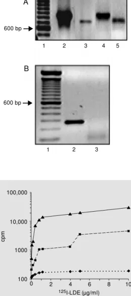
RT-PCR had shown that the expression of rLDL is higher in AML blast cells than in normal bone marrow cells, compared to ß-actin expression (Figure 1A). The second round of PCR amplification confirmed the identity of the rLDL-amplified product ob-tained from the first (Figure 1B).
LD E uptake by K562 ce lls
Figure 2 illustrates the binding (dashed
line) and the uptake (solid line) of 125
I-LDE
by K562 cells. At 4o
C (dotted line) there was no uptake. Under the conditions used, 90%
of 125I-LDE was found in the cytoplasm,
indicating that the emulsion entered the cell by the endocytic pathway.
Cyto to xicity o f the LD E:dauno rubicin
co mple x and fre e dauno rubicin
Figure 3Ashows the cytotoxicity on
nor-mal bone marrow and blast cells of the LDE:daunorubicin complex. A greater cyto-toxic effect of the LDE:daunorubicin com-plex was observed on blast cells than on normal bone marrow cells (P<0.001; Figure 3A). This difference was not observed when the cells were incubated with free daunorubicin (Figure 3B). The apparent
sen-sitivity (log EC50) to the LDE:daunorubicin
complex did not differ between blast cells (-9.60 ± 0.04) and normal bone marrow cells (-9.42 ± 0.08).
D iscussio n
One of the major problems associated with the chemotherapy of cancer is the lack of specificity of the drugs, which may give rise to a variety of side effects. A theoretical possibility of reducing the toxicity and im-proving the therapeutic response is to target
cp
m
100,000
10,000
1000
100
0 2 4 6 8 10
125I-LDE (µg/ml)
Total M embrane
4oC Figure 2. 125I-LDE binding and
uptake by K562 cells. Cells (106) w ere incubated w ith 0.10-10 µg/ ml (lipid concentration) 125I-LDE for 4 h in RPM I 1640 containing 10% FCS at 37oC or 4oC (dotted line). After incubation, the cells w ere w ashed and treated w ith trypsin and centrifuged and the supernatant radioactivity (mem-brane-bound emulsion; dashed line) was measured with a gamma-counter. The cells w ere then lysed and the incorporated radio-activity (solid line) w as deter-mined. Each point represents the mean of 2 experiments.
600 bp
600 bp
A
B
1 2 3 4 5
1 2 3
Figure 1. A, RT-PCR analysis of
low -density lipoprotein (LDL) mRNA in normal bone marrow and acute myelogenous leuke-mia (AM L) blast cells. Lane 1:
100-bp ladder; lanes 2 and 3: 4 µg of normal bone marrow cDNA amplified using ß-actin (lane 2) or LDL receptor (lane 3) primers, respectively; lanes 4 and 5: 1 µg of AM L blast cell cDNA ampli-fied using ß-actin (lane 4) or LDL receptor primers (lane 5), re-spectively. B, The second round
of PCR amplification analysis.
Lane 1: 100-bp ladder; lane 2: 273-bp segment obtained using nested primers; lane 3: negative control.
. . . . .
the cancer cells using a carrier. LDL was proposed as an endogenous carrier in several in vitro studies (6-10,13,21) and proved to
prevent side effects in one in vivo study (7).
Nevertheless, the routine use of LDL might be difficult due to the problems of its purifi-cation.
An alternative to the use of LDL would be an artificial microemulsion (LDE) which resembles its lipid portion (11). Several ex-perimental observations indicate that LDE may be taken up by rLDL and/or by other receptors that may recognize apo E: a) LDE clearance is accelerated in rats pretreated
with 17a-ethynylestradiol, which increases
rLDL expression (12), b) the LDE biodistri-bution in humans is very similar to that of LDL (16), c) LDE removal from plasma is increased in patients presenting AML as oc-curs with native LDL (3,4,16) and after re-mission of AML, LDE clearance in the pa-tients approaches normal values (16), and d) in vitro experiments with normal leukocytes
have shown that LDE competes with LDL for the same receptor (16).
In the present study, we demonstrate that it is possible to incorporate a cytotoxic anthracycline drug into LDE. The LDE:dau-norubicin complex obtained by co-sonica-tion of the drug with LDE possibly is due to the interaction of the ring structure present in the daunorubicin molecule that might inter-act with the interface between the core, com-posed of cholesterol esters and triglycerides, and the surface, composed of phospholipids and cholesterol. The incorporation proce-dure is reproducible and a typical prepara-tion contains 10 nmol of drug per mg of LDE, suggesting a limited number of do-mains for interaction. Since only a small percentage of the initial drug added (0.7%) was found inside highly purified LDE, dau-norubicin should be utilized in preparing the complex for therapeutic use in humans or animals. The low incorporation of daunoru-bicin into LDE observed here was not due to LDE particle characteristics since the same
rate of incorporation has been observed us-ing LDL particles (6).
The aim of the present study was to ana-lyze the cytotoxicity of the LDE:daunorubicin complex on myelogenous blast cells and normal bone marrow hematopoietic cells. Comparison of the cytotoxicity of the LDE: daunorubicin complex against normal bone marrow and AML blast cells clearly showed a lower cytotoxic effect on the former. The RT-PCR results indicate that in AML blast cells there is a higher transcription of rLDL than in normal bone marrow cells.
Further-more, the apparent affinity (EC50) of both
cell types for the LDE:daunorubicin com-plex did not differ, suggesting that both cells express the same receptor population. In addition, the difference in maximal response between normal and blast cells suggests a higher expression of the receptor
respon-%
C
el
l d
ea
th
60.0
40.0
10.0
0.0
0.0 0.5 1.0 1.5 2.0 2.5
LDE:daunorubicin (nM ) 20.0
30.0 50.0
A
AM L blast cells Normal bone marrow cells
Figure 3. A, Cytotoxicity of the LDE:daunorubicin com plex against normal bone marrow cells (squares) and acute mye-logenous leukemia (AM L) blast cells (lozenges). The points rep-resent the mean ± SEM of 7-15 experiments. * P<0.05 and * * P<0.001 compared to normal bone marrow cells (Student t -test). B, Cytotoxicity of free dau-norubicin against normal bone marrow cells (squares) and AM L blast cells (lozenges). The points represent the mean ± SEM of 7-17 experiments.
%
C
el
l d
ea
th
60.0
40.0
10.0
0.0
0.0 0.5 1.0 1.5 2.0 2.5
LDE:daunorubicin (nM ) 20.0
30.0 50.0
B
sible for the uptake of LDE in AML. Taken together, these results indicate that the LDE:daunorubicin complex may use the rLDL endocytic pathway to target AML blast cells. However, the involvement of other receptors cannot be ruled out since it has been shown that cell surface receptors such as rLDL-related protein (14,22) can also mediate specific high affinity degradation of
125
I-labeled LDL.
The complexed drug is as efficient as the free drug in killing blast cells. These results suggest that incorporation does not alter the cytotoxicity of daunorubicin or the receptor binding of LDE. Iwanik et al. (6) showed that the LDL:daunorubicin complex accu-mulates in P388 leukemia cells, indicating that the incorporation of the drug does not affect LDL binding to the receptor. The
in-cubation of free daunorubicin with blast and normal bone marrow cells confirmed the lack of specificity of the free drug.
The LDE:daunorubicin complex has been shown to be effective in protecting normal cells from the toxic effects of the free drug. Highly lipophilic compounds with
promis-ing cytotoxic effects in vitro have never
reached clinical trials because of difficulties in finding a suitable nontoxic solvent. Our results suggest that LDE might be an inter-esting and simple delivery system to admin-ister these compounds to tumor cells exhib-iting high rLDL expression.
Ackno wle dgm e nts
We thank Dr. Regina P. Markus for a critical review of the manuscript.
Re fe re nce s
1. Brow n M S & Goldstein JL (1986). A re-ceptor-mediated pathw ay for cholesterol homeostasis. Science, 232: 34-47.
2. Zannis VI (1989). M olecular biology of hu-man apolipoproteins B and E and associ-ated diseases of lipoprotein metabolism.
Advances in Lipid Research, 32: 1-64. 3. Vitols S, Gahrton G, Öst A & Peterson C
(1984). Elevated low density lipoprotein receptor activity in leukemic cells w ith monocytic differentiation. Blood, 63: 1186-1193.
4. Ho YK, Smith GS, Brow n M S & Goldstein JL (1978). Low density lipoprotein (LDL) receptor activity in human acute myelog-enous leukemia cells. Blood, 52: 1099-1103.
5. M osley ST, Goldstein JL, Brow n M S, Falck JR & Anderson RGW (1989). Tar-geted killing of cultured cells by receptor-dependent photosensitization. Proceed-ings of the National Academy of Sciences, USA, 78: 5717-5721.
6. Iw anik M J, Shaw KV, Ledw ith BJ, Yano-vich S & Shaw M (1989). Preparation and interaction of a low -density lipoprotein: daunorubicin complex w ith P388 leuke-mic cells. Cancer Research, 44: 1206-1215.
7. Vitols S, Söderberg-Reid K, M asquelier M , Sjöström B & Peterson C (1990). Low
density lipoprotein for delivery of a w ater-insoluble alkylating agent to malignant cells. In vitro and in vivo study of a drug-lipoprotein complex. British Journal of Cancer, 62: 724-729.
8. Firestone RA (1994). Low -density lipopro-tein as a vehicle for targeting antitumor compounds to cancer cells. Bioconjugate Chemistry, 5: 105-113.
9. Koller-Lucae SKM , Schot t H & Schw endener RA (1999). Low density lipoprotein and liposome mediated uptake and cytotoxic effect of N4-octadecyl-1 ß-D-arabinofuranosylcytosine in Daudi lym-phoma cells. British Journal of Cancer,
80: 1542-1549.
10. Filipow ska D, Filipow ski T, M orelow ska B, Kazanow ska W, Laudanski T, Lapinjoki S, Åkerlund M & Breeze A (1990). Treat-ment of cancer patients w ith a low -den-sity-lipoprotein delivery vehicle containing a cytotoxic drug. Cancer Chemotherapy and Pharmacology, 29: 396-400.
11. Ginsburg GS, Small DM & Atkinson D (1982). M icroemulsion of phospholipids and cholesterol esters. Protein-free mod-els of low density lipoprotein. Journal of Biological Chemistry, 257: 8216-8227.
12. M aranhão RC, Cesar TB, Pedroso-M ariane SR, Hirata M H & M esquita CH (1993). M etabolic behavior in rats of a nonprotein
microemulsion resembling low -density lipoprotein. Lipids, 28: 691-696. 13. Vitols S, Gahrton G & Peterson C (1984).
Significance of the low density lipopro-tein (LDL) receptor pathw ay for the in vitro accumulation of AD-32 incorporated into LDL in normal and leukemic w hite blood cells. Cancer Treatment and Re-search, 68: 515-520.
14. Rudling M , Gafvels M , Parini P, Gahrton G & Angelin B (1998). Lipoprotein receptors in acute myelogenous leukemia: failure to detect increased low -density lipoprotein (LDL) receptor numbers in cell mem-branes despite increased cellular LDL degradation. American Journal of Pathol-ogy, 153: 1923-1935.
15. M aranhão RC, Roland IA, Toffoletto O, Ramires JA, Gonçalves RP, M esquita CH & Pileggi F (1984). Plasma kinetic behav-ior in hyperlipidemic subjects of a lipidic microemulsion that binds to low density lipoprotein receptors. Lipids, 32: 627-633.
16. M aranhão RC, Garicochea B, Silva EL, Dorlhiac-Llacer P, Cadena SM S, Coelho IJC, M enegheti JC, Pileggi FJC & Cha-mone DAF (1993). Plasma kinetics and biodistribution of a lipid emulsion resem-bling low -density lipoprotein in patients w ith acute leukemia. Cancer Research,
17. Vainchenker W, Testa U, Guichard J, Titeux M & Breton-Gorius J (1981). Het-erogeneity in the cellular commitment of a human leukemic cell line: K562. Blood Cells, 7: 357-375.
18. Langer T, Strober W & Levy RI (1972). The metabolism of low density lipopro-tein in familial type II hyperlipoprolipopro-tein- hyperlipoprotein-emia. Journal of Clinical Investigation, 51: 1528-1536.
19. Dahlin DC, M iw a GT, Lu AY & Nelson SD (1984). N-Acetyl-p-benzoquinone imine: a cytochrome P-450 mediated oxidation product of acetaminophen. Proceedings of the National Academy of Sciences, USA, 81: 1327-1331.
20. Snedecor GW & Cochran WG (1980). Sta-tistical M ethods. 7th edn. Iow a State Uni-versity Press, Ames, IA.
21. Rudling M J, Collins VP & Peterson CO
(1983). Delivery of aclacinomycin A to hu-man glioma cells in vitro by the low -den-sity lipoprotein pathw ay. Cancer Re-search, 43: 4600-4605.