Article
J. Braz. Chem. Soc., Vol. 21, No. 5, 867-871, 2010. Printed in Brazil - ©2010 Sociedade Brasileira de Química 0103 - 5053 $6.00+0.00
*e-mail: [email protected]
New Ceramides from
Acnistus arborescens
Ana Isabel V. Maia,a Maria Leopoldina Veras,a Raimundo Braz-Filho,a
Norberto P. Lopes,b Edilberto R. Silveiraa and Otilia Deusdênia L. Pessoa*,a
aDepartamento de Química Orgânica e Inorgânica, Centro de Ciências, Universidade Federal do
Ceará, CP 12200, 60021-970 Fortaleza-CE, Brazil
bDepartamento de Física e Química, Faculdade de Ciências Farmacêuticas de Ribeirão Preto,
Universidade de São Paulo, 14040-903 Ribeirão Preto-SP, Brazil
Duas novas ceramidas N-(4-hidroxifeniletil)octacosamida (1) e rel-(2S,3S,4R,16E)-2-[(2’R
)-2’-hidroxinonadecanoilamino]-heneicosadec-16-eno-1,3,4-triol (2) foram isoladas do extrato
etanólico de Acnistus arborescens. As estruturas foram elucidadas por métodos espectroscópicos
(experimentos de RMN 1D e 2D, EMAR com ionização por electrospray, EM e IV).
Two new ceramides, N-(4-hydroxyphenethyl)octacosamide (1) and rel-(2S,3S,4R,16E
)-2-[(2’R)-2’-hydroxynonadecanoylamino]-heneicosadec-16-ene-1,3,4-triol (2) were isolated from
the EtOH extract of Acnistus arborescens. The structures were elucidated by spectroscopic (1D
and 2D NMR experiments, HR-ESI-MS, LR-MS and IR) methods.
Keywords: Acnistus arborescens, Solanaceae, ceramides
Introduction
The genus Acnistus (Solanaceae) comprises 50 tropical
American species of shrubs and small trees distributed from Mexico to Southern Argentina.1 Plants of this genus
biosynthesize a complex group of natural C28 steroidal lactones, known as acnistins,2 withanolides,3 and jaborols.4
As part of a collaborative search program to identify novel naturally occurring anticancer agents, we have investigated
Acnistus arborescens (L.) Schlecht.5,6 The plant presents
different therapeutic uses in the traditional medicine, for instance, the hot infusion of leaves and barks is used in the treatment of bruises and sprains, moreover, the leaves have been used to treat liver and spleen diseases, and cancerous growths.1,7,8 Indeed, previous studies on this species have
led to the isolation of several cytotoxic withanolides.7,8
Recently, it was published for the irst time the isolation of withaphysalins, including its cytotoxic effects against several tumor cell lines.5,6
In this paper, the isolation and characterization of two new ceramides from A. arborescens is described. This is
the irst examples of ceramides from a plant of the Acnistus
genus and, to the best of our knowledge, the second
report about such kind of secondary metabolites from the Solanaceae family.9
Ceramides and related compounds have been isolated extensively from fungi10 and several marine organisms
such as sponges,11 tunicates,12 sea stars,13 green algae14
and gorgonians.15 However, more recently, this class of
compound has been isolated from some higher plants.9,16-18
Results and Discussion
The EtOH extracts from leaves and stems of
A. arborescens were fractionated by column chromatography
on silica gel by elution with n-hexane, CH2Cl2, EtOAc
and MeOH. After several chromatographic procedures compound 1 was isolated from the leaves, while compound 2 was obtained from the stems. The two compounds, after
detailed spectroscopic analysis, were identiied as two new ceramides.
Compound 1 was isolated as a white amorphous solid.
Its IR spectrum revealed the absorption bands for either secondary amides or hydroxyl groups at 3302 cm−1, an
amide carbonyl at 1638 cm−1 and skeletal absorptions
at 1547, 1518 and 1464 cm−1 for aromatic rings. The
molecular formula of 1, C36H65NO2, was determined on
New Ceramides from Acnistus arborescens J. Braz. Chem. Soc. 868
the negative mode ([M-H]−) at m/z 542.4907 (C
36H64NO2,
calc. 542.4937). The 1H NMR spectrum of 1 exhibited an
amide proton at d 8.72 (s), two signals, both integrating for two aromatic hydrogens, at d 7.25 (d, J 8.5 Hz,
H-2/H-6) and 7.15 (d, J 8.5 Hz, H-3/H-5), a multiplet at
d 3.76 (2H-8) characteristic of the methylene attached to the amide nitrogen and a triplet at d 2.97 (J 7.5 Hz,
2H-7) for the benzyl methylene. As expected, the COSY spectrum showed vicinal scalar coupling for the former methylene with the benzyl methylene and the amide hydrogen. Additionally, the 1H NMR spectrum showed a
signal at d 2.41 (t, J 7.5 Hz, 2H-2’) typical of methylene
group α to a carbonyl, as well as signals for a terminal
methyl group at 0.89 (t, J 6.5 Hz, 3H-28’) and a series of
aliphatic methylene hydrogens (d 1.39-1.26). The 13C NMR
spectra of 1 showed the presence of a carbonyl signal at
d 174.3 (C-1’) for an amide, signals at d 158.7 (C-4), 131.8 (C-1), 131.7 (C-2/C-6) and 117.6 (C-3/C-5) that justify a para-substituted phenol moiety and two signals
at d 43.0 (C-8) and 36.9 (C-7) for the nitrogenated and benzyl methylene carbons, respectively. The 13C NMR
spectrum also revealed a series of signals at d 33.4-24.2 for the methylene carbons, in addition to a signal at d 15.5 for a terminal methyl group, suggesting that compound
1 possessed a long alkyl chain. The observed HMBC
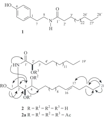
correlations for the methylene protons at d 3.76 (2H-8) with the carbons at d 174.3 (C-1’) and 131.8 (C-1) allowed to determine a N-(4-hydroxyphenethyl)amide
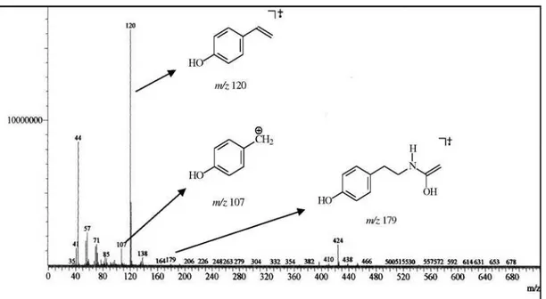
moiety, also justiied by the fragment ion peaks at m/z 179
([p-HO-C6H4-CH2CH2NHCOCH3]+•,deriving from a
McLafferty rearrangement), 120 ([p-HO-C6H4-CH=CH2]+•)
and 107 ([p-HO-C6H4-CH2]+), in the EI-MS spectrum.
Accordingly with the afore mentioned spectral data, the structure of 1 was characterized as the N-(4-hydroxyphenethyl)
octacosamide, an unknown ceramide up to date.
Compound 2 was also isolated as a white amorphous
solid. Its IR spectrum revealed several broad peaks in the range of 3399-3227 cm−1 characteristic of bonded
N-H or O-H stretching, the amide carbonyl at 1621 cm−1,
followed by the N-H bending at 1544 cm−1. A shoulder at
higher frequency than the carbonyl absorption followed by the band at 964 cm−1 is in accordance with a trans
carbon-carbon double bond. The long aliphatic chain was characterized by a band at 723 cm−1. The molecular formula
of 2, C40H79NO5, was determined on the basis of
high-resolution ESI mass spectroscopy in the negative mode ([M-H]−) at m/z 652.5983 (C
40H78NO5, calc. 652.5880).
The 1H NMR spectrum showed characteristic signal for
an amide proton at d 8.60 (d, J 8.9 Hz), resonances for
four hydroxyl groups at d 7.63, 6.72 (integrating for two hydrogens) and 6.10, all appearing as broad singlet; a signal at d 5.15 (m, H-2) for a methine bonded to a nitrogen; signals at d 4.54 (dd, J 10.9 and 4.5 Hz, H-1a) and 4.44
(dd, J 10.9 and 4.8 Hz, H-1b) for a hydroxymethylene, as
well as signals at d 4.64 (H-2´), 4.38 (m, H-3) and 4.32 (m, H-4) corresponding to three oxymethines. Additionally, signals for a double bond at d 5.59 (td, J 15.4 and 5.4 Hz,
H-16) and 5.50 (td, J 15.4 and 5.8 Hz, H-17), two terminal
methyl at d 0.88 (t, J 6.2 Hz, 3H-19´/3H-21) and several
methylene hydrogens at d 2.29-1.28 corresponding to two aliphatic chains were also observed. The COSY spectrum revealed coupling for the methine attached to the nitrogen (H-2) with the oxymethylene (2H-1) and an oxymethine (H-3) protons, and the latter with the oxymethine H-4. As expected, the 13C NMR spectra of 2 exhibited three
downield carbon signals at d 175.7 (C-1’), 131.3 (C-16) and 131.2 (C-17) corresponding to a carbonyl amide and a double bond, respectively. Signals for a nitrogenated methine at d 54.4 (C-2), an oxymethylene at d 62.5 (C-1) and three oxymethynes at d 77.2 (C-3), 73.5 (C-4) and 72.9 (C-2’). In addition, several carbon signals in the range of d
36.2-23.4 related to methylene groups and a carbon signal at 14.8 corresponding to two terminal methyls were also deduced from 13C NMR spectra, suggesting that compound 2 was also a ceramide. The unequivocal positions of the
hydroxyl groups were deduced based on the HMBC spectrum in which the proton signal at d 8.60 (NH) showed correlations with the carbonyl (C-1’) and the nitrogenated methine (C-2), while the proton signal at d 5.15 (H-2) exhibited correlations with the carbon signals at d 62.5
Figure 1. Structures of compounds 1, 2 and the acetylated derivative
Maia et al. 869 Vol. 21, No. 5, 2010
(C-1), 77.2 (C-3) and 73.5 (C-4). Furthermore, HMBC correlation between the proton signal at 4.64 (H-2´) and the carbonyl (C-1’) conirmed the presence of a side chain of a α-hydroxy fatty acid. Subsequently, the four hydroxyl
groups were conirmed through the acetylated compound (2a). The HMQC spectrum showed the obvious additional
methyl signals at dH/dC 2.23/21.2, 2.13/21.1, 2.10/21.5 and
2.08/21.3 characteristic of the four acetyl moieties. Through the HMBC spectra of both compounds 2 and 2a the double
bond was assigned at C-16 and C-17 of the sphingoid chain. HMBC correlations found for 3H-21 (d 0.88) and H-17 (d 5.50) with C-19 (d 32.6) supported this deduction. The
E-coniguration established for C-16/C-17 double bond
was determined through the large vicinal coupling constant (J 15.4 Hz) displayed between H-16 and H-17. The low
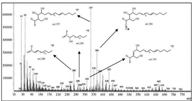
resolution mass spectrum of 2 was crucial to the deinition of
the amide and of the sphingoid portions length. The peaks at
m/z 384, 357 and 339 were in agreement with the sphingoid
moiety when combined with the peaks at m/z 298 and 280,
represented by the structural fragments sketched in Figure S10 (Supplementary Information), were congruent with the proposed structure. After comparison with analogous compounds16-18 the relative stereochemistry inferred for the
stereocenters 2, 3, 4 and 2’ was presumed to be S*, S*, R*
and R*, respectively. On the basis of the above mentioned
data, the structure of compound 2 was established as rel
-(2S,3S,4R,16E)-2-[(2’R
)-2’-hydroxynonadecanoylamino]-heneicosadec-16-ene-1,3,4-triol.
Experimental
General experimental procedures
Melting points were measured on a digital Mettler Toledo FP90 apparatus and are uncorrected. The optical rotations were measured on a Perkin-Elmer 341 digital polarimeter. IR spectra were recorded using a Perkin-Elmer FT-IR 1000 spectrometer. Electrospray ionization-High resolution mass spectra were measured on a quadrupole-time of light instrument (UltrOTOF-Q, Bruker Daltonics, Billerica, MA), while the low resolution Electron ionization mass spectra were acquired on a Shimadzu QP5050A instrument, through direct probe and operating at 70 eV. All NMR experiments were performed on a Bruker Avance DRX-500 spectrometer equipped with a 5 mm inverse detection z-gradient probe. 1H NMR (500.13 MHz) and 13C NMR
(125.77 MHz) spectra were measured at 27 °C using
pyridine-d5 as solvent. Chemical shifts, given on the d scale, were referenced to the residual pyridine [dH 8.74, 7.58, 7.22;
dC 150.35, 135.91, 123.87]. Column chromatography was run using silica gel 60 (70-230 mesh, Vetec; 230-400 mesh,
Merck) and TLC was performed on precoated silica gel polyester sheets (Kieselgel 60 F254, 0.20 mm, Merck) by detection with a spray reagent of vanillin/perchloric acid/ EtOH solution followed by heating at 100 °C.
Plant material
Acnistus arborescens was harvested in August 2006,
in the Pico Alto locality (Guaramiranga Mountain, State of Ceará), at an elevation of 1000 m. The plant material was identiied by Professor Edson P. Nunes. A voucher specimen (No. 30.513) is deposited in the Herbário Prisco Bezera (EAC) of the Departamento de Biologia, Universidade Federal do Ceará.
Extraction and isolation
The leaves (3.7 kg) and stems (3.0 kg) of A. arborescens
were separately soaked with EtOH (2×) at room temperature for 72 h. The extracts were concentrated under vacuum and the residues (leaves = 170 g; stems = 57 g) fractioned over silica gel using n-hexane, CH2Cl2, EtOAc and MeOH
as eluents. The CH2Cl2 fraction (45 g), obtained from the leaf EtOH extract, was subjected to gravity column chromatography over silica gel by elution with n-hexane
and n-hexane:EtOAc in increasing order of polarity. The n-hexane:EtOAc fraction 6:4 (24.1 g) after repeated column
chromatography yielded compound 1 (30 mg) by elution
with CH2Cl2:EtOAc (8:2). The EtOAc fraction (15.5 g), originated from the stem EtOH extract, was subjected to Si-gel column chromatography using gradients of increasing amounts of EtOAc (20-100%) in n-hexane,
followed by EtOAc:MeOH (9:1) to afford 8 fractions (A-H) on the basis of TLC proile. Fraction G (46 mg) was subjected to silica gel lash chromatography using EtOAc:MeOH (9.5:0.5) to afford compound 2 (18 mg).
N-(4-hydroxyphenethyl)octacosamide (1): White
amorphous solid; mp 116.0-119.4 °C; IR (KBr) ν
max/cm−1
3302, 3081, 2919, 2850, 1638, 1547, 1518, 1464, 1250, 1111; HRESIMS m/z 542.4907 [M-H] −; EIMS m/z 543
([M]+, absent), 179 (2), 121 (72), 120 (100), 107 (35); 1H
(500.13 MHz) and 13C NMR (125.77 MHz) data, see Table 1.
r e l ( 2 S , 3 S , 4 R , 1 6 E ) 2 [ ( 2 ’ R ) 2 ’ h y d r o x y -nonadecanoylamino]-heneicosadec-16-ene-1,3,4-triol. (2): White amorphous solid; mp 109.4-111.8 °C; [α]
D20 + 9°
(c 0.2, pyridine); IR (KBr) ν
max/cm−1 3399-3227, 2919, 2850,
1621, 1544, 1467, 1069, 964, 723; HRESIMS m/z 652.5983
[M-H] −; EIMS m/z 653 ([M]+, absent), 408 (9), 384 (15),
New Ceramides from Acnistus arborescens J. Braz. Chem. Soc. 870
279 (7), 265 (12), 97 (29), 83 (35), 57 (47), 43 (100);
1H (500.13 MHz, pyridine-d
5) and 13C NMR (125.77 MHz,
pyridine-d5) data, see Table 1.
Acetylation of 2: Compound 2 (12 mg) was dissolved
in a mixture of pyridine/acetic anhydride 1:2 (1 mL), under catalytic amounts of DMAP, and stirred for 3 h at room temperature. After this, the reaction mixture was neutralized with a solution of HCl 1 mol L−1 (4 drops) and extracted
with CH2Cl2 (3 × 10 mL). The CH2Cl2 layer was evaporated under reduced pressure to yield the peracetylated 2a
(12 mg). White amorphous solid; mp 52.8-54.6 °C; 1H NMR
(500.13 MHz, CDCl3): d 6.64 (1H, d, J 8.9 Hz, NH),
5.48-5.37 (2H, m, H-16 and H-17), 5.15 (1H, m, H-2’), 5.14 (1H, m, H-3), 5.0 (1H, br, d, J 9.4 Hz, H-4), 4.49 (1H, br,
s, H-2), 4.39 (1H, dd, J 11.7, 6.5 Hz, H-1a), 4.06 (1H, dd, J 11.7, 2.8 Hz, H-1b), 2.06-1.90 (4H, m, 2H-15 and 2H-18),
1.85 (4H, m, 2H-3’ and 2H-4’), 1.80-1.60 (2H, m, 2H-5), 1.50-1.20 (56H, m, 2H-6-2H-14, 2H-19, 2H-20 and 2H-5’- 2H-21’), 0.93 (6H, t, J 6.6 Hz, 3H-21 and 3H-22’), 2.23,
2.13, 2.10, 2.08 (s each, 4 × AcO). 13C NMR (125.77 MHz,
CDCl3) d 171.0 (C-1’), 131.6 (C-16), 129.9 (C-17), 74.5 (C-2’), 73.2/73.0 (C-4), 72.7 (C-3), 62.8 (C-1), 48.3 (C-2), 33.0 (C-15), 32.6 (C-18), 32.4 (C-3’), 32.2 (C-19 and C-20’), 30.1-28.6 (C-5, C-7 - C-14, C-5’-C-19’), 25.3 (C-6 and C-4’), 23.1 (C-20 and C-21’), 14.5 (C-21 and C-22’), 171.6/21.5, 171.6/21.3, 170.5/21.2, 170.5/21.1 (4 × AcO). EIMS m/z 864 ([M]+, absent), 790 (93), 776 (60), 761 (41),
748 (100), 733 (41), 705 (12), 691 (29), 583 (29), 483 (10), 440 (47), 310 (15), 261 (23), 139 (17), 56 (35).
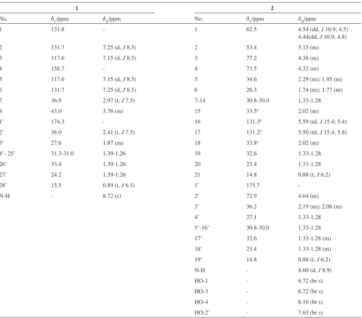
Table 1. 1H and 13C NMR spectral data for compounds 1 and 2, in pyridine-d
5
1 2
No. dC/ppm dH/ppm No. dC/ppm dH/ppm
1 131.8 - 1 62.5 4.54 (dd, J 10.9; 4.5)
4.44(dd, J 10.9; 4.8)
2 131.7 7.25 (d, J 8.5) 2 53.4 5.15 (m)
3 117.6 7.15 (d, J 8.5) 3 77.2 4.38 (m)
4 158.7 - 4 73.5 4.32 (m)
5 117.6 7.15 (d, J 8.5) 5 34.6 2.29 (m); 1.95 (m)
6 131.7 7.25 (d, J 8.5) 6 26.3 1.74 (m); 1.77 (m)
7 36.9 2.97 (t, J 7.5) 7-14 30.8-30.0 1.33-1.28
8 43.0 3.76 (m) 15 33.5a 2.02 (m)
1’ 174.3 - 16 131.3b 5.59 (td, J 15.4; 5.4)
2’ 38.0 2.41 (t, J 7.5) 17 131.2b 5.50 (td, J 15.4; 5.8)
3’ 27.6 1.87 (m) 18 33.8a 2.02 (m)
4’- 25’ 31.3-31.0 1.39-1.26 19 32.6 1.33-1.28
26’ 33.4 1.39-1.26 20 23.4 1.33-1.28
27’ 24.2 1.39-1.26 21 14.8 0.88 (t, J 6.2)
28’ 15.5 0.89 (t, J 6.5) 1’ 175.7
-N-H - 8.72 (s) 2’ 72.9 4.64 (m)
3’ 36.2 2,19 (m); 2.06 (m)
4’ 27.1 1.33-1.28
5’-16’ 30.8-30.0 1.33-1.28
17’ 32.6 1.33-1.28 (m)
18’ 23.4 1.33-1.28 (m)
19’ 14.8 0.88 (t, J 6.2)
N-H - 8.60 (d, J 8.9)
HO-1 - 6.72 (br s)
HO-3 - 6.72 (br s)
HO-4 - 6.10 (br s)
HO-2’ - 7.63 (br s)
Maia et al. 871 Vol. 21, No. 5, 2010
Supplementary Information
Supplementary information for compounds 1 and 2 is
available free of charge as PDF ile at http://jbcs.sbq.org.br
Acknowledgments
The authors thanks to Prof. Edson P. Nunes for the plant identiication and João Carlos Assunção for the LR mass spectra. This work was supported by grants from the Brazilian Governmental Agencies: CAPES, CNPq, PRONEX and FUNCAP.
References
1. Hawkes, J. G.; Lester, R. N.; Nee, M.; Estrada, N.; Solanaceae III: Taxonomy, Chemistry, Evolution. Royal Botanic Gardens, Kew: Great Britain, 1991.
2. Usubillaga, A.; Castellano G.; Zabel, V.; Watson, W. H.; J. Chem. Soc., Chem. Commun.1980, 854.
3. Nittala, S. S.; Lavie, D.; Phytochemistry1981, 20, 2735. 4. Burton, G.; Veleiro, A. S.; Gros E. G.; J. Chromatogr., B1984,
315, 435.
5. Veras, M. L.; Bezerra, M. Z. B.; Lemos, T. L. G.; Uchoa, D. E. A.; Braz-Filho, R.; Chai, H. B.; Cordell, G. A.; Pessoa, O. D. L.; J. Nat. Prod.2004, 67, 710.
6. Veras, M. L.; Bezerra, M. Z. B.; Braz-Filho, R.; Pessoa, O. D. L.; Montenegro, R. C.; Pessoa, Ó. C.; Moraes, M. O.; Costa-Lotufo, L. V.; Planta Med. 2004, 70, 551.
7. Kupchan, S. M.; Anderson, W. K.; Bollinger, P.; Doskotch, R. W.; Smith, R. M.; Renauld, J. A. S.; Schnoes, H. K.; Burlingame, A. L.; Smith, D. H.; J. Org. Chem. 1969, 34, 3858.
8. Minguzzi, S.; Barata, L. E. S.; Shin, Y. G.; Jonas, P. F.; Chai, H. B.; Park, E. J.; Pezzuto, J. M.; Cordell, G. A.; Phytochemistry
2002, 59, 635.
9. Su, B. N.; Misico, R.; Park, E. J.; Santarsiero, B. D.; Mesecar, A. D.; Fong, H. H. S.; Pezzuto, J. M.; Kinghorn, A. D.; Tetrahedron
2002, 58, 3453.
10. Gao, J. M.; Zhang, A. L.; Chen, H.; Liu, J. K.; Chem. Phys. Lipids2004, 131, 205.
11. Nakao, Y.; Takada, K.; Matsunaga, S.; Fusetani, N.; Tetrahedron
2001, 57, 3013.
12. Parrinello, N.; Cammarata, M.; Lipar, L.; Arizza, V.; Dev. Comp. Immunol. 1995, 19, 31.
13. Jim, W.; Rinehart, K. L.; Jares-Erijman, E. A.; J. Org. Chem.
1994, 59, 144.
14. Garg, H. S.; Sharma, M.; Bhakuni, D. S.; Pramanik, B. N.; Bose, A. K.; Tetrahedron Lett. 1992, 33, 1641.
15. Jeong, T. S.; Ahn, J. A.; Kim, Y. K.; Bok, S. H.; Kwon, B. M.; Shin, J.; Seo, Y.; Bioorg. Med. Chem. Lett.1997, 7, 1481. 16. Ramos, F.; Takaishi, Y.; Kawazoe, K.; Osório, C.; Duque, C.;
Acuña. R.; Fujimoto, Y.; Sato, M.; Okamoto, M.; Oshikawa, T.; Ahmed, S.U.; Phytochemistry2006, 67, 1143.
17. Sandjo, L. P.; Hannewald, P.; Yemloul, M.; Kirsch, G.; Ngadjui, B. T.; Helv. Chim. Acta.2008, 91, 1326.
18. Christophe, C. F.; Kouam, S.; Kouam, S. F.; Herve, M. P.; Simo, I. K.; Ngadjui, B. T.; Green, I. R.; Krohn, K.; Biochem. Syst. Ecol.2008, 36, 238.
Received: February 26, 2009
Web Release Date: February 11, 2010
Supplementary Information
J. Braz. Chem. Soc., Vol. 21, No. 5, S1-S8, 2010. Printed in Brazil - ©2010 Sociedade Brasileira de Química 0103 - 5053 $6.00+0.00*e-mail: [email protected]
New Ceramides from
Acnistus arborescens
Ana Isabel V. Maia,a Maria Leopoldina Veras,a Raimundo Braz-Filho,a
Norberto P. Lopes,b Edilberto R. Silveiraa and Otilia Deusdênia L. Pessoa*,a
aDepartamento de Química Orgânica e Inorgânica, Centro de Ciências, Universidade Federal do
Ceará, CP 12200, 60021-970 Fortaleza-CE, Brazil
b Departamento de Física e Química, Faculdade de Ciências Farmacêuticas de Ribeirão Preto,
Universidade de São Paulo, 14040-903 Ribeirão Preto-SP, Brazil
New Ceramides from Acnistus arborescens J. Braz. Chem. Soc.
S2
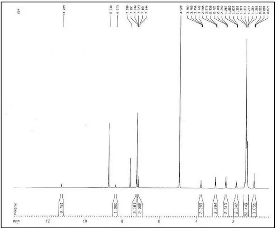
Figure S3.1H NMR spectrum of compound 1 (pyridine-d
5, 500 MHz).
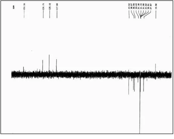
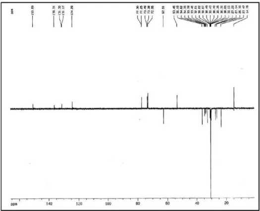
Figure S4.13C NMR spectrum of compound 1 (pyridine-d
Maia et al. S3 Vol. 21, No. 5, 2010
Figure S6.1H, 1H COSY spectrum of compound 1 (pyridine-d 5).
Figure S5.13C NMR - DEPT spectrum of compound 1 (pyridine-d
New Ceramides from Acnistus arborescens J. Braz. Chem. Soc.
S4
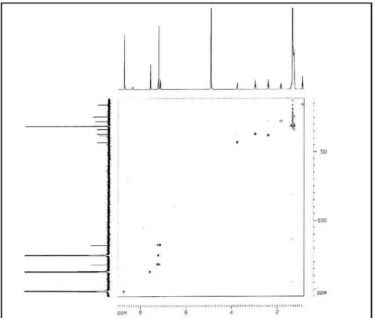
Figure S7. HMQC spectrum of compound 1 (pyridine-d5).
Maia et al. S5 Vol. 21, No. 5, 2010
New Ceramides from Acnistus arborescens J. Braz. Chem. Soc.
S6
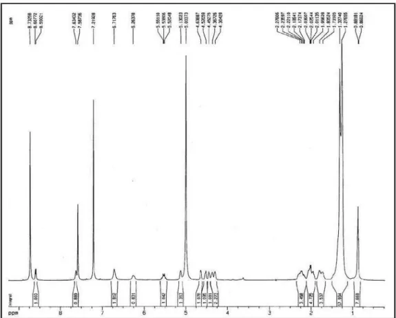
Figure S11.1H NMR spectrum of compound 2 (pyridine-d
5, 500 MHz).
Figure S12.13C NMR spectrum of compound 2 (pyridine-d
Maia et al. S7 Vol. 21, No. 5, 2010
Figure S14.1H, 1H COSY spectrum of compound 2 (pyridine-d 5).
Figure S13.13C NMR - DEPT spectrum of compound 2 (pyridine-d
New Ceramides from Acnistus arborescens J. Braz. Chem. Soc.
S8