Impact of Obstructive Sleep Apnea on the
Levels of Placental Growth Factor (PlGF) and
Their Value for Predicting Short-Term
Adverse Outcomes in Patients with Acute
Coronary Syndrome
Antonia Barcelo1, Josep Miquel Bauça1*, Aina Yañez1, Laura Fueyo1, Cristina Gomez1,
Monica de la Peña1, Javier Pierola1, Alberto Rodriguez1, Manuel Sanchez-de-la-Torre2,
Jorge Abad3, Olga Mediano4, Jose Amilibia5, Maria Jose Masdeu6, Joaquin Teran7, Josep Maria Montserrat8, MercèMayos9, Alicia Sanchez-de-la-Torre2, Ferran Barbé2, Spanish
Sleep Group
1Hospital Universitari Son Espases, Palma, Illes Balears, Spain,2Hospital Universitari Arnau de Vilanova and Santa Maria, IRBLleida, Lleida, Catalonia, Spain,3Hospital Universitari Germans Trias i Pujol, Badalona, Catalonia, Spain,4Hospital Universitario de Guadalajara, Guadalajara, Castilla-La Mancha, Spain,5Hospital Universitario Cruces, Bilbao, Basque Country, Spain,6Hospital Parc Taulí, Sabadell, Catalonia, Spain,7Hospital General Yagüe, Burgos, Castilla-León, Spain,8Hospital Clínic, Barcelona, Catalonia, Spain,9Hospital de la Santa Creu i Sant Pau, Barcelona, Catalonia, Spain
¶ Membership of the Spanish Sleep Group is provided in the Acknowledgments.
*pepmiquel@gmail.com
Abstract
Background
Placental growth factor (PlGF) induces angiogenesis and promotes tissue repair, and plasma PlGF levels change markedly during acute myocardial infarction (AMI). Currently, the impact of obstructive sleep apnea (OSA) in patients with AMI is a subject of debate. Our objective was to evaluate the relationships between PlGF levels and both the severity of acute coronary syndrome (ACS) and short-term outcomes after ACS in patients with and without OSA.
Methods
A total of 538 consecutive patients (312 OSA patients and 226 controls) admitted for ACS were included in this study. All patients underwent polygraphy in the first 72 hours after hos-pital admission. The severity of disease and short-term prognoses were evaluated during the hospitalization period. Plasma PlGF levels were measured using an electrochemilumi-nescence immunoassay.
Results
Patients with OSA were significantly older and more frequently hypertensive and had higher BMIs than those without OSA. After adjusting for age, smoking status, BMI and
OPEN ACCESS
Citation:Barcelo A, Bauça JM, Yañez A, Fueyo L, Gomez C, de la Peña M, et al. (2016) Impact of Obstructive Sleep Apnea on the Levels of Placental Growth Factor (PlGF) and Their Value for Predicting Short-Term Adverse Outcomes in Patients with Acute Coronary Syndrome. PLoS ONE 11(3): e0147686. doi:10.1371/journal.pone.0147686
Editor:Andrea Romigi, University of Rome Tor Vergata, ITALY
Received:September 8, 2015
Accepted:January 7, 2016
Published:March 1, 2016
Copyright:© 2016 Barcelo et al. This is an open access article distributed under the terms of the Creative Commons Attribution License, which permits unrestricted use, distribution, and reproduction in any medium, provided the original author and source are credited.
Data Availability Statement:All relevant data are available on Figshare:https://figshare.com/articles/
PlGF_and_Sleep_Apnea/2113396/1.
Funding:Supported by: ResMed Ltd. (Australia), Fondo de Investigación Sanitaria (PI10/02763 and PI10/02745), the Spanish Respiratory Society (SEPAR), the Catalonian Cardiology Society, Esteve-Teijin (Spain), Oxigen Salud (Spain), and ALLER.
hypertension, PlGF levels were significantly elevated in patients with OSA compared with
patients without OSA (19.9 pg/mL, interquartile range: 16.6–24.5 pg/mL; 18.5 pg/mL,
inter-quartile range: 14.7–22.7 pg/mL; p<0.001), and a higher apnea-hypopnea index (AHI) was
associated with higher PlGF concentrations (p<0.003). Patients with higher levels of PlGF
had also an increased odds ratio for the presence of 3 or more diseased vessels and for a Killip score>1, even after adjustment.
Conclusions
The results of this study show that in patients with ACS, elevated plasma levels of PlGF are associated with the presence of OSA and with adverse outcomes during short-term follow-up.
Trial Registration
ClinicalTrials.govNCT01335087
Introduction
Recent data suggest that obstructive sleep apnea (OSA) is underdiagnosed in patients after
acute myocardial infarction (AMI) [1]. Intermittent episodes of hypoxia and arousals cause an
increase in sympathetic activity, oxidative stress, hypercoagulability and cardiac hyperexcitabil-ity that could aggravate the severhyperexcitabil-ity of AMI and worsen the short-term prognosis of OSA
patients [2–4]. Nevertheless, a cardioprotective role of OSA in the context of AMI, via ischemic
preconditioning, has also been postulated [5]. Such protection would require the activation of
adaptive mechanisms, such as increased recruitment of proliferative and angiogenic
endothe-lial progenitor cells [6].
With the emergence of novel biomarkers, it may be feasible to characterize different aspects
of the pathophysiology of acute coronary syndrome (ACS) [7;8]. Placental growth factor
(PlGF), a member of the vascular endothelial growth factor family (VEGF), is expressed in cells of the cardiovascular system and plays a predominant role in pathological angiogenesis without
affecting quiescent vessels in healthy organs [9;10]. PlGF expression increases in the damaged
human heart, and PlGF levels in blood increase after AMI [11]. Elevated PlGF levels have
emerged as an important, independent marker of short-term adverse outcomes in patients
with ACS [12]. In contrast, PlGF plasma levels in the acute phase after myocardial infarction
(MI) have been found to be positively correlated with the degree of improvement in left ven-tricular function that occurs during the chronic phase of MI; this finding suggests that PlGF
may be involved in repairing injured myocardial tissue [13]. Cardiac PlGF expression is
induced by hypoxia, and it has been suggested that PlGF is a stress-response factor that sup-presses pathological remodeling in the heart by inducing angiogenesis, cardiomyocyte growth and peripheral mobilization of mononuclear cells and bone marrow-derived stem cells towards
ischemic myocardial tissue [11]. Recent evidence demonstrates that PlGF is a crucial mediator
of adaptive cardiac remodeling after myocardial infarction, and it has been suggested that the
effects of PlGF could form the basis of a potential therapeutic strategy in the future [14].
The purpose of this study was to assess the impact of OSA on circulating PlGF levels in patients with ACS and to determine whether PlGF levels have short-term prognostic signifi-cance in patients with OSA compared with patients without OSA.
Materials and Methods
Patients
The Ethics Committee of each participating center approved the study: the Comitè Ètic d’
In-vestigació (Hospital Universitari Son Espases, Palma), the Comité Ético de InIn-vestigación Clín-ica de Euskadi (Hospital de Cruces, Bilbao), the Comité Ético de Investigación ClínClín-ica (Hospital Arnau de Vilanova i Santa Maria, Lleida), the Comitè Ètic d’Investigació Clínica (Hospital Germans Trias i Pujol, Barcelona), the Comité Ético de Investigación Clínica (Hospi-tal General Universitario de Guadalajara, Guadalajara), the Comitè Ètic d’Investigació Clínica (Hospital Parc Taulí, Sabadell), the Comité Ético de Investigación Clínica de Burgos y Soria (Hospital General Yagüe, Burgos), and the Comitè Ètic d’Investigació Clínica (Hospital Clínic, Barcelona). All patients provided written, informed consent.
This is an ancillary study of the ISAACC Study (NCT01335087), a multicenter, open-label,
parallel, prospective, randomized, controlled trial [15]. The ISAACC study is evaluating the
effects of CPAP treatment on the incidence of new cardiovascular events in patients with an episode of ACS and OSA. The ISAACC study includes non-sleepy patients because it is unethi-cal to fail to treat OSA patients with excessive daytime sleepiness. The first patient was included
in April 2011, so in spite of newer guidelines regarding the sleep apnea definitions [16], the
recruitment of patients was consistent as originally established for ISAACC, and published
elsewhere [15;17]. For the present study, we included 538 patients (men and women age18
years old) who were admitted for ACS to coronary care units or cardiology hospitalization wards from fourteen teaching hospitals in Spain. A polygraphic study was performed during the first 48–72 h after admission. The case (n = 312) or control (n = 226) status of each ACS patient was defined using an apnea-hypopnea index (AHI) threshold of 15 or greater. ACS was defined as the acute presentation of coronary disease, with or without ST-elevation, unstable
angina, or MI type 1 [17]. The exclusion criteria for this study were the following: previous
CPAP treatment; psychophysical inability to complete questionnaires; the presence of any
pre-viously diagnosed sleep disorder;>50% of apneas consisting of central apneas or the presence
of Cheyne-Stokes respiration; the presence of daytime sleepiness (Epworth Sleep Scale [ESS]
score>10); the presence of chronic diseases, including neoplasms, renal insufficiency
(GFR<15 mL/min/1.73 m2), severe COPD (FEV1<50%), chronic depression, and other
limit-ing chronic diseases; a medical history that could interfere with the objectives of the study; and
any processes, whether cardiovascular or otherwise, that reduced life expectancy to<1 year;
and patients in cardiogenic shock.
Procedures
The diagnosis of OSA was based on the results of a sleep test, in accordance with the guidelines
of the Spanish national consensus on apnea-hypopnea syndrome [18]. All participating centers
used the same model of polygraph (Embletta; ResMed, Australia) for the diagnosis of OSA. Oronasal flow, thoracoabdominal movements, ECG, and pulse oximetry were recorded. Apnea
is defined as an absence of airflow lasting10 seconds. Hypopnea is defined as a reduction in
airflow lasting10 seconds and is associated with oxygen desaturation. Oxygen desaturation
(ODI) is considered as a decrease in SaO2>4%. The apnea-hypopnea index is defined as the
number of apneas and hypopneas per hour of sleep. The extent of self-reported sleepiness/
drowsiness was analyzed using the Spanish version of the ESS test [19]. Echocardiographic
peak troponin levels, and complications related to the cardiovascular event itself (heart failure, reinfarction, mechanic complications such myocardial rupture, arrhythmia and stroke) and mortality. Other complications related to the ICU admission were excluded, such as infections or venous thrombosis following immobilization, since they were not due to the cardiovascular event for which the patient was admitted.
Blood sampling and analysis
Peripheral blood samples were collected from all subjects after the polygraphic study at the time of randomization. Plasma and serum samples were stored at -80°C until analysis. Cardiac
marker levels were measured by investigators who were blinded to the patients’histories.
Blood chemistry data were measured using commercially available assays. PlGF levels were measured in plasma using an enzyme-based electrochemiluminescence assay (Roche Diagnos-tics, Germany). The lower limit of detection was 3 pg/mL, and the upper limit of detection was 10.000 pg/mL. The intra-assay coefficient of variation was 1.1%, and the inter-assay coefficient of variation 2.6%.
Data analysis
Data on the patients included in this study were incorporated into a password-protected data-base. The mean values (and standard deviations) or frequencies (and percentages) were com-puted to evaluate the differences between the control and OSA patients, and the significance of differences was assessed using Mann-Whitney tests (or t tests, if data were normally distrib-uted) or chi-squared tests, respectively. OSA patients were divided into 3 groups according to AHI, and the Kruskal-Wallis test was used for comparisons among groups. Spearman´s rank correlations were calculated to assess correlations. The associations among PlGF levels, OSA, and variables related to the severity of ACS were assessed using the Mann-Whitney or chi-squared tests and linear or logistic regression models, as appropriate. Additionally, the models were adjusted for smoking status (current or former smoker vs non-smoker), body mass index, age, gender (male vs female), hypertension, diabetes and dyslipidemia.
Odds ratios, their corresponding 95% confidence intervals, and p-values were calculated using logistic regression models; adjusted p-values were also computed. The threshold for
sta-tistical significance was set at p<0.05. SPSS v19 software (IBM Corp., Armonk, NY) was used
for all analyses.
Results
The clinical and sleep-related characteristics of the patients are given inTable 1.
OSA was detected in 58% of the study subjects (312 patients and 226 controls).
Patients with OSA were significantly older and had higher BMIs compared with those without
OSA. No difference was detected between genders (Table 1). The number of hypertensive
patients was significantly higher in the OSA group (Table 1). The number of patients who took
antihypertensive drugs, particularly diuretics and calcium agonists, was higher in the OSA group. PlGF levels were higher in patients with OSA compared with patients without OSA (19.9 pg/mL,
interquartile range: 16.6–24.5 pg/mL; 18.5 pg/mL, interquartile range: 14.7–22.7 pg/mL; p<0.001).
PlGF levels were correlated with age (r = 0.304, p<0.001), AHI (r = 0.142, p = 0.001), Min
SaO2(r = -0.091, p = 0.038), ODI (r = 0.128, p = 0.004), troponin concentration (r = 0.171,
p<0.001) and mean peak troponin level (r = 0.161, p = 0.001). To investigate the strength of
AHI30 group (19.9 pg/mL, interquartile range: 17.5–24.8) than both the AHI 15–29.9 group
(19.3 pg/mL, interquartile range: 16.2–23.5) and the AHI15 group (18.2 pg/mL, interquartile
range: 14.7–22.1); (p<0.003;Fig 1).
To further assess the associations of variables related to sleep and to the severity of ACS with PlGF levels, we classified the study participants into two groups according to PlGF
con-centrations (Table 2). A cutoff value of 20 pg/mL for PlGF levels was used based on prior
studies [20]. Using this cut-off value, we found that up to 156 patients with OSA had PlGF
lev-els>20, which represent 63.7% of patients with PlGF20 pg/mL. By contrast, the number of
non-OSA patients with PlGF levels20 pg/mL was 81, which represent 36.3% of patients with
PlGF20 pg/mL.
There were no differences in the trend for the ACS category and OSA (unstable angina was present in 14.8% of controls and 13.3% of OSA; NSTEMI in 42.6% of controls and 41.5% of OSA; STEMI in 42.6% of controls and 45.2% of OSA). No significant association was detected between ACS category and PlGF levels.
Table 1. Anthropometric, clinical and biochemical variables for patients with obstructive sleep apnea (OSA) and controls.
Controls(n = 226) OSA(n = 312) p-value
Age, years 56.5±11.5 61.2±10.3 <0.001
Male, % 191 (84.5%) 250 (80.1%) 0.192
AHI, h-1 6.3±4.1 35.7±1 7.4 <0.001
ODI, h-1 9.3±17.9 24.9±16.9 <0.001
Mean SaO2, % 93.6±2.2 92.7±2.2 <0.001
Min SaO2, % 86.7±5.2 82.1±7.2 <0.001
Time with SaO2<90%, % 4.2±11.5 11.2±18.0 <0.001
BMI, kg•m-2 26.1±6.1 28.3±6.2 <0.001
Glucose, mg/dL 119±53 128±59 0.078
HDL cholesterol, mg/dL 45±30 40±18 0.058
LDL cholesterol, mg/dL 116±41 110±33 0.103
Triglyceride, mg/dL 154±117 141±79 0.161
Hypertension 87 (38.5%) 178 (57.1%) <0.001
Diabetes mellitus 50 (22.1%) 83 (26.6%) 0.243
Dyslipidemia 108 (47.8%) 134 (42.9%) 0.265
Smoker 0.163
No 57 (25.2%) 103 (32.8%)
Yes 107 (47.3%) 134 (42.7%)
Former smoker 62 (27.4%) 77 (24.5%)
Diuretics 28 (12.5%) 67 (21.4%) 0.008
Anticoagulants 6 (2.7%) 22 (7.1%) 0.025
Antacids 47 (20.9%) 96 (30.8%) 0.011
Hypolipidemic agents 68 (30.4%) 126 (40.4%) 0.017
β-blockers 44 (19.8%) 77 (24.7%) 0.186
Calcium antagonists 9 (4.0%) 44 (14.2%) <0.001
ACEIs 14 (18.9%) 29 (22.5%) 0.050
Insulin 10 (4.5%) 29 (9.3%) 0.034
Oral antidiabetic agents 37 (16.5%) 61 (19.6%) 0.361
Bronchodilators 16 (7.1%) 12 (3.8%) 0.091
Values represent the percentage of the patients and control subjects or means±standard deviations. AHI, apnea-hypopnea index; ODI, oxygen
desaturation index; BMI, body mass index; HDL, high-density lipoprotein; LDL, low-density lipoprotein; ACEIs, angiotensin-converting enzyme inhibitors.
Patients with elevated PlGF levels were more frequently older and hypertensive, and with
respect to ACS severity, the percentages of patients with Killip score>1 and with 3 or more
diseased vessels were higher in the high-PlGF group (Table 2).
In a multivariable analysis, PlGF, together with all baseline characteristics that were found to be predictive in univariate analyses, persisted as independent predictors of severity and Fig 1. Levels of PlGF according to AHI groups.Error bars represent 95% confidence intervals. The Kruskal-Wallis test was used for comparisons among groups.
doi:10.1371/journal.pone.0147686.g001
Table 2. Baseline patient characteristics and variables related to the severity of acute coronary syndrome according to PlGF status.
PlGF<20 pg/mL(n = 301) PlGF20 pg/mL(n = 237) p-value
Age, years 57.0±11.0 61.8±10.5 <0.001
Male, % 245(81.9%) 192 (81.6%) 0.927
OSA 160 (53.3%) 156 (63.7%) 0.015
BMI, kg•m-2 27.3±6.3 27.5±6.2 0.653
Hypertension 135 (44.7%) 132 (55.2%) 0.015
Diabetes mellitus 71 (23.6%) 61 (25.7%) 0.565
Dyslipidemia 167 (55.5%) 129 (54.4%) 0.808
Smoker 0.138
No 86 (28.5%) 75 (31.5%)
Yes 147 (48.7%) 96 (40.3%)
Former smoker 69 (22.8%) 67 (28.2%)
Number of diseased vessels3 48 (17.6%) 65 (30.2%) <0.001
Killip score>1 12 (5.0%) 23 (12.9%) 0.004
Ejection fraction<51.5%* 58 (25.8%) 40 (23.4%) 0.586
Peak troponin level724.5 ng/mL* 57 (24.4%) 45 (26.0%) 0.704
CV complications during hospitalization 16 (5.15%) 23 (9.2%) 0.059
PlGF, placental growth factor; OSA, obstructive sleep apnea; BMI, body mass index; CV, cardiovascular.
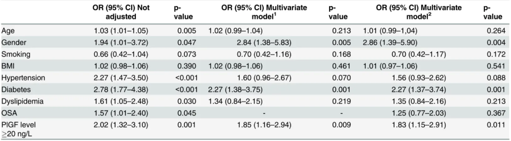
short-term outcome (Tables3and4). Patients with higher levels of PlGF had increased odds
ratios for a Killip classification>1 (Table 3, ORs 3.13, p = 0.009; and ORs 2.67, p = 0.011) and
for the presence of 3 or more diseased vessels (Table 4, ORs 2.02, p = 0.001; and ORs 1.85,
p = 0.009) even after adjustment.
The number of cardiovascular complications (heart failure, reinfarction, mechanic compli-cations, arrhythmia and stroke) tended to be higher in the high-PlGF group. However, the multivariate analysis revealed that despite the fact that patients with OSA had high PlGF levels, when assessing the associations between these ACS severity-related variables and high PlGF
levels, there were no statistically significant differences after adjustment for OSA (Table 3and
Table 4). In addition, no significant interactions were seen between PlGF levels and the effect of OSA with respect to other clinical outcomes such as the length of stay in the coronary unit, length of hospitalization, ejection fraction, number of stents implanted or peak of troponin
(p>0.05).
Table 3. Multivariate adjusted odds ratios for Killip score>1.
OR (95% CI) Not adjusted p-value OR (95% CI) Multivariate model1 p-value OR (95% CI) Multivariate model2 p-value
Age 1.04 (1.01–1.08) 0.013 1.01 (0.97–1.05) 0.576 1.01 (0.97–1.05) 0.593
Gender 0.53 (0.24–1.15) 0.107 0.63 (0.26–1.55) 0.314 0.63 (0.26–1.54) 0.309
Smoking 0.54 (0.26–1.09) 0.087 0.85 (0.36–1.99) 0.702 0.89 (0.38–2.09) 0.781
BMI 1.00 (0.95–1.06) 0.906 0.98 (0.93–1.04) 0.547 0.98 (0.93–1.03) 0.456
Hypertension 3.82 (1.69–8.62) 0.001 2.59 (1.02–6.56) 0.045 2.32 (0.90–5.97) 0.080
Diabetes 2.47 (1.22–5.03) 0.012 1.79 (0.82–3.89) 0.141 1.80 (0.82–3.92) 0.143
Dyslipidemia 1.43 (0.70–2.88) 0.325 1.07 (0.51–2.27) 0.858 1.11 (0.52–2.37) 0.787
OSA 2.07 (0.97–4.41) 0.060 - - 1.83 (0.80–4.20) 0.155
PlGF20 pg/mL 3.13 (1.34–7.34) 0.009 2.67 (1.25–5.69) 0,011 2.70 (1.26–5.77) 0.011
1Multivariate model adjusted for age, gender (male vs female), smoking (current or former smoker vs non-smoker), BMI, hypertension, diabetes,
dyslipidemia.
2Multivariate model adjusted for age, gender (male vs female), smoking (current or former smoker vs non-smoker), BMI, hypertension, diabetes, dyslipidemia and OSA.
doi:10.1371/journal.pone.0147686.t003
Table 4. Multivariate adjusted odds ratios for3 diseased vessels.
OR (95% CI) Not adjusted
p-value
OR (95% CI) Multivariate model1
p-value
OR (95% CI) Multivariate model2
p-value
Age 1.03 (1.01–1.05) 0.005 1.02 (0.99–1.04) 0.213 1.01 (0.99–1,04) 0.264
Gender 1.94 (1.01–3.72) 0.047 2.84 (1.38–5.83) 0.005 2.86 (1.39–5.90) 0.004
Smoking 0.66 (0.42–1.04) 0.073 0.70 (0.42–1.16) 0.168 0.70 (0.42–1.17) 0.172
BMI 1.02 (0.98–1.06) 0.390 1.02 (0.98–1.06) 0.461 1.01 (0.97–1.06) 0.541
Hypertension 2.27 (1.47–3.50) <0.001 1.60 (0.96–2.67) 0.070 1.56 (0.93–2.62) 0.088
Diabetes 2.78 (1.77–4.38) <0.001 2.27 (1.38–3.75) 0.001 2.27 (1.37–3.74) 0.001
Dyslipidemia 1.61 (1.05–2.48) 0.030 1.34 (0.84–2.15) 0.219 1.35 (0.84–2.16) 0.213
OSA 1.57 (1.01–2.40) 0.045 - - 1.25 (0.77–2.03) 0.367
PlGF level
20 ng/L
2.02 (1.32–3.10) 0.001 1.85 (1.16–2.94) 0.009 1.83 (1.15–2.91) 0.011
1Multivariate model adjusted for age, gender (male vs female), smoking (current or former smoker vs non-smoker), BMI, hypertension, diabetes, dyslipidemia.
2Multivariate model adjusted for age, gender (male vs female), smoking (current or former smoker vs non-smoker), BMI, hypertension, diabetes, dyslipidemia and OSA.
Discussion
The results of this study show that in patients with ACS, elevated plasma levels of PlGF are associated with the presence of OSA and with adverse outcomes during short-term follow-up. The present findings suggest that the presence of OSA may affect the clinical significance of PlGF in patients with ACS.
PlGF is an established prognostic marker in ACS. However, no study has specifically exam-ined its prognostic capabilities in OSA patients with ACS.
In a previous study, we observed that OSA influences the severity of ACS and its short-term prognosis. OSA was correlated with an increase in peak plasma troponin levels, with the number
of diseased vessels and with the length of stay in the coronary unit [17]. In this study, we observed
that in patients with ACS, higher PlGF levels were associated with adverse short-term outcomes (greater numbers of diseased vessels, higher Killip scores and a tendency to develop more cardio-vascular complications). These observations suggest that different pathophysiological mecha-nisms may be responsible for the expression of PlGF in patients with and without OSA and that these mechanisms may affect short- and long-term risks after ACS in different ways.
PlGF, a member of the VEGF family of angiogenic proteins, plays an important role in
path-ological angiogenesis [21]. Although PlGF and VEGF activate similar signaling pathways, PlGF
exhibits greater disease-specific activity than VEGF does, while it does not affect quiescent ves-sels in healthy tissues. In addition to enhancing angiogenesis, PlGF is known to improve car-diac performance by promoting cardiomyocyte survival and cardiomyogenesis via recruitment
of bone marrow-derived progenitor cells towards infarcted myocardial tissue [22]. Hypoxia is
an important stimulus of PlGF expression, and hypoxia inducible factor-1-alpha (HIF-1α) can
directly activate its transcription [10;13]. High PlGF release typically accompanies acute
ische-mia and infarction, but elevated levels may reflect underlying acute or chronic hypoxia. Our results demonstrate that OSA is an important determinant of PlGF levels in patients with ACS. Among several confounding factors, OSA seems to mediate a portion of the release of PlGF detected in these patients. In line with this data, PlGF may represent a valuable marker of OSA in patients suspected of having acute coronary syndrome. Furthermore, the presence of OSA may affect the prognostic value of PlGF levels, and this possibility should be considered in studies in which PlGF is used as a clinical biomarker for risk stratification.
There is evidence that OSA may be associated with the activation of cardiovascular adaptive mechanisms. Berger et al reported that the numbers of endothelial progenitor cells are elevated and that angiogenesis increases in patients with AMI and coexistent OSA compared with
patients with AMI without OSA [6]. EPCs are mobilized by signaling pathways, such as the
HIF-1αpathway, which are also activated in OSA [23] [24]. HIF-1αstimulates the production
of VEGF, and several studies have shown that patients with OSA have increased levels of
VEGF [25]. In contrast, another study found that although VEGF expression in monocytes
was found to be higher in patients with AMI and OSA compared with patients with AMI
with-out OSA, no difference was reported for plasma VEGF levels between these groups [6]. On the
other hand, earlier studies, which demonstrated that PlGF levels at presentation are of prog-nostic value for clinical outcomes in patients with ACS, did not find any correlation between
PlGF levels and VEGF levels [12]. Thus far, the effect of OSA on plasma PlGF levels after ACS
has not been investigated. In patients with ACS, plasma levels of PlGF increase acutely and
transiently [12]. A single initial measurement of a patient’s plasma PlGF level appears to extend
the predictive and prognostic information gained from traditional risk markers [26]. The
extent to which PlGF levels are elevated is influenced by the severity of myocardial damage,
and the overall effects of PlGF may vary with disease status and comorbidities [27–29]. In this
given the relationship between PlGF levels after ACS and OSA that was observed in our study. However, it is known that OSA increases the incidence of morning peak of onset in acute myo-cardial infarction. Despite the fact that there are no data on whether PlGF has a diurnal pattern of variation, a perturbation in circadian PlGF balance might be a possible contributor to the
onset of MI [30;31]. Furthermore, elevated PlGF levels may reflect underlying acute or chronic
hypoxia. PlGF has potent angiogenic properties, especially under pathological circumstances,
and increased PlGF levels could counteract the damaging effects of ischemia [21]. Iwama et al
observed that patients with higher plasma PlGF levels on day 3 after AMI showed greater improvement in left ventricular ejection fraction (LEVF) during the chronic phase (6 months post MI) than did patients with lower plasma PlGF levels, and they also observed that patients with improvement in LEVF in the chronic phase had significantly higher plasma levels of PlGF in the acute phase compared with patients without improvement; these findings suggest that
PlGF may be involved in repairing injured myocardial tissue [13]. In addition, experimental
studies have demonstrated that PlGF may serve both as a marker of adaptive cardiac remodel-ing and as a promisremodel-ing novel therapeutic agent for revascularizremodel-ing and regeneratremodel-ing the
infarcted myocardium and for improving its performance after MI [14;22]. Consistent with
these findings, it is possible that the elevated levels of PlGF detected in the group of patients with OSA could exert a beneficial effect that could promote long-term improvement in cardiac function after ACS in these patients.
Limitations
This study has several limitations. First, we excluded patients with daytime sleepiness, which were the patients who exhibited the most severe OSA. Second, OSA was diagnosed based on respiratory polygraphy, which could underestimate the severity of OSA. Third, the high vari-ability in PlGF levels detected both in controls and patients may limit its usefulness. On the other hand, this study was performed only for patients with ACS. Previous clinical studies
including subjects without ACS showed PlGF mean plasma levels of 10 pg/mL [13] and 16.6
pg/mL [31], both of them lower than in our ACS patients groups, either with or without OSA.
Ideally, longitudinal studies measuring PlGF at the time of diagnosis of OSA would add to the verification of PlGF as a biomarker. Fourth, alternative noninvasive biomarkers of cardiac dys-function, such as the brain natriuretic peptide (BNP), were not assessed. As reported elsewhere, PlGF and BNP levels are known to positively correlate in patients with ischemic
cardiomyopa-thy [32]. This demonstrates an increase of PlGF with the severity of heart failure in such
patients, regardless of OSA. Finally, the design of this study does not allow us to evaluate the long-term prognostic role of PlGF in OSA or to draw definitive conclusions.
Conclusions
The results of this study show that in patients with ACS, elevated plasma levels of PlGF are associ-ated with the presence of OSA and with adverse outcomes during short-term follow-up. These findings suggest that different pathophysiological mechanisms might affect the cardiac expression of PlGF after ischemic injury, as well as their predictive role in patients with and without OSA.
Acknowledgments
Complete authors’affiliation: Antonia Barcelo (Servei d’Anàlisis Clíniques, Hospital
Universi-tari Son Espases, Palma, Illes Balears, Spain; Institut d’Investigació Sanitària de Palma (IdISPa), Palma, Illes Balears, Spain; Centro de Investigación Biomédica en Red de Enfermedades Respiratorias (CIBERES), Madrid, Spain), Josep Miquel Bauça (Servei d’Anàlisis Clíniques,
de Palma (IdISPa), Palma, Illes Balears, Spain), Aina Yañez (Institut d’Investigació Sanitària de
Palma (IdISPa), Palma, Illes Balears, Spain), Laura Fueyo (Servei d’Anàlisis Clíniques, Hospital
Universitari Son Espases, Palma, Illes Balears, Spain), Cristina Gomez (Servei d’Anàlisis Clíni-ques, Hospital Universitari Son Espases, Palma, Illes Balears, Spain), Monica de la Peña (Servei de Pneumologia, Hospital Universitari Son Espases, Palma, Illes Balears, Spain; Centro de Investigación Biomédica en Red de Enfermedades Respiratorias (CIBERES), Madrid, Spain), Javier Pierola (Institut d’Investigació Sanitària de Palma (IdISPa), Palma, Illes Balears, Spain), Alberto Rodriguez (Servei de Medicina Intensiva, Hospital Universitari Son Espases, Palma, Illes Balears, Spain), Manuel Sanchez-de-la-Torre (Servei de Pneumologia, Hospital Universi-tari Arnau de Vilanova and Santa Maria, IRBLleida, Lleida, Catalonia, Spain; Centro de Investi-gación Biomédica en Red de Enfermedades Respiratorias (CIBERES), Madrid, Spain), Jorge Abad (Servei de Pneumologia, Hospital Universitari Germans Trias i Pujol, Badalona, Catalo-nia, Spain), Olga Mediano (Servicio de Neumología, Hospital Universitario de Guadalajara, Guadalajara, Castilla-La Mancha, Spain), Jose Amilibia (Servicio de Neumología, Hospital Uni-versitario Cruces, Bilbao, Basque Country, Spain), Maria Jose Masdeu (Servei de Pneumologia, Hospital Parc Taulí, Sabadell, Catalonia, Spain), Joaquin Teran (Servicio de Neumología, Hos-pital General Yagüe, Burgos, Castilla-León, Spain), Josep Maria Montserrat (Servei de Pneu-mologia, Hospital Clínic, Barcelona, Catalonia, Spain), Mercè Mayos (Servei de PneuPneu-mologia, Hospital Universitari Arnau de Vilanova and Santa Maria, IRBLleida, Lleida, Catalonia, Spain), Alicia Sanchez-de-la-Torre (Servei de Pneumologia, Hospital Universitari Arnau de Vilanova and Santa Maria, IRBLleida, Lleida, Catalonia, Spain; Centro de Investigación Biomé-dica en Red de Enfermedades Respiratorias (CIBERES), Madrid, Spain) and Ferran Barbé (Ser-vei de Pneumologia, Hospital Universitari Arnau de Vilanova and Santa Maria, IRBLleida, Lleida, Catalonia, Spain; Centro de Investigación Biomédica en Red de Enfermedades Respira-torias (CIBERES), Madrid, Spain).
This work was supported by ResMed Ltd. (Australia), Fondo de Investigación Sanitaria, Spain, (PI10/02763 and PI10/02745), the Spanish Respiratory Society (SEPAR), the Catalonian Cardiology Society, Esteve-Teijin (Spain), Oxigen Salud (Spain), and ALLER.
The members of the Spanish Sleep and Breathing Group are:Amanda López-Picado, pharmacist, Araba Research Unit, Hospital Universitario Txagorritxu, Vitoria; Erika Miranda-Serrano, statistician, Araba Research Unit, Hospital Universitario Txagorritxu; Cristina Marti-nez-Null, biologist, Sleep Unit, Hospital Universitario, Vitoria and Ciber de Enfermedades Respiratorias (CibeRes); Ramón Rubio, neurologist, Hospital Universitario Txagorritxu, Vito-ria and CibeRes; MaVito-ria Luz Alonso, respiratory physician, Hospital General Yagüe, Burgos and CibeRes; José Cordero, epidemiologist, Hospital General Yagüe, Burgos; Cristobal Esteban, respiratory physician, Hospital de Galdakano, Vizcaya; Antonio Jiménez, respiratory physi-cian, Hospital Universitario Marqués de Valdecilla, Santander; Maria Rosario Carpizo, neuro-physiologist, Hospital Universitario Marqués de Valdecilla, Santander; Gabriel Sanpol,
respiratory physician, Hospital Valle de Hebrón, Barcelona and CibeRes; Jaime Corral, tory physician, Hospital San Pedro de Alcántara, Cáceres and CibeRes; Manola Rubio, respira-tory physician, Hospital San Pedro de Alcántara, Cáceres; Antonia Barceló, clinical analyst, Hospital Son Dureta, Palma de Mallorca and CibeRes; Javier Piérola, biologist, Hospital Son Dureta, Palma de Mallorca and CibeRes; José María Marín, respiratory physician, Hospital Universitario Miguel Servet, Zaragoza and CibeRes.
Author Contributions
Abad OM J. Amilibia MJM JT JMM MM AS. Wrote the paper: AB JMB AY FB. Guarantor and lead investigator of the study: FB.
References
1. Ludka O, Stepanova R, Vyskocilova M, Galkova L, Mikolaskova M, et al.: Sleep apnea prevalence in acute myocardial infarction—the Sleep Apnea in Post-acute Myocardial Infarction Patients (SAPAMI)
Study. Int J Cardiol 2014; 176:13–19. doi:10.1016/j.ijcard.2014.06.020PMID:25064202
2. Sanchez-de-la-Torre M, Campos-Rodriguez F, Barbe F: Obstructive sleep apnoea and cardiovascular disease. Lancet Respir Med 2013; 1:61–72. doi:10.1016/S2213-2600(12)70051-6PMID:24321805
3. Kohler M, Stradling JR: Mechanisms of vascular damage in obstructive sleep apnea. Nat Rev Cardiol 2010; 7:677–685. doi:10.1038/nrcardio.2010.145PMID:21079639
4. Kuniyoshi FH, Garcia-Touchard A, Gami AS, Romero-Corral A, van der WC, Pusalavidyasagar S, et al.: Day-night variation of acute myocardial infarction in obstructive sleep apnea. J Am Coll Cardiol 2008; 52:343–346. doi:10.1016/j.jacc.2008.04.027PMID:18652941
5. Shah N, Redline S, Yaggi HK, Wu R, Zhao CG, Ostfeld R, et al.: Obstructive sleep apnea and acute myocardial infarction severity: ischemic preconditioning? Sleep Breath 2012.
6. Berger S, Aronson D, Lavie P, Lavie L: Endothelial progenitor cells in acute myocardial infarction and sleep-disordered breathing. Am J Respir Crit Care Med 2013; 187:90–98. doi:
10.1164/rccm.201206-1144OCPMID:23155141
7. Ramasamy I: Biochemical markers in acute coronary syndrome. Clin Chim Acta 2011; 412:1279–1296.
doi:10.1016/j.cca.2011.04.003PMID:21501603
8. Kehl DW, Iqbal N, Fard A, Kipper BA, De La Parra LA, Maisel AS: Biomarkers in acute myocardial injury. Transl Res 2012; 159:252–264. doi:10.1016/j.trsl.2011.11.002PMID:22424429
9. Carmeliet P, Moons L, Luttun A, Vincenti V, Compernolle V, De MM, et al.: Synergism between vascular endothelial growth factor and placental growth factor contributes to angiogenesis and plasma extrava-sation in pathological conditions. Nat Med 2001; 7:575–583. PMID:11329059
10. Liu X, Claus P, Wu M, Reyns G, Verhamme P, Pokreisz P, et al.: Placental growth factor increases regional myocardial blood flow and contractile function in chronic myocardial ischemia. Am J Physiol Heart Circ Physiol 2013; 304:H885–H894. doi:10.1152/ajpheart.00587.2012PMID:23316060
11. Carnevale D, Lembo G: Placental growth factor and cardiac inflammation. Trends Cardiovasc Med 2012; 22:209–212. doi:10.1016/j.tcm.2012.07.022PMID:22925712
12. Heeschen C, Dimmeler S, Fichtlscherer S, Hamm CW, Berger J, Simoons ML, et al.: Prognostic value of placental growth factor in patients with acute chest pain. JAMA 2004; 291:435–441. PMID:
14747500
13. Iwama H, Uemura S, Naya N, Imagawa K, Takemoto Y, Asai O, et al.: Cardiac expression of placental growth factor predicts the improvement of chronic phase left ventricular function in patients with acute myocardial infarction. J Am Coll Cardiol 2006; 47:1559–1567. PMID:16630991
14. Accornero F, van Berlo JH, Benard MJ, Lorenz JN, Carmeliet P, Molkentin JD: Placental growth factor regulates cardiac adaptation and hypertrophy through a paracrine mechanism. Circ Res 2011; 109:272–280. doi:10.1161/CIRCRESAHA.111.240820PMID:21636802
15. Esquinas C, Sanchez-de-la Torre M, Aldoma A, Flores M, Martinez M, Barcelo A, et al.: Rationale and methodology of the impact of continuous positive airway pressure on patients with ACS and nonsleepy OSA: the ISAACC Trial. Clin Cardiol 2013; 36:495–501. doi:10.1002/clc.22166PMID:23843147
16. Berry RB, Budhiraja R, Gottlieb DJ, Gozal D, Iber C, Kapur VK, et al.: Rules for scoring respiratory events in sleep: update of the 2007 AASM Manual for the Scoring of Sleep and Associated Events. Deliberations of the Sleep Apnea Definitions Task Force of the American Academy of Sleep Medicine. J Clin Sleep Med 2012; 8:597–619. doi:10.5664/jcsm.2172PMID:23066376
17. Barbe F, Sanchez-de-la-Torre A, Abad J, Duran-Cantolla J, Mediano O, Amilibia J, et al.: Effect of obstructive sleep apnoea on severity and short-term prognosis of acute coronary syndrome. Eur Respir J 2015.
18. Spanish national Consensus in sleep Apnea-hypopnea syndrome(SAHS): Arch Bronconeumol 2005; 41:7–9.
19. Chiner E, Arriero JM, Signes-Costa J, Marco J, Fuentes I: [Validation of the Spanish version of the Epworth Sleepiness Scale in patients with a sleep apnea syndrome]. Arch Bronconeumol 1999; 35:422–427. PMID:10596338
diagnosis in patients with suspected acute myocardial infarction. Eur Heart J 2011; 32:326–335. doi:
10.1093/eurheartj/ehq429PMID:21138939
21. Accornero F, Molkentin JD: Placental growth factor as a protective paracrine effector in the heart. Trends Cardiovasc Med 2011; 21:220–224. doi:10.1016/j.tcm.2012.05.014PMID:22902069
22. Iwasaki H, Kawamoto A, Tjwa M, Horii M, Hayashi S, Oyamada A, et al.: PlGF repairs myocardial ische-mia through mechanisms of angiogenesis, cardioprotection and recruitment of myo-angiogenic compe-tent marrow progenitors. PLoS One 2011; 6:e24872. doi:10.1371/journal.pone.0024872PMID: 21969865
23. Arnardottir ES, Mackiewicz M, Gislason T, Teff KL, Pack AI: Molecular signatures of obstructive sleep apnea in adults: a review and perspective. Sleep 2009; 32:447–470. PMID:19413140
24. de la Peña M, Barcelo A, Barbe F, Pierola J, Pons J, Rimbau E, et al.: Endothelial function and circulat-ing endothelial progenitor cells in patients with sleep apnea syndrome. Respiration 2008; 76:28–32.
PMID:17921670
25. Gozal D, Lipton AJ, Jones KL: Circulating vascular endothelial growth factor levels in patients with obstructive sleep apnea. Sleep 2002; 25:59–65. PMID:11833862
26. Bui AH, Bonaca MP, Sabatine MS, Ray KK, Rifai N, Cannon CP, et al.: Elevated concentration of pla-cental growth factor (PlGF) and long term risk in patients with acute coronary syndrome in the PROVE IT-TIMI 22 trial. J Thromb Thrombolysis 2012; 34:222–228. doi:10.1007/s11239-012-0704-zPMID:
22446996
27. Tarnow L, Astrup AS, Parving HH: Elevated placental growth factor (PlGF) predicts cardiovascular mor-bidity and mortality in type 1 diabetic patients with diabetic nephropathy. Scand J Clin Lab Invest Suppl 2005; 240:73–79. PMID:16112962
28. Theilade S, Lajer M, Jorsal A, Tarnow L, Parving HH, Rossing P: Evaluation of placental growth factor and soluble Fms-like tyrosine kinase 1 as predictors of all-cause and cardiovascular mortality in patients with Type 1 diabetes with and without diabetic nephropathy. Diabet Med 2012; 29:337–344.
doi:10.1111/j.1464-5491.2011.03482.xPMID:21988672
29. Lenderink T, Heeschen C, Fichtlscherer S, Dimmeler S, Hamm CW, et al.: Elevated placental growth factor levels are associated with adverse outcomes at four-year follow-up in patients with acute coro-nary syndromes. J Am Coll Cardiol 2006; 47:307–311. PMID:16412852
30. Nakashima H, Henmi T, Minami K, Uchida Y, Shiraishi Y, Nunohiro T, et al.: Obstructive sleep apnoea increases the incidence of morning peak of onset in acute myocardial infarction. Eur Heart J Acute Car-diovasc Care 2013; 2:153–158. doi:10.1177/2048872613478557PMID:24222825
31. Bagai K, Muldowney JA III, Song Y, Wang L, Bagai J, Artibee KJ, et al.: Circadian variability of fibrino-lytic markers and endothelial function in patients with obstructive sleep apnea. Sleep 2014; 37:359–
367. doi:10.5665/sleep.3414PMID:24497664
32. Nakamura T, Funayama H, Kubo N, Yasu T, Kawakami M, Momomura S, et al.: Elevation of plasma placental growth factor in the patients with ischemic cardiomyopathy. Int J Cardiol 2009; 131:186–191.