Age-related changes in bone architecture
Alterações na estrutura óssea relacionadas à idade
Vincenzo Giordano, acBc-rJ1; José sérGio Franco2; Hilton auGusto KocH3; Pedro José laBronici4; roBinson esteVes s. Pires5; ney
PeceGueirodo amaral1.
INTRODUCTION
T
he use of bone graft procedure is common in current orthopedic practice. Although in our literature there are no data on the number of grafts performed each year, Heppenstall estimated that about 200,000 bone grafting were performed each year in the United States in the ear-ly 80’s1. Clinical conditions such as delayed consolidation, non-union, large bone defects after tumor resection or infections are frequent indications of bone graftsapplica-tion2-4. Historically, the use of autografts is the first option in such cases. Their osteogenic (cellularity), osteoinduc-tive (growth factors and bone differentiation) and osteo-conductive (extracellular matrix) properties are extremely important in this choice, since to date no existing bone substitute could display all these features5,6. Nevertheless, several authors have pointed out disadvantages andcom-plications related to the use of autografts7,8. The sources and the amount of grafts are limited and the morbidity in the donor site is frequent, ultimately exerting profound effect on treatment outcome. Furthermore, recent stud-ies have shown reduced osteogenic potential in some clinical situations, such as diabetes mellitus, advanced age and after chronic use of corticosteroids, nicotine and alcohol9-11.
The purpose of this study was to evaluate the histologic and morphometric characteristics of bone bi-opsies from the anterior iliac crest of patients of different age groups, using brightfield optical microscopy.
METHODS
In a period of six months, we collected 57 bone fragments of the anterior iliac crest of patients
undergo-1 - Service of Orthopedics and Traumatology Professor Nova New Monteiro, Miguel Couto County Hospital, Rio de Janeiro, RJ, Brazil. 2 - Department of Traumatology and Orthopedics, Faculty of Medicine, Federal University of Rio de Janeiro, Rio de Janeiro, RJ, Brazil. 3 - Department of Radiology, Faculty of Medicine, Federal University of Rio de Janeiro, Rio de Janeiro, RJ, Brazil. 4 - Department of Orthopedics and Traumatology Professor Donato D’Angelo, Santa TeresaHospital, Petrópolis, RJ, Brazil. 5 - Department of Orthopedics and Traumatology, Federal University of Minas Gerais, Belo Horizonte, MG, Brazil.
A B S T R A C T
Objective: to evaluate the histologic and morphometric characteristics of bone biopsies of the anterior iliac crest of patients of different age groups. Methods: we studied 30 bone samples from the iliac crest, using brightfield optical microscopy. We divided the samples by donors’ age groups in three groups: Group 1 (n = 10), subjects aged between 25 and 39 years; Group 2 (n = 10), subjects aged between 40 and 64 years; Group 3 (n = 10), individuals aged 65 years and over. We randomly divided the samples into two sets with 15 specimens. In the first study segment (n = 15), we used histological to assess the osteogenic property of the graft, through the analysis of cell reserve in the periosteum, the number of osteocytes in the lacunae and the number of Haversian and Volkmann’s canals. In the second study segment (n = 15), we investigated the morphology of osteoconductive property of the graft, through quantification of the trabecular meshwork (Vv) and trabecular area (Sv). Results: histologically, we observed degeneration of bone occurring with age, characterized by thinning of the periosteum, with gradual replacement of the steogenic layer by fibrous tissue, small amount of Haversian and Volkmann’s canals, osteocyte lacunae voids and fine spongy bone trabeculae, allowing ample medullary space, usually occupied by fat cells and adipocytes. Morpholog-ically, with respect to the quantification of the trabecular meshwork (Vv), we found statistically significant differences between Groups 1 and 3 and between Groups 2 and 3, with reduction of the trabecular meshwork of about 45% in the elderly over 65 years old ; there was no statistically significant difference between Groups 1 and 2. There was also no statistical difference between the Groups regarding Sv. Conclusion: the results of this experiment suggest that, in the elderly (over 65 years old), the osteogenic property of autologous bone graft decreases and the osteoconductive property is compromised.
ing orthopedic surgery in the orthopedics service of the lead author. All patients signed an informed consent and the Review Committee of the lead author Institution ap-proved the study.
Of the 57 biopsies performed, we selected the 30 best specimens for microscopic analysis; of these, we randomly selected 15 for histological analysis and 15 for morphometric analysis. We divided the material by do-nors’ age group in three groups: Group 1 (n=10), subjects aged less than 39 years; Group 2 (n=10), subjects aged between 40 and 64 years; Group 3 (n=10), individuals aged 65 years and over. Tables 1 and 2 show the pa-tients’ demographic data.
We operated all patients on in the supine po-sition. To remove the graft,we performeda curvilinear access on the anterior iliac crest of approximately 5cm by dissection of planes till reaching the periosteum. We withdrew a 1cm³ corticocancellous block, preserving the
periosteum. We used electrocautery during the bone bi-opsy. We placed the material in vials containing 3ml of buffered 10% paraformaldehyde for five days and then sent them for histological and morphometric studies.
Histological Analysis: After fixation, the frag-ments were decalcified in 5% nitric acid for five days, dehydrated in alcohol, cleared and embedded in paraffin. We used a Spencer® microtome (American Optical, USA) to make 5μmthick sections, sagittal to the longitudinal plane of the bone block. The sections were stained with hematoxylin-eosin (H&E), according to the methodology described by Bancroft and Cook12. We used a brightfield optical microscope (Olympus BHs-RFCA, Japan).
The same researcher performed the histolog-ical readings, blindly, systemathistolog-ically, and according to a pre-defined script (Table 3).
Morphometric analysis: After fixation, the fragments were decalcified in 5% nitric acid for five
Table 1. Demographics of the patients used for histological analysis.
GENDER AGE
(in years) DIAGNOSIS
SURGERY
PERFORMED COMORBIDITIES
M 35 L ankle arthrosis Tibio-tarsal
arthrodesis ALC, SMK
M 21 R femur PA ORIF
-M 22 R femur PA ORIF
-M 26 2nd Metacarpus fracture (R) ORIF ALC
M 30 PA of upper arms (R and L) ORIF
-M 57 L humerus PA ORIF
-F 48 R humerus PA ORIF
-F 56 Fracture of proximal third of humerus (R) ORIF
-M 48 Fracture of neck of humerus (R) ORIF
-F 57 R tibia PA ORIF HypothyroidismDM, SMK,
F 77 R tibial plateau fracture ORIF ALC
F 72 Supracondylar fracture of femur (R) ORIF ALC
M 84 Fracture of proximal third of humerus (R) ORIF HAS
F 73 Fracture of distal third of tibia(L) ORIF
-F 72 Diaphyseal fracture of femur (L) ORIF
-Source: SOT, 2015
days, dehydrated in alcohol, cleared and embedded in paraffin. We used a Spencer® microtome (American Optical, USA) to make 10μmthick cuts in the cancellous bone, transversely to the longitudinal axis of the bone block. The sections were stained with H&E and studied by brightfield optical microscopy (Olympus® BHs-RFCA, Japan)12. The same researcher performed the morpho-metric analysis, blindly. We calculated the amount of trabecular meshwork (Vv) and the area of the trabecu-lar meshwork (Sv) of cancellous bone according to the method used by Tabor, and we statistically treated the results with significance level = 0.0513. We used the ANOVA test for comparison between groups and the multiple comparison test of Newman-Keuls for paired comparisons14-17.
RESULTS
HISTOLOGICAL ANALYSIS
Periosteum
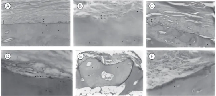
In young subjects (Group 1), the periosteum had become quite thickened and adhered, with a quite visible osteogenic layer, showing great amount of osteo-genic cells (pre-osteoblasts and osteoblasts). In its bone para-cortical surface, we observed the basophilic cement-ing line, with numerous restcement-ing surface osteoblasts (line cells) and in some areas the presence of remodeling gaps, with aggregates of highly secreting osteoblasts depositing new osteoid matrix (Figures 1A and 1B). In subjects from Group 2 (between 40 and 64 years of age), the perios-teum had become partially detached, irregular and thin. Table 2. Demographic data of the patients used for morphometric analysis.
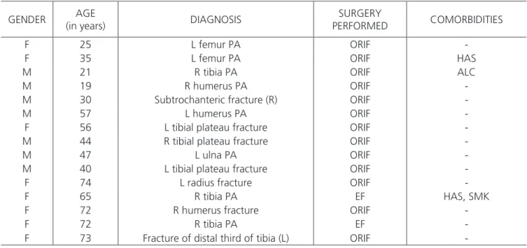
GENDER AGE
(in years) DIAGNOSIS
SURGERY
PERFORMED COMORBIDITIES
F 25 L femur PA ORIF
-F 35 L femur PA ORIF HAS
M 21 R tibia PA ORIF ALC
M 19 R humerus PA ORIF
-M 30 Subtrochanteric fracture (R) ORIF
-M 57 L humerus PA ORIF
-F 56 L tibial plateau fracture ORIF
-M 44 R tibial plateau fracture ORIF
-M 47 L ulna PA ORIF
-M 40 L tibial plateau fracture ORIF
-F 74 L radius fracture ORIF
-F 65 R tibia PA EF HAS, SMK
F 72 R humerus fracture ORIF
-F 72 R tibia PA EF
-F 73 Fracture of distal third of tibia (L) ORIF
-Source: SOT, 2015
Subtitles: M-male; F-female; L-left; R-right; PA-pseudarthrosis; ORIF-open reduction and internal fixation; EF-external fixation; ALC-alcoholism; SMK--smoking; HAS-hypertension
Table 3. Histological analysis.
Periosteum Cortical bone Cancellous bone
Cellularity (osteogenic layer)
Cellularity
(osteocytes in osteoplasts) Bone marrow
External cementing line (presence of acid proteoglycans)
Bone thickness Thickness of bone trabeculae
The osteogenic layer was visible. The external cementing line was irregular and basophilic, with various resting sur-face osteoblasts (Figures 1C and 1D). We hardly observed remodeling gaps. In Group 3 (elderly), we found the peri-osteum partially detached and thin. The osteogenic lay-er was extremely small, with few osteogenic cells, with mixed cellularity in some areas (pre-osteoblasts, fibro-blasts and osteofibro-blasts). The external cementing line was frankly basophilic, very irregular, with some osteoblasts aggregates that had no sign of being secreting new ma-trix (Figure 1E and 1F).
Cortical bone
in Group 1 (individuals under 39 years old), the cortical bone was thick, with numerous Haversian chan-nels of large diameter and concentric lamellae. There was a great number of osteocytes in the gaps (average of six per field, H&E, 400X), not counting the empty gaps. There was an average of 1.6 Volkmann’schannels per field (H&E 100X) (Figure 2A). In subjects between 40 and 64 years of age (Group 2), the cortical bone was thinner
than in the young (Group 1), occupying a smaller area and demonstrating a smaller diameter and number of Haversian and Volkmann’s channels (mean 0.6 per field, H&E, 100X). There were an average of four osteocytes per field (H&E 400X) (Figure 2B). In individuals over 65 years of age (Group 3), the cortical bone was extremely thin, with small amount of Haversian channels. In certain areas,the decrease in collagen matrix was evident. Almost all the gaps were empty, averaging two osteocytes per field (H&E, 400X) (Figure 2C). There was an average of 0.4 Volkmann channels per field (H&E 100X).
Cancellous bone
In the young (Group 1), the bone marrow found was intact, generally presenting small areas of necrosis in the periphery and, less often, bleeding ar-eas. The bone trabeculae were clearly visible, quite thick, making networks with continuity aspect (Figure 3A). There were no osteoclasts. In subjects in Group 2 (between 40 and 64 years), the bone marrow was full, with peripheral areas of necrosis and hemorrhage. The
bone trabeculae were thinner than in the young (Group 1), generally parallel, but still with a net aspect (Figure 3B). In one biopsy, we observed the presence of osteo-clasts (MLCC, 57 years old). In the elderly (Group 3), the bone trabeculae were very thin, forming a network, al-lowing large medullary spaces occupied by fat cells and adipocytes (Figure 3C). The bone marrow was scarce and there were loads of necrotic areas on the periphery. There were no osteoclasts.
MORPHOMETRIC ANALYSIS
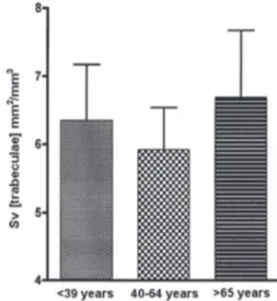
The amount of trabecular meshwork (Vv) was 52.2 ±5.0 (mean ± SD) in Group 1, 52.1 ± 13.3 in Group 2 and 2.9 ± 28.9 in Group 3. There was signifi-cant loss of Vv between the groups (p < 0.05, ANOVA), with the elderly population (Group 3) displaying a re-duction of 45% of the trabecular meshwork in the can-cellous bone (p < 0.01, Newman-Keuls). We observed no statistically significant difference with respect to Vv between Groups 1 and 2 was (p>0.05, Newman-Keuls) (Figure 4).
The area of the trabecular meshwork (Sv) was 6.35 ± 0.82 (mean ± SD) in Group 1, 5.96 ± 0.62 in Group 2 and 6.69 ± 0.98 in Group 3. There was no sta-tistical difference between the groups (p > 0.05, ANOVA and Newman-Keuls) (Figure 5).
DISCUSSION
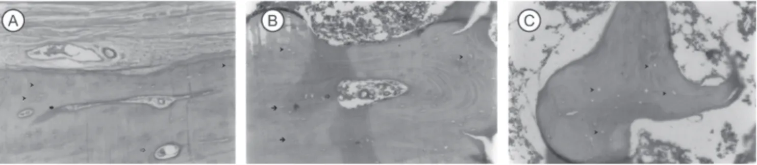
The development of new biomaterials to mimic the characteristics of autologous bone graft has advanced in recent years. In vitro and clinical investiga-tions have suggested that some of these bone substi-tutes may actually stimulate consolidation18,19. Howev-er, the great diversity among the biomaterials available and poor understanding of the mechanisms by which these substances participate in the bone repair process limit their application2,18. Even today, the use of the au-tograft is the best solution for reconstruction of large bone defects and osteogenic stimulating bone heal-ing2-5. Its unique structure provides an excellent mech-anism of self-regulation and functional adaptability. Its Figure 2. Photomicrograph of histological sections of the anterior iliac crest biopsies of adults – CORTICAL BONE. A (Group 1) – Mostly filled
osteoplasts (). Note the presence of Haversian () and Volkmann’s () channels (H&E, 100X); B (Group 2) – filled osteoplasts () and empty osteoplasts (). Note Haversian channel in the center () (H&E, 100X); C (Group 3) – Mostly empty osteoplasts () (H&E, 100X).
solid matrix facilitates the exchange of biomechanical, biochemical and electromechanical signals, endogenous and exogenous of system to which it is exposed to,to the cells responsible for bone modeling6,20. The sum of these interactions determines the success or failure of the grafting procedure. In general, the less biologically active is the graft, the more dependent on the receptor site it becomes2,6. This aspect of bone physiology gains more importance from the moment that recent studies have shown that complex changes occur in the skeletal microarchitecture throughout life, with reduction main-ly of the volume and density of the trabecular bone and hematopoietic tissue21-24 .
Birkenhäger-Frenkel et al.used electron mi-croscopy to investigateiliac biopsies of 94 human spec-imens between 20 and 80 years of age21. They noted that both the trabecular bone area and the number of trabeculae decrease with age in areas commonly used as donor sites for bone graft. Burkhardt et al.used op-tical microscopy to retrospectively analyze 81 biopsies of the iliac crest and 400 samples of iliac crest, ster-num, lumbar vertebra, calcaneus and radius distal third taken between two and 12 hours postmortem of 79 normal individuals of different age groups (one at 96 years old)22. They observed that the volumes of
trabecu-lar bone, osteoid matrix and hematopoietic tissues and the cell number are contingent on age, with a gradu-al decrease in older individugradu-als. Their results indicate a possible role of microcirculation in the genesis of these changes, since the reduction in the number of sinusoids is common in the geriatric population, always accompa-nied by aplasia of hematopoietic marrow and increase in the number of fat cells. Rehman et al. applied semi-au-tomated analysis to ileum biopsies images of 234 sub-jects between 16 and 100 years of age. They found that the trabecular bone volume decreases with age in both genders, reaching statistically significant values between 61 and 70 years in women (56% reduction) and be-tween 81 and 90 years in men (34% reduction)24.
Based on these authors’ findings21-24, in this ex-periment we studied histologic and morphometric char-acteristics of bone biopsies from the anterior iliac crest. We divided patients into three groups of different age groups. With regard to Groups 1 and 2, we set the di-vision based on the hormonal decline and consequent deterioration of bone tissue microstructure that occur around 40 years of age, especially in women. There is a direct relationship between low bone density and patho-logic fractures25. The inclusion of Group 3 followed the guidelines of the World Health Organization, which con-siders “elderly” individuals aged 65 years and over26.
Histologically, we observed that bone tissue degeneration occurs with age. In Groups 1 (age under Figure 4. Amount of trabecular meshwork (Vv) – There was a
signifi-cant loss of Vv between the groups (p<0.05, ANOVA), the elderly population (Group 3) displaying a reduction of 45% of the trabecular meshwork in the cancellous bone (p<0.01, Newman-Keuls). There was no statistically significant diffe-rence with respect to Vv between Groups 1 and 2 (p>0.05, Newman-Keuls).
40 years) and 2 (between 40 and 64 years of age), the osteogenic periosteum was very thickened, rich in bone lineage cells (pre-osteoblasts and osteoblasts). In the cor-tical layer, there were loads of nutritious (Haversian) and connecting (Volkmann’s) channels and most osteoblasts were occupied. The trabeculae of the cancellous bone were clearly visible, thick (which became more evident in the Group 1) and arranged like a net. In Group 3 (over 65 years), the periosteum was thin, low in osteogenic cells, though displaying intense basophilia in the cementing line, reflecting increased synthesis of acid proteoglycans. Gradual replacement of the osteogenic layer of fibrous tissue, bone turnover imbalance and reduction of osteo-blastic activity may be associated with the presence of mixed cellularity and decreased collagen matrix observed in these individuals27. In the cortical bone, there was a small amount of Haversian and Volkmann’s channels and almost all osteoblasts were empty. Several authors have observed that the number of occupied osteoblasts falls from 95% at ten years of age to about 70% at 40 years28-30. Parfitt showed that the number of osteocytes significantly reduced with age, with decline in overall density and in the ratio of occupied gaps, particularly in the deeper layers of the cortical bone30. Ultimately, the osteocytes deficiency may contribute to the observed bone fragility of the elderly31. Finally, in the cancellous bone the trabeculae were thin, allowing ample medullary space, often occupied by fat cells and adipocytes.
Since the structure of the cancellous bone is determinant of bone strength, the adoption of proce-dures for quantitation of trabecular bone has been classi-cally proposed32-39. Croucher et al. showed a strong cor-relation between different rates of assessment of bone structure, such as analysis of the ultrastructure, starring volume and pattern of bone trabecular factor32. Kubik et al. confirmed the value of such methods, especially in the description of age-related changes in the trabecular bone in individuals over 50 years34. Vesterby showed in-creased starry medullary space volume in the iliac crest and the first lumbar vertebra in ten human cadavers aged 27 to 87 years, suggesting that the reduction in trabec-ular bone occurs in all bony structures of the elderly39. In this experiment, we used the amount of trabecular
mesh-work (Vv) and the area of trabecular meshmesh-work (Sv). We observed a statistically significant difference in Groups 1 and 2 with respect to Group 3 as for Vv, with reduction of 45% in the elderly, but not between Groups 1 and 2. There was no statistical difference between the groups regarding the Sv. The interpretation of these results clear-ly shows that the resorption of trabecular bone occurs with age, manifesting itself clinically by increased fracture risk in older individuals40.
We can extrapolate theseresults for the quality of the bone tissue taken from the anterior iliac crest and its use as a graft in situations where there is a need for structural support (e.g. in tibial plateau or vertebral body fractures) or osteogenic stimulus (such as delayed union or avascular pseudarthrosis) in the elderly population. At least two of its fundamental properties, i.e., osteogenic and osteoconductive, are definitely committed in this age group. The reduction in the trabeculae thickness, the in-crease in the intertrabecular distance and the osteocytes numerical reduction potentially make the elderly patients’ iliac bones less resistant and of low quality. Thus, we be-lieve that the bone graft taken from the iliac crest should be avoided in the elderly, and other donor sources be considered. Papavero and Santin demonstrated that the removal of autologous bone graft from the distal third of the femur and proximal third of the tibia is a good option for these individuals5. Another good alternative is the use of the Reamer Irrigator Aspirator (RIA) system in the long bones of the lower limbs41-43. Henrich et al. showed that, compared with the graft from the iliac crest, the aspirate obtained from the femur using RIA has a higher concen-tration of CD34+ lineage osteogenerating cells and mes-enchymal stem cells43.
REFERENCES
1. Heppenstall RB. Bone grafting in fracture treatment
and healing. Philadelphia: W.B. Saunders; 1980.
2. Bauer TW, Muschler GF. Bone graft materials. An
overview of the basic science. Clin Orthop Relat Res. 2000;(371):10-27.
3. Friedlænder GE. Bone grafts. The basic science
ratio-nale for clinical applications. J Bone Joint Surg Am. 1987;69(5):786-90.
4. Netto HD, Olate S, Klüppel L, do Carmo AM, Vásquez
B, Albergaria-Barbosa J. Histometric analyses of can-cellous and cortical interface in autogenous bone grafting. Int J Clin Exp Pathol. 2013;6(8):1532-7.
5. Papavero A, Santin RAL. Retirada percutânea de
enx-erto ósseo autólogo. Rev Bras Ortop. 2003;38(4):213-20.
6. Stevenson S, Arnoczky SP. Transplantation of
muscu-loskeletal tissues. In: Buckwalter JA, Einhorn TA, Si-mon SR, editors. Orthopaedic basic science: biology and biomechanics of the musculoskeletal system. Chi-cago: American Academy of Orthopaedic Surgeons; 2000. p. 567-79.
7. Almaiman M, Al-Bargi HH, Manson P.
Complica-tion of anterior iliacbone graft harvesting in 372 adult patients from may 2006 to may 2011 and a literature review. Craniomaxillofac Trauma Reconstr. 2013;6(4):257-66.
8. Goulet JA, Senunas LE, DeSilva GL, Greenfield ML.
Autogenous iliac crest bone graft. Complications and functional assessment. Clin Orthop Relat Res. 1997;(339):76-81.
9. Bonfante S, Bosco AF, Luize DS, de Almeida JM,
Ce-stari TM, Taga R. Influence of nicotine on healing pro-cess of autogenous bone block grafts in the mandible: a histomorphometric study in rats. Int J Oral Maxillo-fac Implants 2008;23(3):437-44.
10. Mendes PHB, Scofano Jr AR, Silva MG, Souza I, Silva
Filho NM, Abreu AV, et al. Consolidação da fratura após o uso prolongado de corticóide: estudo exper-imental em ratos. Rev Bras Ortop. 2001;36(9):345-51.
11. Padula EOC, Andrade ML, Giordano V, Ramalho
MV. Aspectos morfológicos do processo de consol-idação de fratura em ratos diabéticos. Rev Bras Or-top. 2003;38(3):127-36.
12. Bancroft JD, Cook HC. Manual of histological
tech-niques and their diagnostic application. New York: Churchill Livingstone; 1994.
13. Tabor Z, Rokita E. Comparison of trabecular bone
architecture in young and old bones. Med Phys. 2000;27(5):1165-73.
14. Niemcryk SJ, Kraus TJ, Mallory TH. Empirical
con-siderations in orthopaedic research design and data analysis. Part II: the application of data analytic tech-niques. J Arthroplasty. 1990;5(2):105-10.
15. Santner TJ. Fundamentals of statistics for
orthopae-dists: Part I. J Bone Joint Surg Am. 1984;66(3):468-71. R E S U M O
Objetivo: avaliar as características histológicas e morfométricas de biópsias ósseas da região anterior da crista ilíaca de pacientes de diferentes faixas etárias. Métodos: foram estudadas 30 amostras de osso da crista ilíaca, utilizando-se microscopia óptica de campo claro. As amostras foram divididas pela faixa etária dos doadores em três grupos: Grupo 1 (n = 10), indivíduos com idade entre 25 e 39 anos; Grupo 2 (n = 10), indivíduos com idade entre 40 e 64 anos; Grupo 3 (n = 10), indivíduos com idade igual ou superior a 65 anos. As amostras foram separadas aleatoriamente em dois conjuntos com 15 peças. No primeiro segmento do estudo (n = 15), foi avaliada histologicamente a propriedade osteogênica do enxerto, através da análise da reserva celular no periósteo, do número de osteócitos nas lacunas e da quantidade de canais de Havers e de Volkmann. No segundo segmento do estudo (n = 15), investigou-se morfologicamente a propriedade osteocondutora do enxerto, através da quantificação da rede trabecular (Vv) e da área trabecular (Sv). Resultados: histo-logicamente, observou-se que ocorre degeneração do tecido ósseo com a idade, caracterizada pelo adelgaçamento do periósteo, com substituição gradual da camada osteogênica por tecido fibroso, pequena quantidade de canais de Havers e de Volkmann, osteoplastos vazios e trabéculas finas de osso esponjoso, permitindo amplo espaço medular, em geral ocupado por células lipídicas e adipócitos. Morfologicamente, com relação à quantificação da rede trabecular (Vv), foi observada diferença estatisticamente significante entre os Grupos 1 e 3 e entre os Grupos 2 e 3, com redução da rede trabecular de cerca de 45% no idoso acima de 65 anos de idade; não foi observada diferença estatisticamente significante entre os Grupos 1 e 2. Não foi observada diferença estatística entre os grupos quanto à Sv. Conclusão:os achados do presente experimento sugerem que nos indivíduos idosos (acima de 65 anos de idade), a propriedade osteogênica do enxerto ósseo autólogo diminui e a propriedade osteocondutora está comprometida.
16. Santner TJ, Burstein AH. Fundamentals of statistics
for orthopaedists: Part II. J Bone Joint Surg Am. 1984;66(5):794-9.
17. Santner TJ, Wypij D. Current concepts review.
Fun-damentals of statistics for orthopaedists: Part III. J Bone Joint Surg Am. 1984;66(8):1309-18.
18. Emara KM, Diab RA, Emara AK. Recent biological
trends in management of fracture non-union. World J Orthop. 2015;6(8):623-8.
19. Öhman C, Unosson J, Carlsson E, Ginebra MP,
Pers-son C, Engqvist H. Porosity prediction of calcium phosphatecements based on chemical composition. J Mater Sci Mater Med. 2015;26(7):210. Epub 2015 Jul 14.
20. Knothe Tate ML. “Whither flows the fluid in
bone?” An osteocyte’s perspective. J Biomech. 2003;36(10):1409-24.
21. Birkenhäger-Frenkel DH, Courpron P, Hüpscher
EA, Clermonts E, Coutinho MF, Schmitz PI, et al. Aged-related changes in cancellous bone structure. A two-dimensional studying the tran-siliac and iliac crest biopsy sites. Bone Miner. 1988;4(2):197-216.
22. Burkhardt R, Kettner G, Böhm W, Schmidmeier
M, Chlag R, Frisch B, et al. Changes in trabecular bone, hematopoiesis and bone marrow vessels in aplastic anemia, primary osteoporosis, and old age: a comparative histomorphometric study. Bone. 1987;8(3):157-64.
23. Kerndrup G, Pallesen G, Melsen F, Mosekilde L.
Histomorphometrical determination of bone mar-row cellularity in iliac crest biopsies. Scand J Haema-tol. 1980;24(2):110-4.
24. Rehman MT, Hoyland JA, Denton J, Freemont AJ.
Age related histomorphometric changes in bone in normal British men and women. J Clin Pathol. 1994;47(6):529-34.
25. Wasnich RD. Epidemiology of osteoporosis. In:
Favus MJ, Christakos S, Goldring SR, Hend6y GN, Holick MF, Kaplan F, et al, editors. Primer on the metabolic bone diseases and disorders of mineral metabolism. 3rd ed. Philadelphia: Lippincott-Raven; 1996. p. 249-51.
26. World Health Organization. Ageing and life-course
[Internet]. Genebra; 2015. [acesso em 2015 dez 01]. Disponível em: http://www.who.int/hpr/ageing/in-dex.htm
27. Donahue HJ, Zhou Z, Li Z, McCauley LK.
Age-relat-ed decreases in stimulatory G protein-couplAge-relat-ed ad-enylate cyclase activity in osteoblastic cells. Am J Physiol. 1997;273(4 Pt 1):E776-81.
28. Busse B, Djonic D, Milovanovic P, Hahn M, Püschel
K, Ritchie RO, et al. Decrease in the osteocyte nar density accompanied by hypermineralized lacu-nar occlusion reveals failure and delay of remodeling in aged human bone. Aging Cell. 2010;9(6):1065-75. Epub 2010 Oct 28.
29. Gabet Y, Bab I. Microarchitectural
chang-es in the aging skeleton. Curr Osteoporos Rep. 2011;9(4):177-83.
30. Parfitt AM. Life history of osteocytes: relationship
to bone age, bone remodeling, and bone fragility. J Musculoskelet Neuron Interact 2002;2(6):499-500.
31. Noble BS, Reeve J. Osteocyte function, osteocyte
death and bone fracture resistance. Mol Cell Endo-crinol. 2000;159(1-2):7-13.
32. Croucher PI, Garraham NJ, Compston JE.
Assess-ment of cancellous bone structure: comparison of strut analysis, trabecular bone pattern factor, and marrow space star volume. J Bone Miner Res. 1996;11(7):955-61.
33. Genant HK, Gordon C, Jiang Y, Lang TF, Link TM,
Majumdar S. Advanced imaging of bone macro and micro structure. Bone.1999;25(1):149-52.
34. Kubik T, Pasowicz M, Tabor Z, Rokita E. Optimizing
the assessment of age-related changes in trabecular bone. Phys Med Biol. 2002;47(9):1543-53.
35. Mackie EJ. Osteoblasts: novel roles in
orchestra-tion of skeletal architecture. Int J Biochem Cell Biol. 2003;35(9):1301-5.
36. Matsubara M, Morita S, Shinomiya K, Kawamata K,
37. Saha PK, Gomberg BR, Wehrli FW.
Three-dimen-sional digital topological characterization of cancel-lous bone architecture. Int J Imaging Syst Technol. 2000;11(1):81-90.
38. Smit TH, Schneider E, Odgaard A. Star length
dis-tribution: a volume-based concept for the char-acterization of structural anisotropy. J Microsc. 1998;191(3):249-57.
39. Vesterby A. Star volume of marrow space and
tra-beculae in iliac crest: sampling procedure and cor-relation to star volume of first lumbar vertebra. Bone. 1990;11(3):149-55.
40. Lewiecki EM. Bone density measurement and
as-sessment of fracture risk. Clin Obstet Gynecol. 2013;56(4):667-76.
41. Cobbs KF. RIA use in a community orthopedic
trau-ma practice: applying technology, respecting biolo-gy. Injury. 2010;41 Suppl 2:S78-84.
42. Giannoudis PV, Suk M, Pape HC. RIA: the
jour-ney just started but what the future holds? Injury. 2010;41 Suppl 2:S1-3.
43. Henrich D, Seebach C, Sterlepper E, Tauchmann C,
Marzi I, Franck J. RIA reamings and hip aspirate: a comparative evaluation of osteoprogenitor and endothelial progenitor cells. Injury. 2010;41 Suppl 2:S62-8.
Received in: 05/04/2016
Accepted for publication: 13/07/2016 Conflict of interest: none.
Source of funding: none.
Mailing address:
Vincenzo Giordano