353
Revista da Sociedade Brasileira de Medicina Tropical 45(3):353-356, may-jun, 2012
INTRODUCTION
Article/Artigo
1. Programa de Pós-Graduação em Medicina Tropical, Núcleo de Medicina Tropical, Universidade de Brasília, Brasília DF. 2. Unidade de Clínica Médica, Hospital do Paranoá, Secretária de Saúde do Distrito Federal, Brasília, DF. 3. Ex-Professor titular do Depto de Clínica Médica, Universidade Federal de Goiás, Goiânia, GO. 4. Professor Honoris Causa, Universidade de Brasília, Brasília, DF. †In memorian. Address to: Prof. Cleudson Castro. Depto de Clínica Médica/NMT/UnB. Caixa Postal 4517, 70904-970 Brasília, DF, Brasil.
Fax: 55 61 3307-1154 e-mail: [email protected] Received in 06/06/2011 Accepted in 29/07/2011
Occurrence of dolichocolon without megacolon in chronic Chagas
disease patients
Ocorrência de dolicocólon sem megacólon em chagásicos crônicos
Cleudson Castro
1, Esperanza Bernal Hernandez
2, Jofre Rezende
3,4and Aluizio Prata
†ABSTACT
Introduction: Since 1970, lengthening of the rectosigmoid has been suspected to be a solitary manifestation of Chagas colopathy. Methods: To test this hypothesis, opaque enema was administered on 210 seropositive and 63 seronegative patients, and radiographs in the anteroposterior and posteroanterior positions were examined blind to the serological and clinical indings. he distal colon was measured using a lexible ruler along the central axis of the image from the anus to the iliac crest. Results: Dolichocolon was diagnosed in 31 (14.8%) seropositive and 3 (4.8%) seronegative patients. he mean length was 57.2 (±12.2)cm in seropositive patients and 52.1 (±8.8)cm in the seronegative patients (p = 0.000), that is, the distal colon in Chagas patients was, on average, 5.1cm longer. Seropositive female patients presented a mean length of 58.8 (±12.3)cm, and seronegative female patients presented 53.2 (±9.1)cm (p = 0.002). Seropositive male patients had a mean length of 55 (±11.6)cm, and seronegative male patients had 49.9 (±7.8)cm (p = 0.02). Among 191 patients without megacolon and suspected megacolon, the mean length was 56.3 (±11.6)cm in seropositive individuals and 52 (±8.8)cm in seronegative patients (p = 0.003). Among individuals with distal colon ≥70cm, there were 31 Chagas patients with mean length of 77.9 (±7.1)cm and three seronegative with 71.3 (±1.1)cm (p = 0.000). Among 179 with distal colon <70cm, seropositive individuals had a mean length of 53.6 (±8.8)cm, and seronegative patients had 51.2 (±7.8)cm (p = 0.059). Serological positive women had longer distal colon than men (p = 0.02), whereas the mean length were the same among seronegative individuals (p = 0.16). Conclusions: In endemic areas of Brazil Central, solitary dolichocolon is a radiological Chagas disease signal.
Keywords: Dolichocolon. Solitary dolichocolon. Chagas disease. Lengthening of the sigmoid. Length of the sigmoid.
RESUMO
Introdução: Desde 1970, suspeita-se que o alongamento do retossigmoide pode ocorrer como manifestação isolada da colopatia chagásica. Métodos: Para testar esta hipótese, 210 pacientes soropositivos e 63 soronegativos izeram enema opaco e as radiograias nas posições ântero-posterior e póstero-anterior foram lidas sem conhecimento dos dados clínicos e sorológicos. O comprimento do cólon distal foi medido com curvímetro, percorrendo-se o eixo central da imagem, do ânus à crista ilíaca. Resultados: O diagnóstico de dolicocólon foi estabelecido em 31 (14,8%) pacientes soropositivos e 3 (4,8%) soronegativos. O comprimento médio nos pacientes soropositivos foi de 57,2 (±12,2)cm, enquanto nos soronegativos foi de 52,1 (±8,8)cm (p=0,000), isto é, os chagásicos apresentaram o cólon distal em média 5,1cm maior. Os indivíduos do sexo feminino soropositivos exibiram comprimento médio de 58,8 (±12,3)cm, e os soronegativos de 53,2 (±9,1)cm, (p=0,002). Nos pacientes do sexo masculino soropositivos, o comprimento médio foi de 55 (±11,6)cm, enquanto nos soronegativos foi de 49,9 (±7,8)cm (p=0,02). Nos 191 pacientes, sem megacólon e suspeitos de megacólon, o comprimento médio foi de 56,3 (±11,6) cm nos soropostivos e 52 (±8,8)cm nos soronegativos (p=0,003). Dos indivíduos com cólon distal ≥70cm, os 31 chagásicos tiveram comprimento médio de 77,9 (±7,1)cm, enquanto nos três não chagásicos foi de 71,3 (±1,1)cm, (p=0,000). Nos 179 com cólon distal <70cm, os soropositivos tiveram em média 53,6 (±8,8)cm, e os soronegativos 51,2 (±7,8)cm, (p=0,059). Dentre os com sorologia positiva, as mulheres apresentaram cólon distal maior que os homens (p=0,02), enquanto naqueles com sorologia negativa o comprimento médio foi igual (p=0,16). Conclusões: Nas áreas endêmicas do Brasil Central, o dolicocólon solitário é um sinal radiológico da doença de Chagas.
Palavras-chaves: Dolicocólon. Dolicocólon solitário. Doença de Chagas. Alongamento do sigmoide. Comprimento do sigmoide.
Carlos Chagas suspected that the digestive tract participated in the disease that came to bear his name1, through his acceptance that megaesophagus,
common in areas that were endemic for Chagas disease, could be one of its manifestations. During the same era, Neiva and Pena observed hundreds of cases of megas on a journey through the interior of Brazil, especially in the State of Goiás2. Nevertheless,
for a long time, Chagas disease and megas were considered to be diferent entities3. In the 1950s,
Sabino Vieira de Freitas studied the two most important hypotheses relating to the likely etiology of megas (vitamin B1 deiciency and Chagas disease) and concluded reluctantly that the etiology still could not be established. He took the view that the link to Chagas disease was doubtful3. At that time,
renowned professors thought that the two entities had diferent etiologies3.
At the Eighth Medical Congress of the Triângulo Mineiro and Central Brazil, held in Uberaba in 1956, Pedreira de Freitas declared that, despite the great reactions against accepting the link between megas and Chagas disease among Brazilian researchers, there already was a solid basis for establishing such a connection3. In 1955, Koeberle
and Nador had established the pathogenetic basis for megaesophagus in Brazil4. In the following year,
based on extensive clinical experience, Rezende5
354
METHODS
RESULTS Castro C et al - Solitary dolichocolon in chronic Chagas disease
dolichocolon. he speciic literature relating to dolichocolon is sparse, both in Portuguese and in English, and is limited to reports on cases associated with megacolon and Hirschprung’s disease and, more rarely, to cases of acquired dolichocolon of uncertain cause. he aim of the present study was to present radiological and statistical data that favor including cases of solitary dolichocolon as part of Chagas disease.
Patients studied
his radiological study was conducted between May 2000 and August 2001 among the population in the municipality of Mambaí, State of Goiás, which is an area endemic for Chagas disease. A list of individuals within the Mambaí project was prepared and handed over to the physician who was coordinating the work in the municipality. This physician was unaware of the clinical and serological status of the patients. he patients included were adults with six serologically positive reactions for Chagas disease who presented the indeterminate, cardiac, or digestive form of the disease and individuals with six negative serological reactions who had constipation as a symptom. Cases of severe cardiopathy and pregnancy were excluded. he task of locating the patients was undertaken by a health agent who lived in the municipality and had been working in the project since 1975. he patients were transported by car from their homes to Damianópolis, and an average of six examinations per day was performed. At the hospital, before undergoing the examination, the patients answered a standard questionnaire that sought identification, age and sex data, and information on the symptom of constipation.
Radiological study and parameters
The test used was opaque enema, by means of the method recommended by Ximenes et al.6, with small modiications that, in
brief, did away with colon preparation and introduced air for double contrast. he examinations were performed in the municipality of Damianópolis, which is a town 18km from the main setlement of Mambaí that had an X-ray machine appropriate for the procedure. A radiology technician with experience of performing opaque enemas went from Brasília to train the technician who was based in Damianópolis, and they carried out the first examinations together. Wearing a hospital gown, the patients were positioned in dorsal decubitus on the X-ray table, and a simple radiograph of the abdomen was produced irst to rule out the presence of fecaloma. Immediately before the examination, a suspension of 150ml of barium sulfate in 1,000ml of water was prepared. he enema was applied with the patient in the let lateral decubitus, and the receptacle containing the contrast medium was positioned 1.5m above the plane of the examination table. Two radiographs were produced, in anteroposterior and posteroanterior views, at 80 to 100 kilovolts and 0.30 to 0.40 milliamperes (mAs). he ilms were developed immediately ater the examinations. he time taken to carry out one examination, from administering the questionnaire to producing the radiographs, was approximately 40min. he radiographs were taken to Goiânia and interpreted by one of the authors ( JR), who was unaware of the patients’ data. he interpretation was performed by visual observation of the radiographs on an X-ray ilm viewer and by measuring the length of the distal colon, which was taken
here to mean the sigmoid loop, rectum, and anal canal. According to Testut’s Anatomy7, the length of the rectum in adults ranges from
12 to 15cm, and that of the sigmoid loop is from 30 to 50. hus, measurements on the segment from the sigmoid loop to the rectum range between 42 and 65cm. Ater adding 2 to 3cm for the anal canal, the total length of the distal colon, from the anus to the transition to the descending colon, reaches 45 to 68cm. his measurement was made using a lexible ruler, which was laid out along the central axis of the radiological image in the ascending direction, going from the anus to the level of the let iliac crest. he measurement obtained had relative value because of the irregular and variable layout of the sigmoid loop, which included segments going in an anteroposterior direction, thereby diminishing their length on the radiological projection. Lengths greater than or equal to 70cm were deined as dolichocolon.
Statistical analysis
he information was stored in a database. he analysis was carried out with the aid of the SPSS sotware, version 9.0, for Windows. For all the tests used, the signiicance level was set at less than 0.05. Student’s t test was used to determine the signiicance of diferences that were found in the means of the quantitative variables.
Ethical considerations
he population of Mambaí had been undergoing tests since 1974. his project was approved by the ethics commitee of the School of Health Sciences, University of Brasília (UnB), and was conducted in accordance with the standards of Resolution 196/96. he aims of the study and the type of examination that would be performed were explained to each patient. Writen consent was requested, and the examinations were only carried out ater receiving the patients’ agreement. Patients who needed specialized treatment were referred to the University Hospital of Brasília. During the study period, the population was atended at a health clinic maintained through the project.
Of the 303 individuals who were examined radiologically, 30 were excluded: four had no available serological tests, two had doubtful serological tests, and 24 had distal colon length that was impossible to measure. hus, 273 patients were evaluated, of whom, 210 were seropositive for Chagas disease, and 63 were seronegative. he patients’ ages ranged from 27 to 87 years; the mean age of the patients examined was 48.9 ± 12.4 years, and the mean age of the seropositive patients was 49.3 ± 12.4 years.
he radiographs were analyzed by one of the authors ( JR), who was unaware of the patients’ serological and clinical status. In addition to visual analysis of the radiographs on an X-ray ilm viewer, the largest diameters of the rectum and sigmoid8 and the length of the distal
colon were measured. To make these measurements, preference was given to anteroposterior radiographs, whereas posteroanterior views were only used when no measurement could be obtained using the anteroposterior view.
355
Rev Soc Bras Med Trop 45(3):353-356, may-jun, 2012
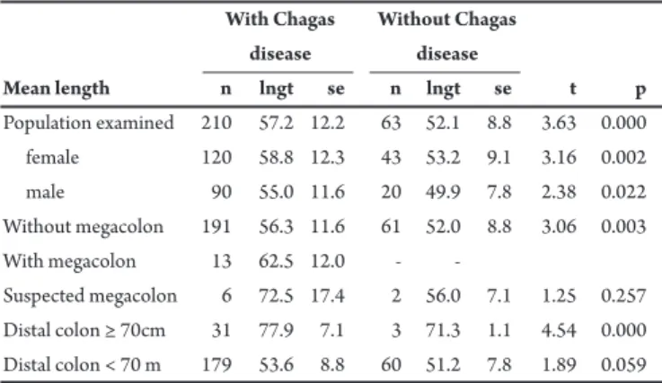
TABLE 1 - Mean length of the distal colon in centimeters, in patients with and without Chagas disease.
With Chagas Without Chagas
disease disease
Mean length n lngt se n lngt se t p
Population examined 210 57.2 12.2 63 52.1 8.8 3.63 0.000 female 120 58.8 12.3 43 53.2 9.1 3.16 0.002 male 90 55.0 11.6 20 49.9 7.8 2.38 0.022 Without megacolon 191 56.3 11.6 61 52.0 8.8 3.06 0.003 With megacolon 13 62.5 12.0 - -
Suspected megacolon 6 72.5 17.4 2 56.0 7.1 1.25 0.257 Distal colon ≥ 70cm 31 77.9 7.1 3 71.3 1.1 4.54 0.000 Distal colon < 70 m 179 53.6 8.8 60 51.2 7.8 1.89 0.059 n: number of patients; lngt: length; se: standard error; t: Student’s t test; p: probability.
TABLE 2 - Mean length of the sigmoid loop, rectum, and anal canal, in centimeters, in relation to gender and serological status for Chagas disease.
Male Female
Serological status n lngt se n lngt se t p
Positive 90 55.0 11.6 120 58.8 12.3 2.28 0.02 Negative 20 49.9 7.8 43 53.2 9.1 - 1.39 0.16 n: number of patients; lngt: length; se: standard error; t: Student’s t test; p: probability.
TABLE 3 - Results from radiological examinations on the colon among patients known to have the acute form of Chagas disease, in diferent places, and over diferent lengths of evolution.
Examinations Years of Colon (%)
Locality (n) evolution normal lengthened megacolon
Bahia 16 3.1 100.0 0.0 0.0
Uberaba 32 8.1 78.1 15.6 6.3
Goiânia 17 11.1 88.4 5.8 5.8
Bambuí 95 17.0 71.6 14.7 13.7
those of the noninfected patients. Among the female patients, analysis on the mean length of the distal colon showed that the women with Chagas disease had distal colon that were longer (58.8 ± 12.3cm) than those of the noninfected patients (53.2 ± 9.1cm; p = 0.002). hus, the women with Chagas disease had distal colons that, on average, were 5.6cm longer than those of the non-infected patients. his also was noted in relation to the mean length of the distal colon in the men: those with Chagas disease had a mean length of 55 ± 11.6cm, whereas the length in the noninfected men was 49.9 ± 7.8cm (p = 0.02). hus, the men with Chagas disease presented distal colons that, on average, were 5.1cm longer than those in men without infection. Ater subtracting 13 patients with evident megacolon and another six with megacolon suspected from the visual inspection (19 patients) from the total of 273 patients, the mean measurement among the remaining 191 seropositive patients was 56.3 ± 11.6cm, and that among the 61 seronegative patients was 52 ± 8.8cm (p = 0.003). herefore, ater excluding the Chagas patients with a dilated colon, the remainder presented a distal colon that was, on average, 4.3cm longer than that among the noninfected patients.
he mean length of the distal colon in individuals of diferent genders varied according to the serological status for Chagas disease. Among the seropositive individuals, female distal colons were signiicantly longer (58.8 ± 12.3cm) than male distal colons (55 ± 11.6cm; p = 0.02). Among the seronegative individuals, the female distal colon continued to be longer (53.2 ± 9.1cm) than the male distal colon (49.9 ± 7.8cm) but without statistical signiicance (p = 0.16) (Table 2).
Of the 34 patients with dolichocolon, 31 had Chagas disease, and their mean distal colon length was 77.9 ± 7.1cm, whereas among the three individuals without Chagas disease, it was 71.3 ± 1.1cm (t = 4.54; p = 0.000). he diference was signiicant, that is, the Chagas dolichocolon was, on average, 6.6cm longer.
Ater excluding the patients with distal colons measuring 70cm or longer, the remaining patients consisted of 179 seropositive individuals, with a mean distal colon length of 53.6 ± 8.8cm, and 60 seronegative individuals, with a mean length of 51.2 ± 7.8cm (p = 0.059). In this analysis, the distal colon in the Chagas patients was, on average, 2.4cm longer, with a strong trend toward having a longer intestinal segment but without statistical signiicance.
Among the 31 seropositive patients with dolichocolon, the longest measurement was 100cm, whereas among the three seronegative patients with dolichocolon, the longest measurement was 72cm. Among the seropositive individuals, there were ive with dolichomegacolon and four with possible megacolon. Among the 24 individuals for whom measurement of the distal colon was impossible, 15 were seropositive, and nine were seronegative (Table 3).
DISCUSSION
Summary data from this investigation were published in 20039,
and the data are detailed in the present study. he speciic literature relating to dolichocolon in Brazilian gastroenterological publications is sparse. In other Latin American publications, dolichocolon is mentioned in connection with studies on Chagas disease and megacolon10-13. When dolichocolon appears as the only radiological
inding, it has been considered to be of congenital origin or unknown cause. When diagnosed in individuals who are seropositive for Chagas disease, there is some doubt regarding the true cause, although data allowing linkage with American trypanosomiasis exist. Since 1970, when a group of Brazilian researchers presented information regarding the evolution of Chagas disease10, at a congress in Porto
Alegre, it has become possible to link dolichocolon to Chagas disease from the data presented. In Table 3, which summarizes the indings from this investigation, it can be seen that the lengthening of the colon occurs more frequently than does megacolon10. Dias11 reinforced the
impression that dolichocolon is the initial stage of Chagas colopathy, and Rezende et al.14 brought in new support through radiological
imaging measurements from an opaque enema that showed that the mean length of the distal colon in Chagas patients without megacolon was greater than that in seronegative controls.
Among the 273 patients examined in the present study, 34 (12.5%) presented dolichocolon, among whom, 31 (14.8%) were seropositive, and 3 (4.8%) were among the 63 seronegative individuals in this study. Also, among these individuals, 14 (6.2%) cases of megacolon or dolichomegacolon were diagnosed8. his
356
constipation, although Dias11 observed that patients in the Bambuí
project were symptomatic. here are no data on the symptom of constipation among patients exclusively presenting dolichocolon.
Recent studies have taken up this topic again, with large samples, thereby reinforcing the opinion of Rezende et al.14. he data from the
present study and those obtained by Lopes15 point in this direction.
In the study by Lopes15, all the 72 patients with megacolon presented
lengthening of the rectosigmoid. All the patients with a possible diagnosis of megacolon presented rectosigmoid length greater than the established limits, and the median rectosigmoid length among the Chagas patients without megacolon was signiicantly greater than among the non-Chagas individuals15.
All the indications are that the morphological changes to the colon occur at a time that difers from when the changes to the esophagus occur. In the esophagus, the lengthening appears at the end (in megaesophagus group IV), whereas in the colon, it is the irst change and precedes megacolon.
Pathological anatomy does not seem to be an adequate method for studying the lengthening of the rectosigmoid. Adad16 studied surgical
and necropsy specimens from individuals with unquestionable clinical and radiological diagnoses of Chagas colopathy and did not ind any diference in sigmoid length between Chagas patients and seronegative controls. Even when dealing with organs that have been radiologically deined as dolichomegacolon, pathologists are oten unable to conirm the diagnosis16.
he present study was conducted blindly by a single local team that did not know the results from the serological tests, and the radiographs were analyzed and measured by a researcher 500km away, who did not know anything about the patients.
The diagnosis of dolichocolon is essentially radiological. Assessment of the image by a radiologist or clinician seals the diagnosis even in the absence of symptoms. According to Lopes15,
who is a radiologist, the feature that draws atention most in the diagnosis is the lengthening of the rectosigmoid. He did not make any distinction between dolichocolon and dilatation of the colon and considered both to be megacolon, within the concept of the greater colon. In his view, megacolon mostly starts with lengthening of the sigmoid, whereas the lengthening is associated with increasing diameter at advanced stages. The present study demonstrated that even among individuals with distal colon length within the normal maximum limit (i.e., less than 70cm), those with American trypanosomiasis present a strong tendency to have longer distal colons than seen in seronegative controls.
Old autopsy and radiological studies conducted outside Latin America revealed that dolichocolon or redundant colon was found in 14 to 16% of the population17. To the best of our knowledge, such data
do not exist in Brazilian setings, where Chagas disease predominates. With the exception of Andean dolichocolon18, which is found in the
Andes Mountains above the altitude of 3,000m and is atributable to dietary causes, it is possible that on endemic areas in Latin America, the main cause of solitary dolichocolon is Chagas disease.
In conclusion, the study conducted in the 1970s10, the
information from Dias11 relating to the Bambuí project, the study
he authors declare that there is no conlict of interest. CONFLICT OF INTEREST
REFERENCES
1. Chagas C. Trypanosomiase americana. Forma aguda da moléstia. Mem Inst Oswaldo Cruz 1916; 8:37-60.
2. Neiva A, Pena B. Viagem cientifica pelo norte da Bahia, sudoeste de Pernambuco, sul do Piauhi e de norte a sul de Goiás . Mem Inst Oswaldo Cruz 1916; Tomo III fasc 3:75-224.
3. Porto C, Porto CC. Historia do megaesôfago nos congressos no Brasil Central. Rev Goiana Méd 1970; 16:117-136.
4. Koeberle F, Nador E. Etiologia e Patogenia do megaesôfago no Brasil. Rev Paulista Med 1955; 47: 643.
5. Rezende JM. Megaesôfago por doença de Chagas. Rev Goiana Med 1956; 2:297-314.
6. Ximenes CA, Rezende JM, Moreira H, Vaz MGM. Técnica simpliicada para diagnóstico radiológico do megacólon chagásico. Rev Soc Bras Med Trop 1984; 17 (supl): 23.
7. Testut L. Traité D’anatomie humaine, 8º ed. Paris: Gaston Doien & Cie; 1931. 8. Castro C, Hernandez EB, Rezende J, Prata A. Estudo radiológico do
megacólon em área endêmica de doença de Chagas. Rev Soc Bras Med Trop 2010; 43:562-566.
9. Hernandez EBR, Rezende JM, Macedo V, Castro C. Estudo radiológico do cólon através da técnica de Ximenes em área endêmica de Chagas: prevalência de dolicocólon. Rev Soc Bras Med Trop 2003; 36 (supl II):73-74.
10. Prata AR, Dias JCP, Ferreira HO, Rassi A, Macedo VO, Rezende JM, et al. Grupo de estudos sobre a evolução da doença de Chagas. Porto Alegre: Resumos do VI Congresso da Sociedade Brasileira de Medicina Tropical; 1970.
11. Dias JCP. Doença de Chagas em Bambuí Minas Gerais, Brasil. Estudo clínico epidemiológico a partir da fase aguda, entre 1940 e 1982. [Tese de doutorado]. [Belo Horizonte]: Universidade Federal de Minas Gerais; 1982. 376 p. 12. Calderon C, Aldana M. Estudio del colon en pacientes chagasicos asintomaticos
y sintomaticos. Parasitol al Dia 1987; 11:65-71.
13. Calderon C. Estudios digestivo y cardiologico simultaneos en pacientes chagasicos asintomaticos. Rev Med Chile 1992; 120:43-47.
14. Rezende JM, Ximenes CA, Moreira H, Vaz MGM, Luqueti AO. Alongamento do cólon distal em pacientes com a forma digestiva da doença de Chagas. Paper presented at: Resumos do XXXI Congresso Brasileiro de gastroenterologia e VII Congresso Brasileiro de Endoscopia digestiva; Belém, Brazil; 1990.
15. Lopes GP. Estudo radiológico do comprimento e calibre do retosigmóide em pacientes chagásicos e em controles procedentes de diferentes altitudes. [Dissertação de Mestrado]. [Uberaba]: Universidade Federal do Triângulo Mineiro; 2003. 86 p.
16. Adad SJ. Contribuição ao estudo da anatomia patológica e patogênese do megacólon chagásico. [Doctor Dissertation]. [Uberaba]: Faculdade de Medicina do Triângulo Mineiro; 1996. 212 p.
17. Bockus HL. Developmental anomalies of the colon. In: Bockus HL, editor. Gastroenterology. Vol II. Saunders: Company; 1949. p. 391.
18. Franco FE. El colon íleo-pelvico en los peruanos. [Doctor Dissertation]. [Lima]: Universidad Nacional Mayor de San Marcos; 1965.
Castro C et al - Solitary dolichocolon in chronic Chagas disease
by Rezende et al.14 on patients in hospitals in Goiânia, the data from
the present study obtained from Mambaí, and the study by Lopes in Uberaba15 constitute a robust data set that, in endemic areas of Brazil