CLINICAL SCIENCE
I Department of Dermatology, Faculdade de Medicina da Universidade de
São Paulo - São Paulo/SP, Brazil.
II Department de Patology, Faculdade de Ciências Médicas da Santa Casa
de São Paulo - São Paulo/SP, Brazil.
III Department of Histopathology, University College Hospital - London/
United Kingdom.
Email: [email protected] Tel.: +55 11 3031.0093
Received for publication on September 14, 2009 Accepted for publication on October 22, 2009
POLYMERASE CHAIN REACTION-BASED
CLONALITY ANALYSIS OF CUTANEOUS B-CELL
LYMPHOPROLIFERATIVE PROCESSES
Claudia Z Melotti,I Maria Fernanda Carriel Amary,II Miriam Nacagami Sotto,I
Timothy Diss,III Jose Antonio SanchesI
doi: 10.1590/S1807-59322010000100009
Melotti CZ, Amary MFC, Sotto MN, Diss T, Sanches Jr. JA. Polymerase chain reaction-Based clonality analysis of cutaneous b-cell lymphoproliferative processes. Clinics.2010;65(1):53-60.
INTRODUCTION: The differential diagnosis of B-cell lymphoproliferative processes remains a challenge for pathologists, der-matologists and oncologists, despite advances in histology, immunohistochemistry and molecular biology.
OBJECTIVE: Evaluate aid and limitations of clonality analysis in the diagnosis of primary cutaneous B-cell lymphomas and B-cell pseudolymphomas.
METHODS: This study included 29 cases of B-cell lymphoproliferative processes classiied as primary cutaneous B-cell lym-phomas (13), B-cell pseudolymlym-phomas (6) and inconclusive cases (10) using histology and immunohistochemistry. The clonality analysis was performed by polymerase chain reaction analysis of immunoglobulin light chain and heavy chain rearrangements.
RESULTS: DNA quality was shown to be generally poor; eight samples were inadequate for polymerase chain reaction analysis. The results showed monoclonality in eight of the primary cutaneous B-cell lymphomas and polyclonality in four of the B-cell pseudolymphomas. In addition, monoclonality was shown in two of the inconclusive cases by histology and immunohistochemistry, demonstrating the utility of polymerase chain reaction as an ancillary diagnostic tool for primary cutaneous B-cell lymphomas.
DISCUSSION: The low quality DNA extracted from these cases demanded the use of an IgH protocol that yielded small fragments and IgK. Both methods used together improved detection.
CONCLUSION: Use of the two protocols, immunoglobulin heavy chain FR3-trad and immunoglobulin light chain-Kappa Biomed protocols for clonality analysis improved diagnostic accuracy.
KEYWORDS: Cutaneous lymphoma; Polymerase chain reaction; Gene rearrangement; Clonality; B-cell; Lymphoproliferative processes.
INTRODUCTION
B-cell lymphoproliferative processes, including primary cutaneous B-cell lymphomas (PCBCLs) and B-cell
pseudolymphomas (B-PSLs), have been exhaustively studied in the last few decades.1,2 Nevertheless, differential diagnosis between the two entities remains a challenge for dermatopathologists, hematologists, dermatologists a n d o n c o l o g i s t s , d e s p i t e a d va n c e s i n h i s t o l o g y, immunohistochemistry and molecular biology.3-6
of clonality through investigation of immunoglobulin light and heavy chain gene rearrangements has been used to supplement the diagnosis of PCBCL.9,11,12 Usually, polymerase chain reaction (PCR) is the molecular technique of choice due to higher sensitivity and lower cost when compared with Southern blot. As an additional advantage, this tool offers the possibility to work with formalin-ixed and parafin-embedded tissue.12-14
PCR has been very useful in the diagnosis of PCBCL when used to investigate rearrangement of the IgH gene located on chromosome 14 (14q32).15,16 The rearrangement of IgH genes is the irst rearrangement event to take place in the differentiation of B cells, preceding that of the IgL genes, and it is present in the majority of PCBCLs.17 As several regions (FR3, FR2, and FR1) of IgH genes can be evaluated, a number of protocols have been developed. These include the Biomed-2 protocols and more traditional ones.14,18,19
Sensitivity can vary from less than 20% to more than 90%, depending on the employed method. 9,13,20 In some cases, it is not possible to amplify the IgH gene because primers fail to bind to the rearrangement that occurred in the neoplastic B lymphocytes or an inadequate DNA sample. Rearrangement of the IgL-K (2q12) and IgL lambda (22q11) genes can also be investigated and may increase the diagnostic sensitivity up to 93%, particularly when combined with IgH analysis.11, 20, 21
In the present study, the impact of the analysis of IgH and IgL-K gene rearrangement in the diagnosis of B-cell lymphoproliferative cutaneous processes and the limitations of these methods are assessed.
DIAGNOSTIC METHOD
Twenty-nine patients with B-cell lymphoproliferative processes were evaluated. Clinical data were retrieved from the clinical records, and all cases were classiied according to the histological and immunohistochemical characteristics. We included cases diagnosed by two dermatopathologists who were experts in cutaneous lymphoma without prior knowledge of the clinical setting. The pathologists established a diagnosis of PCBCL (13 cases) or B-PSL (6 cases). Whenever there was a discrepancy between the diagnoses of both pathologists or when they could not deine the diagnosis, the case was considered inconclusive (10 cases) (Table 1). When the diagnosis of PCBCL was established, the cases were classiied according to the WHO-EORTC 2005 Classiication.22
Clonality was investigated by IgH gene rearrangement using protocols for IgH FR2 (Biomed IgH tube B), IgH FR3 (Biomed IgH tube C) and a traditional method for IgH FR3 (IgH-trad), which yields smaller fragments than the Biomed
protocols.14,23 The investigation of immunoglobulin light chain (IgL) gene rearrangement was carried out using the IgL Kappa protocols (Biomed Igkappa tubes A and B).
DNA extraction (crude lysate method): Five sections of 10 µm formalin-ixed and parafin-embedded cutaneous
biopsies were deparafinized in xylene and ethanol. Tissue digestion was carried out in 100 µl of a solution containing 200 µg/ml proteinase K (Sigma, UK) in 1 X PCR buffer (Sigma, UK). The material was incubated at 37°C for 24 hours and heated at 95°C for 15 minutes to deactivate the proteinase K. A total of 1 µl of the supernatant was used as template for DNA ampliication.
DNA ampliication A mastermix solution composed of 1 X Buffer II, 200 µM each dNTP (Promega, UK), 1.5 mM
MgCl2, 1 unit Amplitaq Gold (Applied Biosystems), and 10 pmol each speciic primer (MWG, Germany) was added to a reaction microtube (Table 2). A total of 1 µl of DNA was added to the mastermix. Cycling parameters were as follows: 95°C, 7 min, 1 cycle; 93°C, 45 s; 60°C, 45 s; 72°C, 1 min 30 s, 40 cycles; 72°C, 5 min, 1 cycle. For the FR3-trad protocol, the cycling parameters were as follows: 95°C, 7 min, 1 cycle; 93°C, 45 s; 55°C, 45 s; 72°C, 1 min 30 s, 40 cycles; 72°C, 5 min, 1 cycle.
PCR Analysis The IgH, IgL kappa and TCR-gamma genes are shown in Table 2. A negative control (no template) and a positive control (B-cell lymphoma) were used in each reaction. Ampliication of non-rearranging genes to yield products of 100-400 bp was performed as a DNA quality control for each sample.14
Electrophoresis The PCR products (8 µl) were run on 6% or 8% polyacrylamide gels. Electrophoresis was performed at 200 V for 40 minutes, gels were stained with 0.5 µg/mL ethidium bromide for 5 minutes and the DNA bands were documented by use of the Gel Documentation System 1000 (Bio-Rad).
Evaluation of the Band Patterns The band patterns were analyzed, and each case was classiied as follows: (i) Monoclonal (when one or two dominant bands of the same size were visualized in duplicate ampliications); (ii) Polyclonal (when a smear or variable ladder was detected); and (iii) Non-informative (when absence of bands or weak, non-reproducible patterns were observed) (Figure 1A).
Table 1 - Clinical, histological and immunohistochemical data for 29 cases with B-cell lymphoproliferative processes
I G A Dist Localization Morphology Diam N HE IHC IHC HE+IHC L, M, V Stage Status
1 M 54 reg face, neck,
trunk, back papule, nodule 1 10 PCBCL
Anti-CD20 + CD10+,
BCL-2-, CD23- PCBCL PCBCL Y,Y,Y system DWD (36)
2 F 57 loc back plaque 6 2 INC Anti-CD20 + CD10 -,
BCL-2 +, CD23+ INC INC N,N,N cut ativ AWD (78)
3 M 76 loc LL nodule, plaque 16 1 PCBCL(+) Anti-CD20 + CD10 -,
BCL-2 +, CD23- B-PSL(-) PCBCL N,N,N cut ativ DWD (20)
4 F 63 diss UL, back,
palate nodule, plaque 10 10 PCBCL(+)
Anti-CD20 + CD10 +,
BCL-2 +, CD23+ B-PSL(+) INC N,N,N cut ativ AWD (182)
5 M 50 loc anterior trunk plaque 5 1 INC(-) Anti-CD20 + CD10 +,
BCL-2 -, CD23+ B-PSL(+) B-PSL N,N,N cut ativ AWD (135)
6 M 71 reg face, neck,
trunk, back nodule, plaque 3 20 B-PSL(-)
Anti-CD20 + CD10 -,
BCL-2 +, CD23+ INC(+) INC N,N,N cut ativ DWD (32)
7 M 67 loc Back plaque 3 1 B-PSL(-) Anti-CD20 + CD10 +,
BCL-2 +, CD23- PCBCL(+) PCBCL N,N,N cut ativ AWD (66)
8 F 36 reg UL e LL papule, nodule 2 15 B-PSL Anti-CD20 + CD10 -,
BCL-2 +, CD23- B-PSL B-PSL N,N,N cut ativ AWD (43)
9 F 37 diss anterior trunk,
back, LL
papule, nodule,
plaque 2 20 PCBCL
Anti-CD20 + CD10 -,
BCL-2 -, CD23- PCBCL PCBCL N,N,N cut ativ AWD (172)
10 F 62 loc back nodule 1 2 PCBCL(+) Anti-CD20 + CD10 -,
BCL-2 +, CD23+ B-PSL(+) INC N,N,N cut rem AND (199)
11 F 43 loc face papule, nodule 0,5 5 B-PSL(-) Anti-CD20 + CD10 +,
BCL-2 -, CD23+ PCBCL(+) PCBCL N,N,N cut rem AND (138)
12 F 26 loc back nodule 8 1 INC Anti-CD20 + CD10 -,
BCL-2 +, CD23+ INC INC N,N,N cut rem AND (33)
13 M 74 loc anterior
trunk papule, plaque 5 7 PCBCL
Anti-CD20 + CD10 +,
BCL-2 -, CD23+ PCBCL PCBCL N,N,N cut ativ AWD (42)
14 M 29 loc back papule, nodule 2 8 PCBCL Anti-CD20 + CD10 -,
BCL-2 -, CD23- PCBCL PCBCL N,N,N cut rem AND (179)
15 M 40 loc back plaque 15 1 PCBCL Anti-CD20 + CD10 -,
BCL-2 +, CD23- PCBCL PCBCL N,N,N cut rem AND (101)
16 F 40 loc UL papule 1 3 INC(-) Anti-CD20 + CD10 -,
BCL-2 +, CD23+ B-PSL(+) B-PSL N,N,N cut rem AND (47)
17 F 62 loc gluteal
region nodule 3 1 INC
Anti-CD20 + CD10 -,
BCL-2 +, CD23+ INC INC N,N,N cut ativ AWD (102)
18 F 32 loc UL nodule 1 1 B-PSL Anti-CD20 + CD10 -,
BCL-2 +, CD23+ B-PSL B-PSL N,N,N cut rem AND (102)
19 M 65 loc scalp plaque 5 1 PCBCL Anti-CD20 + CD10 +,
BCL-2 -, CD23+ PCBCL PCBCL N,N,N cut ativ AWD (36)
20 M 85 loc face papule, nodule 5 3 B-PSL Anti-CD20 + CD10 -,
BCL-2 -, CD23- B-PSL B-PSL N,N,N cut ativ AWD (66)
21 M 51 loc face papule, nodule 3 3 B-PSL(-) Anti-CD20 + CD10 +,
BCL-2 +, CD23+ INC(+) INC N,N,N cut ativ AWD (96)
22 F 68 loc scalp nodule 3 2 PCBCL Anti-CD20 + CD10 -,
BCL-2 -, CD23- PCBCL PCBCL N,N,N cut ativ AWD (62)
23 M 64 loc back nodule 5 1 PCBCL(+) Anti-CD20 + CD10 -,
BCL-2 +, CD23+ B-PSL(+) INC N,N,N cut rem AND (60)
24 M 23 loc UL plaque 1 1 PCBCL(+) Anti-CD20 + CD10 +,
BCL-2 +, CD23+ B-PSL(+) INC N,N,N cut ativ AWD (96)
25 M 45 diss trunk,
UL, LL papule, nodule 2 9 PCBCL(+)
Anti-CD20 + CD10 -,
BCL-2 +, CD23- INC( -) PCBCL N,N,N cut ativ AWD (40)
26 M 36 diss trunk,
UL, LL
macula, papule,
plaque 3 6 INC
Anti-CD20 + CD10 +,
BCL-2 +, CD23+ INC INC N,N,N cut ativ AWD (92)
27 M 45 loc scalp nodule 1 1 PCBCL Anti-CD20 + CD10 -,
BCL-2 +, CD23- PCBCL PCBCL N,N,N cut ativ AWD (50)
28 M 30 loc scalp nodule 2 1 PCBCL Anti-CD20 + CD10 -,
BCL-2 +, CD23- PCBCL PCBCL N,N,N cut rem AND (60)
29 M 31 loc scalp nodule 2 1 B-PSL Anti-CD20 + CD10 -,
BCL-2 +, CD23+ B-PSL B-PSL N,N,N cut rem AND (55)
primers. The reactions were repeated separately or in duplicate (two identical reactions using two independent samples of DNA from the same case) for result conirmation in each case (Figure 2).
RESULTS
Overall, monoclonality was observed in 10 cases, polyclonality in 12, and 7 cases were considered non-informative (Tables 3 and 4). The DNA of all samples showed low to moderate quality. When a housekeeping gene was analyzed, 8 cases yielded bands of only 100 bp, 15 cases yielded bands of 100 and 200 bp, and 6 cases yielded bands of 100, 200 and 300 bp. Bands of 400 bp were not observed (Figure 1B, Table 3). TCR-gamma rearrangement analysis did not reveal evidence of T cell monoclonality in any of the 29 cases studied.
Analyses of the IgH gene rearrangements in all cases were non-informative using the Biomed IgH FR2 method, Figure 1A - Clonality analysis of a polyacrylamide gel following electro-phoresis of PCR products using the IgL -K protocol. (12) Polyclonality (broad band, similar to a smear), (13) Monoclonality (well-deined, dominant band) and (14) Not deined (absence of bands or bands without deinition)
Figure 1B - DNA control: ampliication of housekeeping gene fragments of 100, 200, 300 and 400 bp; lane 1: case 1 presenting a band of 100 bp; lanes 2, 3 and 4: cases 2, 3 and 4, respectively, each presenting bands of 100 and 200 bp; lane 5: case 5 showing 3 bands of 100, 200 and 300 bp
Table 2 - Sequences of primers used in the different DNA ampliication protocols
Protocol and Size of Ampli-ied Products
Primers- sequences (5’-3’)
IgH FR2 Biomed Tube B 250 – 295 bp
VH1-FR2 CTGGGTGCGACAGGCCCCTGGACAA VH2-FR2 TGGATCCGTCAGCCCCCAGGGAAGG VH3-FR2 GGTCCGCCAGGCTCCAGGGAA VH4-FR2 TGGATCCGCCAGCCCCCAGGGAAGG VH5-FR2 GGGTGCGCCAGATGCCCGGGAAAGG VH6-FR2 TGGATCAGGCAGTCCCCATCGAGAG VH7-FR2 TTGGGTGCGACAGGCCCCTGGACAA JH CTTACCTGAGGAGACGGTGACC
IgH FR3 Biomed 100 – 170 bp
VH1-FR3 TGGAGCTGAGCAGCCTGAGATCTGA VH2-FR3 CAATGACCAACATGGACCCTGTGGA VH3-FR3 TCTGCAAATGAACAGCCTGAGAGCC VH4-FR3 GAGCTCTGTGACCGCCGCGGACACG VH5-FR3 CAGCACCGCCTACCTGCAGTGGAGC VH6-FR3 GTTCTCCCTGCAGCTGAACTCTGTG VH7-FR3 CAGCACGGCATATCTGCAGATCAG JH CTTACCTGAGGAGACGGTGACC
IgH FR3-trad 80 – 120 bp
FR3 CCGAGGACACGGCCGTGTATTACTG JH AACTGCTGAGGAGACGGTGACC
Ig kappa 130 – 150 bp
TUBE A
VK1f/6 TCAAGGTTCAGCGGCAGTGGATCTG VK2f GGCCTCCATCTCCTGCAGGTCTAGTC VK3f CCCAGGCTCCTCATCTATGATGCATCC VK4 CAACTGCAAGTCCAGCCAGAGTGTTTT VK5 CCTGCAAAGCCAGCCAAGACATTGAT VK7 GACCGATTTCACCCTCACAATTAATCC JK1-4 CTTACGTTTGATCTCCACCTTGGTCCC JK5 CTTACGTTTAATCTCCAGTCGTGTCCC TUBE B
VK1f/6 TCAAGGTTCAGCGGCAGTGGATCTG VK2f GGCCTCCATCTCCTGCAGGTCTAGTC VK3f CCCAGGCTCCTCATCTATGATGCATCC VK4 CAACTGCAAGTCCAGCCAGAGTGTTTT VK5 CCTGCAAAGCCAGCCAAGACATTGAT VK7 GACCGATTTCACCCTCACAATTAATCC KDE CCTCAGAGGTCAGAGCAGGTTGTCCTA INTR CGTGGCACCGCGAGCTGTAGAC
TCR-gamma tubes A and B 145-255 bp (A) and 80-220 bp (B)
Vγ1f (A) GGAAGGCCCCACGCRTCTT Vγ10 (A) AGCATGGGTAAGACAAGCAA Vγ9 (B) CGGCACTGTCAGAAAGGAATC Vγ11 (B) CTTCCACTTCCACTTTGAAA JG11/21 TTACCAGGCGAAGTTACTATGAGC JG13/23 GTGTTGTTCCACTGCCAAAGAG
Housekeeping genes - Biomed 100, 200, 300 and 400bp
TBXASI-US GCCCGACATTCTGCAAGTCC TBXASI-DS GGTGTTGCCGGGAAGGGTT RAGI-US TGAGCTGCAAGTTTGGCTGAA RAGI-DS TGTTGACTCGATCCACCCCA PLFZ-US TGCGATGTGGTCATCATGGTG PLFZ-DS CGTGTCATTGTCGTCTGAGGC AF4-US CCGCAGCAAGCAACGAACC AF4-DS GCTTTCCTCTGGCGGCTCC From: van Dongen JJ et al. Leukemia 2003 Dec;17(12):2257-317 and Diss TC et al. Mol Pathol. 2002 Apr;55(2):98-101.
Figure 2 - Clonality analysis of a polyacrylamide gel following electro-phoresis of PCR products using the IgH Nizet protocol. Lane 1: positive control; lanes 2, 3, 4 and 5: polyclonal results (cases 2 and 4 in duplicate);
and only three cases could be evaluated by the Biomed FR3 protocol (monoclonality in one case that was considered inconclusive at HE and IHC, and polyclonality in two cases considered PCBCL and 1 B-PSL, respectively, at HE and IHC). The IgH FR3-trad was the most useful protocol
as 21 cases were conclusively classiied. Monoclonality was observed in ive cases (3 PCBCL and 2 considered inconclusive at HE and IHC), and polyclonality was observed in 16 cases (6 PCBCL, 4 B-PSL and 6 considered inconclusive at HE and IHC) (Tables 3 and 4).
Table 3 - Clonality analysis of the 29 cases with B-cell lymphoproliferative processes
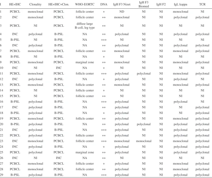
I HE+IHC Clonality HE+IHC+Clon. WHO-EORTC DNA IgH F3 Nizet IgH F3
Biomed IgH F2 IgL kappa TCR
1 PCBCL monoclonal PCBCL follicle center + ND NI NI monoclonal NI
2 INC monoclonal PCBCL follicle center ++ monoclonal NI NI polyclonal polyclonal
3 PCBCL NI PCBCL diffuse large
B-cell, leg type ++ NI NI NI NI NI
4 INC polyclonal B-PSL NA ++ polyclonal NI NI polyclonal polyclonal
5 B-PSL NI B-PSL NA +++ NI NI NI NI NI
6 INC polyclonal B-PSL NA ++ polyclonal NI NI polyclonal polyclonal
7 PCBCL monoclonal PCBCL follicle center ++ monoclonal NI NI monoclonal polyclonal
8 B-PSL NI B-PSL NA + NI NI NI NI NI
9 PCBCL monoclonal PCBCL marginal zone ++ monoclonal NI NI monoclonal polyclonal
10 INC NI INC NA + NI NI NI NI NI
11 PCBCL monoclonal PCBCL follicle center +++ polyclonal polyclonal NI monoclonal polyclonal
12 INC polyclonal B-PSL NA + polyclonal NI NI polyclonal NI
13 PCBCL monoclonal PCBCL follicle center ++ monoclonal NI NI monoclonal polyclonal
14 PCBCL NI PCBCL follicle center + NI NI NI NI NI
15 PCBCL NI PCBCL follicle center ++ NI NI NI NI NI
16 B-PSL polyclonal B-PSL NA +++ polyclonal NI NI polyclonal NI
17 INC polyclonal B-PSL NA ++ polyclonal NI NI NI polyclonal
18 B-PSL polyclonal B-PSL NA + polyclonal NI NI NI polyclonal
19 PCBCL monoclonal PCBCL follicle center ++ polyclonal NI NI monoclonal polyclonal
20 B-PSL polyclonal B-PSL NA ++ polyclonal polyclonal NI polyclonal polyclonal
21 INC polyclonal B-PSL NA +++ polyclonal NI NI polyclonal polyclonal
22 PCBCL polyclonal PCBCL follicle center ++ polyclonal NI NI polyclonal polyclonal
23 INC monoclonal PCBCL follicle center +++ monoclonal monoclonal NI monoclonal polyclonal
24 INC polyclonal B-PSL NA + polyclonal NI NI polyclonal polyclonal
25 PCBCL polyclonal PCBCL marginal zone ++ polyclonal NI NI polyclonal polyclonal
26 INC NI INC NA ++ NI NI NI NI NI
27 PCBCL monoclonal PCBCL follicle center + polyclonal NI NI monoclonal polyclonal
28 PCBCL monoclonal PCBCL follicle center ++ polyclonal NI NI monoclonal polyclonal
29 B-PSL polyclonal B-PSL NA +++ polyclonal NI NI polyclonal polyclonal
I: identiication number of the case; HE: hematoxylin-eosin; IHC: immunohistochemistry; Clon: Clonality; PCBCL: primary cutaneous B-cell lympho-ma; INC: inconclusive; NI: non-informative; NA: not applicable; B-PSL: B-cell pseudolympholympho-ma; DNA: quality control of the DNA in each sample; (-) absence of DNA in the sample; (+) presence of DNA with 100 bp; (++) presence of DNA with 100 and 200 bp; (+++) presence of DNA with 100, 200 and 300 bp; TCR: T-cell receptor; NR: number of reactions.
Table 4 - Summary of clonality analysis results of the 29 cases with B-cell lymphoproliferative processes
HE+IHC Samples Monoclonal Polyclonal Non-informative
PCBCL 13 8 2 3
B-PSL 6 0 4 2
INC 10 2 6 2
Analysis of clonality through investigation of IgL-K gene rearrangements yielded results in 20 cases. Monoclonality was detected in 9 cases (8 PCBCL and 1 considered inconclusive at HE and IHC) and polyclonality in 11 cases (2 PCBCL, 3 B-PSL and 6 considered inconclusive at HE and IHC) (Tables 3 and 4).
Among the 10 cases considered inconclusive by morphology and IHC, monoclonality was demonstrated in two cases (20%) by investigation of IgH and IgL kappa gene rearrangements (Tables 3 and 4). Despite the small number of PCBCL cases, it was possible to observe monoclonality in 7 out of 10 cases that had been classiied as primary cutaneous follicle center lymphoma and in 1 out of 2 cases that had been classified as primary cutaneous marginal zone B-cell lymphoma, according to the WHO-EORTC classiication. It was not possible to deine clonality in the single case of cutaneous diffuse large B-cell lymphoma, leg-type, by any of the protocols used in the present report.
The differential diagnosis between PCBCL and B-PSL is relevant for an appropriate follow up and treatment of patients with B-cell lymphoproliferative processes. However, the differentiation between such entities is complex and challenging.24 Since, to date, there is no “gold-standard” method that could be autonomously performed for such differentiation, this present study evaluated the diagnostic contribution of clonality analysis by investigating IgH and IgL clonality as an ancillary method to diagnose PCBCL.
The importance of the histological and immuno-histochemical evaluation in the determination of PCBCL is incontestable, even though many cases may remain without diagnosis regardless the use of several immunohistochemical markers.25,26 In our study, 10 cases were considered inconclusive by histology and immunohistochemistry, which demonstrates the necessity of additional ancillary diagnostic methods. New markers such as IRTA-1 and MUM 1, tissue microarrays, chromosomal translocation analysis and luorescence in situ hybridization (FISH) have been used to
increase the diagnostic accuracy of PCBCL.27-33
The analysis of clonality by PCR-based tests has played an important diagnostic role in PCBCL, but appropriate protocols must be selected and optimized to ensure the improved diagnostic sensitivity of these techniques. DNA quality in the samples is of crucial importance and skin biopsies are notoriously poor in this respect. The process of tissue ixation in formalin and parafin embedding variably damages the DNA by fragmentation or other mechanisms, thus impairing the sensitivity of the test and making it dificult to amplify relatively large fragments.14,20,34 These observations could explain the low to moderate quality of DNA found in our samples and the dificulty in amplifying the DNA, especially using the IgH FR2 protocol. This
protocol ampliies a product of 250-295 bp, requiring a non-fragmented DNA sample. The same observation was also highlighted by other groups.14,19,35
In the present study, we noticed higher sensitivity of the IgH FR3 Nizet protocol compared to others (IgH FR3 Biomed and Ig FR2 Biomed) when the rearrangement of IgH genes was investigated. This protocol corroborated the diagnosis of three of the PCBCL and four of the B-PSL cases. The present inding can also be explained by the smaller size of the target DNA. The size of the target product in the IgH FR3 Nizet is of 80-120 bp, compatible with the DNA size present in the samples, while IgH FR3 Biomed and FR2 Biomed amplify larger nucleotide fragments (100-170 bp and 250-295 bp), requiring well-preserved DNA.
The main clinical contribution of our molecular investigation was the classification of two out of ten cases that had been previously considered inconclusive by histological and immunohistochemical methods. After determination of monoclonality by IgH gene rearrangement, these cases could be diagnosed as PCBCL. Although “false-positive” clonality results in benign cutaneous lesions are described in the literature, those two cases presented infiltrated lesions on the back that were clinically very suggestive of PCBCL (cases 2 and 23). Thus, the clonality analysis is of extreme relevance, mainly when analyzed in the context of the, histological and immunohistochemical data 36
Of subsidiary relevance, but still important, investigation of the Ig kappa gene rearrangements supported the previous diagnosis of eight of the PCBCL and three of the B-PSL cases. Amongst all cases shown to be monoclonal in the present study, this result was obtained only by Ig kappa gene rearrangement in ive cases. It was striking that those ive cases had a polyclonal result using IgH protocols. This inding indicates the need for analysis of both Ig heavy and Ig kappa genes.11,19
Based upon these findings, in our experience, the two most eficient molecular methods in this study, IgH FR3-trad and Ig kappa Biomed protocols, for analysis of formalin–ixed, parafin-embedded samples could facilitate
a differential diagnosis between PCBCL and B-PSL. Finally, interpretation of the clonality analysis in a clinical, histological and immunohistochemical context can be critical to the diagnosis of PCBCL.
REFERENCES
1. Burg G, Kerl H, Przybilla B, Braun-Falco O. Some statistical data, diagnosis, and staging of cutaneous B-cell lymphomas. J Dermatol Surg Oncol. 1984;10:256-62.
2. Cerroni L. Lymphoproliferative lesions of the skin. J Clin Pathol. 2006;59:813-26.
3. Cerroni L, Kerl H. Diagnostic immunohistology: cutaneous lymphomas and pseudolymphomas. Semin Cutan Med Surg. 1999;18:64-70. 4. Holm N, Flaig MJ, Yazdi AS, Sander CA. The value of molecular
analysis by PCR in the diagnosis of cutaneous lymphocytic iniltrates. J Cutan Pathol. 2002;29:447-52.
5. Rijlaarsdam U, Bakels V, van Oostveen JW, Gordijn RJ, Geerts ML, Meijer CJ, et al. Demonstration of clonal immunoglobulin gene rearrangements in cutaneous B-cell lymphomas and pseudo-B-cell lymphomas: differential diagnostic and pathogenetic aspects. J Invest Dermatol. 1992;99:749-54.
6. Willemze R, Beljaards RC, Meijer CJ, Rijlaarsdam JR. Classiication of primary cutaneous lymphomas. Historical overview and perspectives. Dermatology. 1994;189 Suppl 2:8-15.
7. Bogle MA, Riddle CC, Triana EM, Jones D, Duvic M. Primary cutaneous B-cell lymphoma. J Am Acad Dermatol. 2005;53:479-84.
8. Swerdlow SH, Campo E, Harris NL, Jaffe ES, Pileri SA, Stein H, Thiele J, Vardiman JW. Classiication of tumours, pathology & genetics, tumours of hematopoietic and lymphoid tissues. 4th Edition ed. Lyon: IARC; 2008.
9. Bagg A, Braziel RM, Arber DA, Bijwaard KE, Chu AY. Immunoglobulin heavy chain gene analysis in lymphomas: a multi-center study demonstrating the heterogeneity of performance of polymerase chain reaction assays. J Mol Diagn. 2002;4:81-9.
10. Magro C, Crowson AN, Porcu P, Nuovo GJ. Automated kappa and lambda light chain mRNA expression for the assessment of B-cell clonality in cutaneous B-cell iniltrates: its utility and diagnostic application. J Cutan Pathol. 2003;30:504-11.
11. Diss TC, Liu HX, Du MQ, Isaacson PG. Improvements to B cell clonality analysis using PCR ampliication of immunoglobulin light chain genes. Mol Pathol. 2002;55:98-101.
12. Hughes J, Weston S, Bennetts B, Prasad M, Angulo R, Jaworskit R, et al. The application of a PCR technique for the detection of immunoglobulin heavy chain gene rearrangements in fresh or parafin-embedded skin tissue. Pathology. 2001;33:222-5.
13. Nikiforova MN, Hsi ED, Braziel RM, Gulley ML, Leonard DG, Nowak JA, et al. Detection of clonal IGH gene rearrangements: summary of molecular oncology surveys of the College of American Pathologists. Arch Pathol Lab Med. 2007;131:185-9.
14. van Dongen JJ, Langerak AW, Bruggemann M, Evans PA, Hummel M, Lavender FL, et al. Design and standardization of PCR primers and protocols for detection of clonal immunoglobulin and T-cell receptor gene recombinations in suspect lymphoproliferations: report of the BIOMED-2 Concerted Action BMH4-CT98-3936. Leukemia. 2003;17:2257-317.
15. Diss TC, Ashton-Key M, Pan LX, Isaacson PG. Clonality analysis of B-cell lymphomas. Hum Pathol. 1995;26:1046.
16. Korsmeyer SJ, Hieter PA, Ravetch JV, Poplack DG, Waldmann TA, Leder P. Developmental hierarchy of immunoglobulin gene rearrangements in human leukemic pre-B-cells. Proc Natl Acad Sci U S A. 1981;78:7096-100.
17. van der Burg M, Tumkaya T, Boerma M, de Bruin-Versteeg S, Langerak AW, van Dongen JJ. Ordered recombination of immunoglobulin light chain genes occurs at the IGK locus but seems less strict at the IGL locus. Blood. 2001;97:1001-8.
18. Diss TC, Pan L, Peng H, Wotherspoon AC, Isaacson PG. Sources of DNA for detecting B cell monoclonality using PCR. J Clin Pathol. 1994;47:493-6.
19. Nihal M, Mikkola D, Wood GS. Detection of clonally restricted immunoglobulin heavy chain gene rearrangements in normal and lesional skin: analysis of the B cell component of the skin-associated lymphoid tissue and implications for the molecular diagnosis of cutaneous B cell lymphomas. J Mol Diagn. 2000;2:5-10.
20. Gong JZ, Zheng S, Chiarle R, De Wolf-Peeters C, Palestro G, Frizzera G, et al. Detection of immunoglobulin kappa light chain rearrangements by polymerase chain reaction. An improved method for detecting clonal B-cell lymphoproliferative disorders. Am J Pathol. 1999;155:355-63. 21. Amara K, Trimeche M, Ziadi S, Sriha B, Mokni M, Korbi S. PCR-based
clonality analysis of B-cell lymphomas in parafin-embedded tissues: diagnostic value of immunoglobulin kappa and lambda light chain gene rearrangement investigation. Pathol Res Pract. 2006;202:425-31. 22. Willemze R, Jaffe ES, Burg G, Cerroni L, Berti E, Swerdlow SH, et al.
WHO-EORTC classiication for cutaneous lymphomas. Blood. 2005 15;105:3768-85.
23. Nizet Y, Van Daele S, Lewalle P, Vaerman JL, Philippe M, Vermylen C, et al. Long-term follow-up of residual disease in acute lymphoblastic leukemia patients in complete remission using clonogeneic IgH probes and the polymerase chain reaction. Blood. 1993;82:1618-25. 24. Pimpinelli N. New aspects in the biology of cutaneous B-cell
lymphomas. J Cutan Pathol. 2006;33 Suppl 1:6-9.
26. Kodama K, Massone C, Chott A, Metze D, Kerl H, Cerroni L. Primary cutaneous large B-cell lymphomas: clinicopathologic features, classiication, and prognostic factors in a large series of patients. Blood. 2005;106:2491-7.
27. Ashton-Key M, Diss TC, Isaacson PG, Smith ME. A comparative study of the value of immunohistochemistry and the polymerase chain reaction in the diagnosis of follicular lymphoma. Histopathology. 1995;27:501-8. 28. Child FJ, Scarisbrick JJ, Calonje E, Orchard G, Russell-Jones R, Whittaker SJ. Inactivation of tumor suppressor genes p15(INK4b) and p16(INK4a) in primary cutaneous B cell lymphoma. J Invest Dermatol. 2002;118:941-8.
29. Hallermann C, Niermann C, Fischer RJ, Schulze HJ. New prognostic relevant factors in primary cutaneous diffuse large B-cell lymphomas. J Am Acad Dermatol. 2007;56:588-97.
30. Hsi ED, Mirza I, Gascoyne RD. Absence of t(14,18) chromosomal translocation in primary cutaneous B-cell lymphoma. Br J Dermatol. 2002;146:1110-1; author reply 1-2.
31. Streubel B, Scheucher B, Valencak J, Huber D, Petzelbauer P, Trautinger F, et al. Molecular cytogenetic evidence of t(14;18)(IGH;BCL2) in a substantial proportion of primary cutaneous follicle center lymphomas. Am J Surg Pathol. 2006;30:529-36.
32. Sundram U, Kim Y, Mraz-Gernhard S, Hoppe R, Natkunam Y, Kohler S. Expression of the bcl-6 and MUM1/IRF4 proteins correlate with overall and disease-speciic survival in patients with primary cutaneous large B-cell lymphoma: a tissue microarray study. J Cutan Pathol. 2005;32:227-34.
33. Wiesner T, Streubel B, Huber D, Kerl H, Chott A, Cerroni L. Genetic aberrations in primary cutaneous large B-cell lymphoma: a luorescence in situ hybridization study of 25 cases. Am J Surg Pathol. 2005;29:666-73.
34. Zsikla V, Baumann M, Cathomas G. Effect of buffered formalin on ampliication of DNA from parafin wax embedded small biopsies using real-time PCR. J Clin Pathol. 2004;57:654-6.
35. Pai RK, Chakerian AE, Binder JM, Amin M, Viswanatha DS. B-cell clonality determination using an immunoglobulin kappa light chain polymerase chain reaction method. J Mol Diagn. 2005;7:300-7. 36. Flaig MJ, Schuhmann K, Sander CA. Impact of molecular analysis in
the diagnosis of cutaneous lymphoid iniltrates. Semin Cutan Med Surg. 2000;19:87-90.