Case Report
Keywords
Amiodarone; thyrotoxicosis; thyroiditis.
Amiodarone-induced thyroid dysfunction has been reported to affect 2-24% of users. Despite the easy management of amiodarone-induced hypothyroidism, the development of thyrotoxicosis leads to a difficult approach in most cases.
The aim of this study is to describe three different cases of patients with amiodarone-induced thyrotoxicosis and discuss the clinical and laboratorial aspects, and the different approaches to them.
It is essential to carefully evaluate patients before and during amiodarone therapy, since the prompt diagnosis and treatment of this condition is essential in patients with high cardiovascular risk.
Amiodarone and Thyrotoxicosis: Case Reports
Ana Beatriz Winter Tavares, Simone Kalil de Paula, Mario Vaisman, Patrícia de Fátima dos Santos Teixeira
Serviço de Endocrinologia do Hospital Universitário Clementino Fraga Filho - UFRJ - Rio de Janeiro, RJ - Brazil
Mailing address: Ana Beatriz Winter Tavares •
Rua Barão de Lucena, 135/202 - Botafogo - 22260-020 - Rio de Janeiro, RJ - Brazil
E-mail: [email protected]
Manuscript received December 13, 2008; revised manuscript received June 09, 2009; accepte 23/06/09.
Levothyroxine (LT4) replacement is the treatment of choice for hypothyroidism and amiodarone withdrawal is not always necessary2.
Amiodarone may also induce thyrotoxicosis, more commonly found in iodine deficient areas3,4. It may occur 4 months to 3 years after initiating therapy or after drug withdrawal and is not related to cumulative drug dosage3.
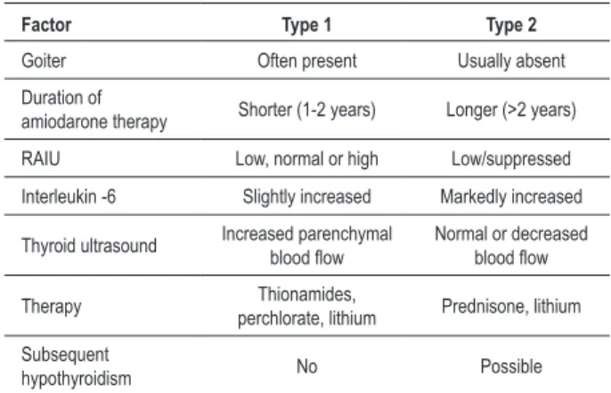
There are two types of amiodarone-induced thyrotoxicosis (AIT). Type 1 AIT is defined as iodine-induced hyperthyroidism, developing in individuals with underlying thyroid disease or positive circulating thyroid peroxidase antibodies (TPOAb) and is due to increased synthesis and release of thyroid hormone (Jod-Basedow effect). Type 2 AIT is a drug-induced destructive thyroiditis that occurs in individuals with no underlying thyroid disease and is more frequent in iodine sufficient areas3 - table 2. However, distinguishing one type from the other may be troublesome, and some cases may in fact represent mixed forms, where individuals may have characteristics of both AIT subtypes. Because of this heterogeneity, AIT poses a difficult diagnostic and therapeutic challenge2,5.
Treatment of type 1 AIT includes the use of antithyroid drugs (methimazole or propylthiouracil) and discontinuation of amiodarone is necessary, if possible2,3. In type 2 AIT, treatment must be done with glucocorticoids (prednisone)2,3. Cases of mixed AIT may not respond to monotherapy with either antithyroid drugs or glucocorticoids but may respond to both agents together. Another strategy could be starting all patients on both antithyroid drug and prednisone daily. If there is a very rapid response (i.e., within 1-2 weeks), then the patient is very likely to have type 21 AIT.
The aim of this study is to describe three different clinical cases of patients with amiodarone-induced thyrotoxicosis and discuss the clinical and laboratorial aspects, as well as different approaches to them.
Case reports
Patient 1
A sixty year-old man developed subclinical hyperthyroidism in the fourth month of amiodarone use, which was prescribed for ventricular tachycardia and electrical instability due to Chagas heart disease. There were no symptoms of thyroid dysfunction, no goiter. Serum TSH level was <0.01 mUI/ ml (reference range - RR: 0.4-4.0 mUI/ml), negative thyroperoxidase antibody (TPOAb) and normal serum values of FT4 (0.8-1.9 ng/dl). Iodine uptake (RAIU) in 24 hours was 1%. A reduction in the amiodarone dosage was performed and the following serum TSH was 0.25 mUI/ml. After twelve months of follow-up, serum TPOAb became positive, and
Introduction
Amiodarone is a Type-III anti-arrhythmic agent that blocks myocardial potassium channels and has some beta-blocking properties. Each molecule of amiodarone has a significant structural resemblance to the thyroid hormones and contains two iodine atoms, which constitute 37.5% of its mass. Hence, a patient taking a 200 mg daily dose of amiodarone leads an amount of free iodine into circulation that is 20-40 times higher than the daily iodine intake in the general population1.
This excessive load of iodine generates important adjustments in hormone metabolism and physiological alterations in serum thyroid function tests - table 11.
Amiodarone has a half-life of approximately 100 days, mainly due to its storage in adipose tissue, and its toxic effects may persist or even occur after its discontinuation1.
Thyroid dysfunction has been reported to affect 2-24% of amiodarone users2. Amiodarone-induced hypothyroidism occurs typically between 6-12 months of treatment3. It may be a consequence of Wolff-Chaikoff effect with blockage of hormone secretion or a consequence of chronic autoimmune thyroiditis induced by iodine excess2.
Case Report
Tavares et al Amiodarone and thyrotoxicosis
Arq Bras Cardiol 2010; 95(5): e122-e124
Table 1 - Physiological effects of amiodarone on thyroid function tests
Serum hormone level
Acute effects (< 3 months)
Chronic effects (> 3 months)
FT4* and total T4 ↑ 50% ↑ 20-40% of baseline values
T3 ↓ 15-20% remains in low-normal
range
rT3** ↑ 200% ↑ 150%
TSH ↑ 20-50%, transient
(however < 20mUI/ml) normal
Adapted from ref 1.* FT4 - free thyroxine; ** rT3 - reverse triiodothyronine.
Table 2 - Comparison of AIT type 1 and 2
Factor Type 1 Type 2
Goiter Often present Usually absent
Duration of
amiodarone therapy Shorter (1-2 years) Longer (>2 years)
RAIU Low, normal or high Low/suppressed
Interleukin -6 Slightly increased Markedly increased
Thyroid ultrasound Increased parenchymal blood low Normal or decreased blood low
Therapy Thionamides,
perchlorate, lithium Prednisone, lithium
Subsequent
hypothyroidism No Possible
Adapted from refs 2, 3.
patient developed thyrotoxicosis, with FT4=1.9 ng/dl and TSH=0.20 mUI/ml. Despite an initial prescription of 40 mg/ day of prednisone for 2 months, the patient still presented thyrotoxicosis (FT4= 1.9 ng/dl and TSH=0.069 mUI/ml). Propylthiouracil was added to the prescription and patient restored euthyroidism after 5 months (FT4=1.58 ng/dl), still in use of amiodarone. Afterwards, cardiologists were able to take out amiodarone and the patient maintained euthyroidism. After that, it was also possible to withdraw prednisone and propylthiouracil from prescription.
Patient 2
A fifty-four-year-old woman developed thyrotoxicosis in the fifth year of amiodarone use (FT4=2.1 ng/dl; TSH=0.232 mUI/ml; negative TPOAb). Amiodarone was prescribed for ventricular tachycardia and electrical instability due to Chagas heart disease. Thyroid was enlarged (approximately 50 g) and RAIU in 24 hours <1%. Amiodarone was maintained because of the risk of sudden death. The patient received methimazole (30 mg/day) and prednisone (20 mg/day). Euthyroidism was restored after 3 months and prednisone was suspended after that. The patient needed 5mg/day of methimazole to maintain euthyroidism for two years, when she died from Chagas heart disease complications, still in use of amiodarone.
Patient 3
A sixty-five-year-old man developed thyrotoxicosis after three months of amiodarone use, prescribed for atrial fibrillation (FT4=2.4 ng/dl; TSH=0.083 mUI/ml; negative TPO-Ab). Thyroid was enlarged, with a palpable nodule. Amiodarone was then suspended. Thyroid ultrasound disclosed a nodule expanding to both lobes, measuring 6.1 cm, with peripheral and intrinsic flow. The first RAIU was <1%. However, after 12 months of amiodarone withdrawal, the thyroid scintillography showed a predominant uptake in the left lobe, with the aspect of multinodular goiter and RAIU of 7.63% (normal). Cytopathology of the nodule demonstrated benign nodular goiter. Euthyroidism was achieved 10 months after the suspension of amiodarone, without any complementary treatment.
Discussion
We describe three cases of thyrotoxicosis in patients taking amiodarone: in the first two cases the severity of diseases and the possibly consequent morbidity associated to the thyroid hormone excess justify the initial approach with corticosteroids and antithyroid drugs. The combined treatment must be the initial choice in patients with severe cardiovascular disease. Furthermore, they have characteristics that may misdiagnosis the specific type of AIT, which is a common difficulty seen in daily clinical practice.
In case 1, some findings are suggestive of type 1 AIT: amelioration of thyrotoxicosis only when propylthiouracil was added to prescription, RAIU of 1% (where we expected a null uptake); and also suggestive of type 2: development of circulating antibodies during the evaluation, still in use of amiodarone, defining an AIT with mixed pattern.
Case 2 may also present a mixed AIT, however the goiter and the long-time use of methimazole suggest type 1 predominance, despite RAIU < 1%. Since the RAIU interpretation must be difficult in these patients, the Doppler ultrasound would be appropriate (table 2).
Case 3 demonstrates a patient with a big thyroid nodule, which might develop autonomous function, precipitated by the use of amiodarone; due to Jod-Basedow phenomenon. This phenomenon is more common in patients with previous thyroid diseases, especially nodular disease and in iodine deficient areas of the world.
Even knowing the effects of amiodarone in the thyroid gland, many physicians do not proceed to an adequate evaluation. Our group detected that 33.9% of patients taking amiodarone had thyroid dysfunction (1.8 and 3.6% had clinical and subclinical hyperthyroidism, respectively, and 10.7 and 17.9% had clinical and subclinical hypothyroidism, respectively). However, only 49.2% of the cardiologists used to follow thyroid function frequently6.
It is essential to carefully evaluate patients before and during amiodarone therapy. Careful thyroid gland examination and baseline TSH, FT4 and TPOAb determinations are recommended. Serum TSH and FT4 should be measured after 3 months of therapy and semiannually after that 6 months7. A deterioration of cardiac function implies the
Case Report
Tavares et al
Amiodarone and thyrotoxicosis
Arq Bras Cardiol 2010; 95(5): e122-e124
References
1. Basaria S, Cooper DS. Amiodarone and the thyroid. Am J Med. 2005; 118 (7): 706-14.
2. Martino E, Bartalena L, Bogazzi F, Braverman LE. The effects of amiodarone on the thyroid. Endocr Rev. 2001; 22 (2): 240-54.
3. Pavan R, Jesus AMX, Maciel LMZ. A amiodarona e a tireóide. Arq Bras Endocrinol Metab. 2004; 48 (1): 176-81.
4. Schaan BD, Cunha CP, Francisconi A, Zotiis B, Brum G, Bruch RS, et al. Amiodarone-induced thyroid dysfunction in a tertiary center in South Brazil. Arq Bras Endocrinol Metab. 2005; 49 (6): 916-22.
5. Bartalena L, Bogazzi F, Martino E. Amiodarone-induced thyrotoxicosis: a difficult diagnostic and therapeutic challenge. Clin Endocrinol. 2002; 56 (1): 23-4.
6. Fuks AG, Vaisman M, Buescu A. Disfunção tireoidiano e conduta dos cardiologistas em pacientes usando amiodarona. Arq Bras Cardiol. 2004; 82 (6): 523-7.
7. Goldschlager N, Epstein AE, Naccarelli G, Olshansky B, Singh B. Practical guidelines for clinicians who treat patients with amiodarone. Practice Guidelines Subcommittee, North American Society of Pacing and Electrophysiology. Arch Intern Med. 2000; 160 (12): 1741-8.
suspicion of associated thyroid dysfunction, even in the absence of classic symptoms.
Potential Conflict of Interest
No potential conflict of interest relevant to this article was reported.
Sources of Funding
There were no external funding sources for this study.
Study Association
This study is not associated with any post-graduation program.